Table of Contents
Definition / general | Case reports | Gross description | Microscopic (histologic) description | Microscopic (histologic) images | Positive stains | Negative stains | Electron microscopy description | Molecular / cytogenetics descriptionCite this page: Jain D. Conjunctival oncocytoma. PathologyOutlines.com website. https://www.pathologyoutlines.com/topic/eyeoncocytomaconjunc.html. Accessed December 21st, 2024.
Definition / general
- Usually caruncle (Arch Ophthalmol 1977;95:474, Ophthal Plast Reconstr Surg 2012;28:14)
- Often elderly (mean age 73), more common in women
- Proposed origin from lacrimal and accessory lacrimal glands based on cytokeratin profile (Acta Ophthalmol 2011;89:263)
Case reports
- 51 year old woman with tumor of caruncle (Int Ophthalmol 2004;25:321)
- 60 year old woman with tumor of caruncle (Klin Monatsbl Augenheilkd 2005;222:733)
- 72 year old woman with tumor of bulbar conjunctiva (Klin Monatsbl Augenheilkd 1996;209:176)
- 75 year old woman with tumor of caruncle (Indian J Ophthalmol 2002;50:60)
Gross description
- Red-orange or yellow-tan mass
Microscopic (histologic) description
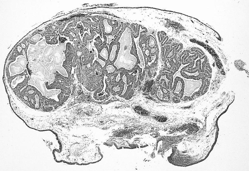
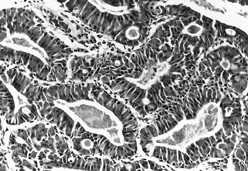
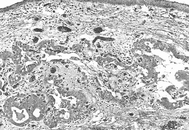
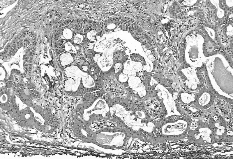
- Solid nests and cords of polyhedral cells with abundant, finely granular acidophilic cytoplasm and round / oval paracentral nuclei, usually with one prominent nucleolus
- May have microcystic areas with occasional goblet cells (Am J Dermatopathol 2007;29:279), may have malignant histology and behavior
Microscopic (histologic) images
AFIP images
Images hosted on other servers:
Negative stains
Electron microscopy description
- Large numbers of mitochondria (Br J Ophthalmol 1980;64:935)
Molecular / cytogenetics description
- Mitochondrial DNA mutations (Arch Ophthalmol 2011;129:664)


