
Eye
Globe
Phthisis bulbi
Author: Nat Pernick, M.D.

Last author update: 1 February 2014
Last staff update: 28 December 2020
Copyright: 2004-2024, PathologyOutlines.com, Inc.
PubMed Search: Phthisis bulbi [title] globe
Table of Contents
Definition / general | Microscopic (histologic) description | Microscopic (histologic) imagesCite this page: Pernick N. Phthisis bulbi. PathologyOutlines.com website. https://www.pathologyoutlines.com/topic/eyeglobephthisisbulbi.html. Accessed April 19th, 2024.
Definition / general
- Degenerative change of globe involving all tissues
- Usually takes several years to develop
- Often due to accidental or post-surgical trauma
- Also found in eyes removed for blindness, pain, glaucoma, inflammation
- Due to reduced production of aqueous humor causing reduced intraocular pressure (hypotony) and shrinkage of globe
- Also due to organization of inflammatory exudate
- Degenerative changes and degree of shrinkage are variable in different tissues
- May be calcification and ossification with bone marrow
- Note: must decalcify globe
Microscopic (histologic) description
- Typically disorganization of intraocular contents, opaque media, corneal scars, exudate in anterior and posterior chambers, advanced cataracts, destruction of vitreous, scleral thickening, cyclitic membrane extends from one ciliary body behind the lens to the other ciliary body, complete detachment of retina; also ossification or bone formation
- Usually histology does not disclose initial condition leading to phthisis bulbi
- Occasionally occult melanoma or lymphoma is found
Microscopic (histologic) images
AFIP images
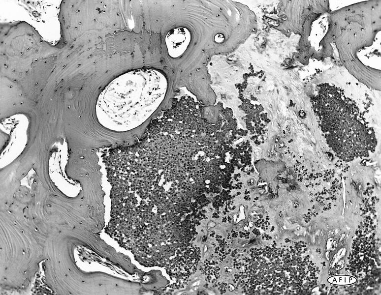
Regressed retinoblastoma
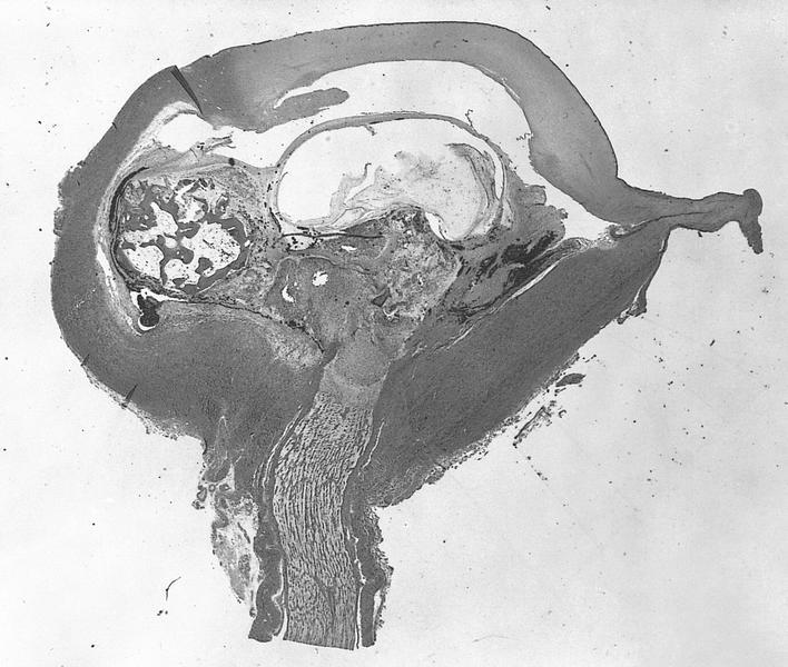
Disorganized intraocular contents with ossification and calcification
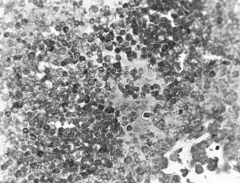
Fossilized tumor cells
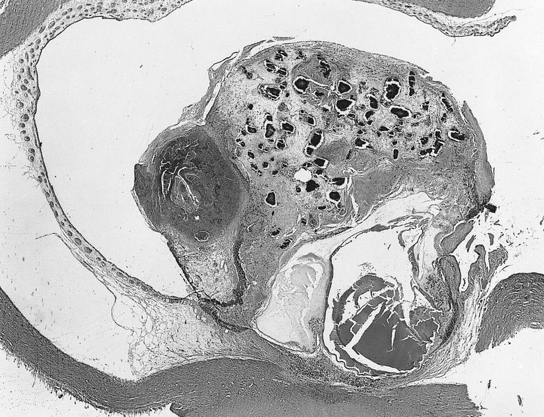
Massive retinal gliosis
