
Eye
General
Anatomy & histology-cornea
Author: Nat Pernick, M.D.

Last author update: 1 October 2013
Last staff update: 17 April 2024
Copyright: 2004-2024, PathologyOutlines.com, Inc.
PubMed Search: Cornea anatomy and histology
Table of Contents
Definition / general | Hasall-Henle bodies (warts) | Hyperopia | Laser assisted in situ keratomileusis (LASIK) | Limbus | Myopia | Schlemm canal and trabecular meshwork | Vasculature | Microscopic (histologic) images | Drawings | Positive stains | Negative stains | Electron microscopy description | Additional referencesCite this page: Pernick N. Anatomy & histology-cornea. PathologyOutlines.com website. https://www.pathologyoutlines.com/topic/eyecorneaanatomy.html. Accessed April 23rd, 2024.
Definition / general
- Wider than tall (11.7 mm horizontally vs. 10.6 mm vertically)
- Thickness varies from 0.5 mm (central) to 0.67 mm (peripheral)
- Cornea and overlying tear film are major refractive surface of eye, not the lens
- 6 distinct layers (outside to inside):
- Outer epithelium: stratified squamous, nonkeratinized, 5 layers thick centrally, thicker peripherally, polygonal at basal layer but flatten as they approach surface; basal cells may have mitotic figures; Langerhans cells are CD1a+; note: layers often rubbed off while grossing specimen
- Epithelial basal lamina (basement membrane): highlighted with PAS stain
- Bowman layer: most anterior stroma, acellular, 8 - 14 microns thick, not a true basement membrane, composed of randomly oriented delicate collagen fibers, does not regenerate
- Stroma: also called substantia propria, no blood vessels or lymphatics, 90% of cornea's thickness, contains regularly spaced collagen fibrils; normally separated by glycoprotein and mucoprotein which makes cornea transparent; normally see stromal lamellae separated by clefts, a processing artifact, absence of clefts is caused by stroma edema (causes corneal clouding), due to damage of endothelium
- Descemet [pronounced DEZMET] membrane: a true basal lamina produced by underlying corneal endothelial cells; 3 - 4 microns at birth, 10 - 12 microns in adults; does not regenerate; site of copper deposition in Kayser-Fleisher ring of Wilson disease
- "Endothelium": single layer of very flat cells, does not regenerate, functions as pump to keeps cornea dehydrated and transparent
- Neural crest origin (S100+); does not line blood vessels or lymphatic spaces; directly contacts aqueous humor of anterior chamber, often rubbed off while grossing specimen
Hasall-Henle bodies (warts)
- Focal excrescences that form on peripheral Descemet membrane with normal aging
- Not seen in surgically excised corneal buttons because are too peripheral in location
Hyperopia
- Eye too short for its refractive power
Laser assisted in situ keratomileusis (LASIK)
- Sculpt cornea and change its refractive properties to eliminate need for glasses
Limbus
- Junction of peripheral cornea and anterior sclera, 1.5 to 2.0 mm wide
- Not a distinct anatomic site but a significant clinical landmark
- Composed of conjunctiva (epithelium and stroma), cornea and scleral stroma, episclera, Tenon capsule (fibrous tissue that covers the globe)
- Descemet membrane terminates at limbus and gives rise to Schwalbe ring
- 15% have prominent area of thickening at this site
- Contains trabecular meshwork and Schlemm canal
- Site of incisions for surgery on anterior eye
- Restricts deeper extension of superficial tumors
Myopia
- Eye too long for its refractive power
Schlemm canal and trabecular meshwork
- Schlemm canal
- Anterior and superficial to trabecular meshwork
- Endothelial lined venous canal that completely encircles limbus
- Separated from trabecular meshwork by thin connective tissue and separate endothelial linings
- Trabecular meshwork
- With Schlemm canal, are apparatus for removal of aqueous from eye
- Collection of finely branching and delicately pigmented connective tissue bands
- Lining cells are continuous with corneal endothelium
- Posteriorly, trabecular meshwork extends to scleral connective tissue called scleral spur
Vasculature
- No blood vessels or lymphatics within cornea
- Arterial plexus is present at junction of cornea and sclera
- Is also nourished by aqueous humor of anterior chamber
Microscopic (histologic) images
Images hosted on other servers:
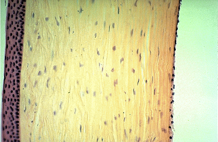
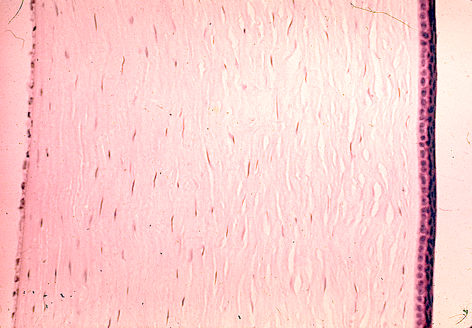
Full thickness
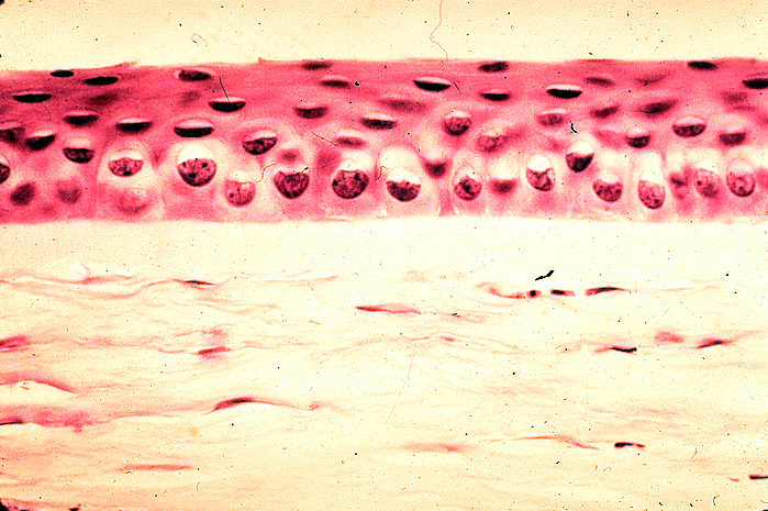
Epithelium and Bowman layer
Drawings
Images hosted on other servers:

Cornea
Positive stains
Negative stains
Electron microscopy description
- Schlemm canal endothelial cells contain giant cytoplasmic vacuoles
Additional references
