
Eye
Conjunctiva
Melanocytic tumors
Melanoma-conjunctiva
Author: Nat Pernick, M.D.

Last author update: 1 April 2014
Last staff update: 26 May 2022
Copyright: 2004-2025, PathologyOutlines.com, Inc.
PubMed Search: Melanoma conjunctiva

Table of Contents
Definition / general | Epidemiology | Prognostic factors | Clinical features | Case reports | Treatment | Clinical images | Gross description | Gross images | Whole mount images | Microscopic (histologic) description | Microscopic (histologic) images | Cytology images | Positive stains | Negative stains | Differential diagnosis | Additional referencesCite this page: Pernick N. Melanoma-conjunctiva. PathologyOutlines.com website. https://www.pathologyoutlines.com/topic/eyeconjmelanoma.html. Accessed April 3rd, 2025.
Definition / general
- #2 malignancy of conjunctiva after squamous cell carcinoma
- 2% of ocular malignancies, 5% of ocular melanomas
- Due to primary acquired melanosis, nevi (20 - 30%) or no apparent precursor lesion (18 - 25%)
- Usually fair complexioned individuals age 40+ years
- Be wary of diagnosis in children as it is very rare (J Pediatr Ophthalmol Strabismus 2007;44:277)
Epidemiology
- 0.012 cases/100K in US, 0.024/100K in Sweden, 0.052/100K in Denmark
- Rare in blacks
Prognostic factors
- Excellent if small, localized and bulbar
- Intermediate if diffuse and bulbar
- Poor prognostic factors:
- Fornix, caruncle, plica semilunaris or palpebral conjunctiva
- Tumor thickness > 4 mm
- Epithelioid cells or 5+ mitotic figures / 10 HPF (Br J Ophthalmol 1994;78:252)
- Prognosis not related to nature of initial lesion, although acquired melanosis cases are often multicentric
Clinical features
- Metastases to parotid or submandibular lymph nodes, but uncommon if primary tumor less than 1.5 cm
- May extend directly into orbit, eyelids, sinuses, anterior chamber (Graefes Arch Clin Exp Ophthalmol 2007;245:431)
- Often recurs locally
- Overall mortality 25% - 32% (J Fr Ophtalmol 1999;22:315)
Case reports
- 32 year old man with amelanotic tumor (Cutis 2006;77:377)
- 61 year old woman with prior primary acquired melanosis (University of Iown Health Care: Conjunctival Melanoma arising from PAM [Accessed 30 April 2018])
- 79 year old man with metastasis to parotid gland (Br J Ophthalmol 2003;87:1428)
Treatment
- Complete excision or radical surgery, depending on extent of disease
- Cryotherapy for margins and base
- Also topical mitomycin C
- Recommended to avoid incisional biopsy (Trans Am Ophthalmol Soc 2000;98:471)
Clinical images
AFIP images
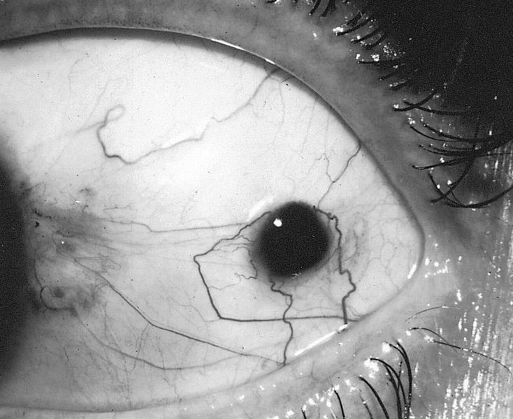
Pigmented lesion
adjacent to primary
acquired melanosis
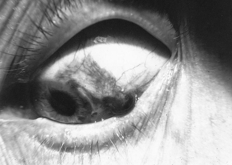
Elevated melanotic nodule
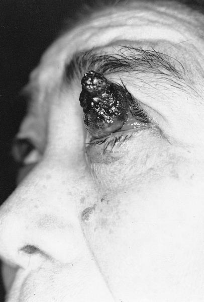
Large neglected melanoma
Gross description
- Vascular, pigmented, nodular
Gross images
AFIP images
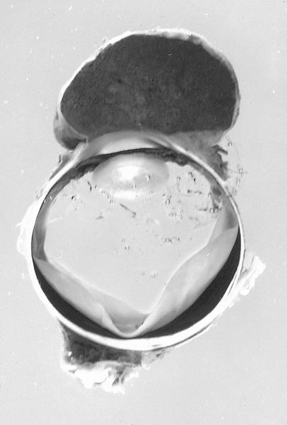
Large heavily pigmented nodule covers cornea
Whole mount images
AFIP images
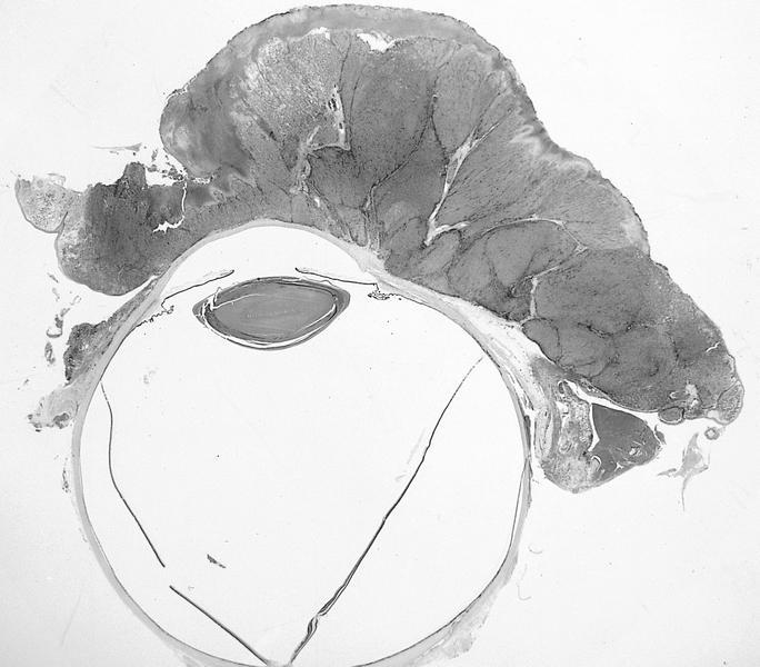
Exophytic tumor covers conjunctiva and cornea
Microscopic (histologic) description
- Invasion of atypical melanocytes into epithelial connective tissue
- Usually thin surface epithelium
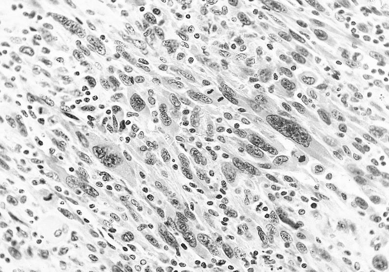
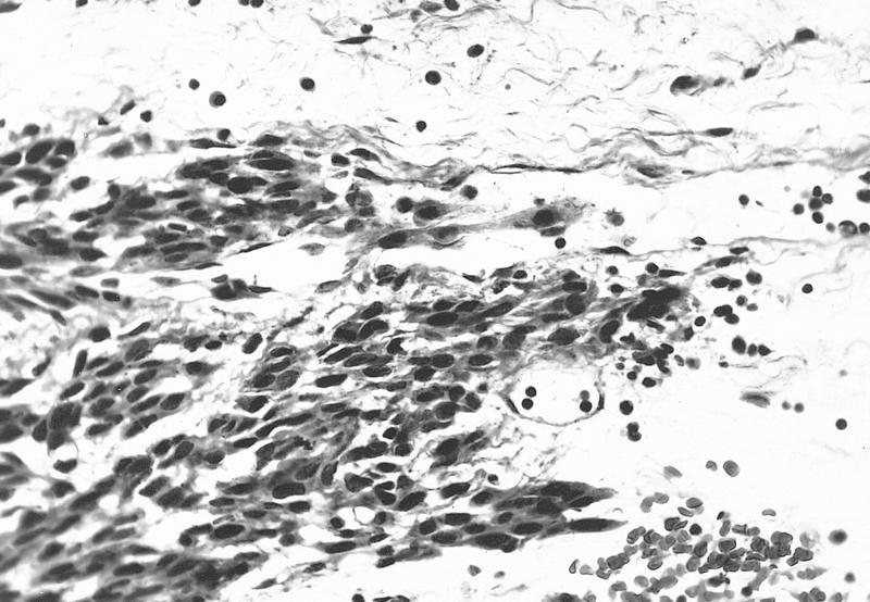
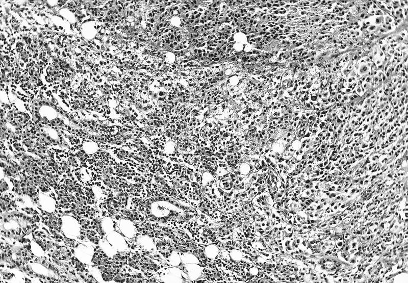
- Bizarre polygonal epithelioid cells with eosinophilic cytoplasm, large atypical nuclei, prominent eosinophilic nuclei
- Also spindle cells, smaller cells, balloon cells containing lipid
- Often lymphocytes at base or tumor margins
- Report: presence of primary acquired melanosis or nevi, presence of pagetoid spread at edge of excision, atypical intraepithelial melanocytes, nevus cells; also tumor thickness from surface of lesion to deepest margin using calibrated micrometer
Microscopic (histologic) images
AFIP images
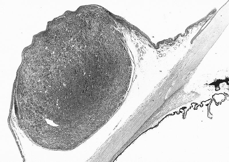
Nodular tumor at limbus
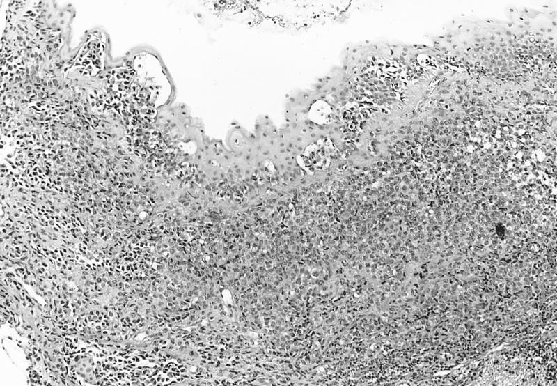
Anaplastic melanocytes within epithelial nests
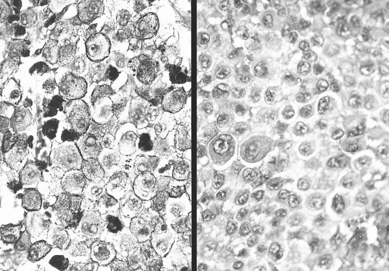
Pigmented epithelioid cells with prominent nucleoli
Pleomorphic tumor cells
Malignant spindled melanocytes
Malignant epithelioid melanocytes
Contributed by John Irlam, D.O.
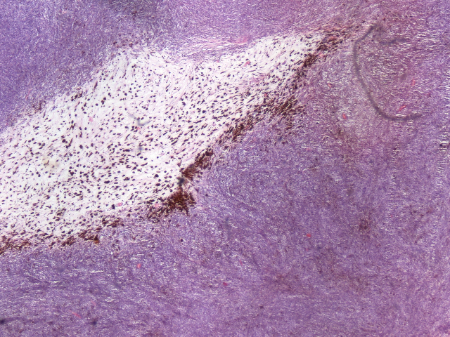
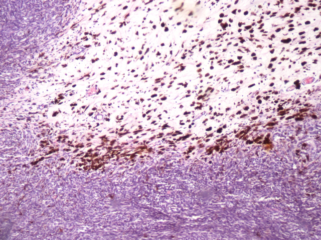
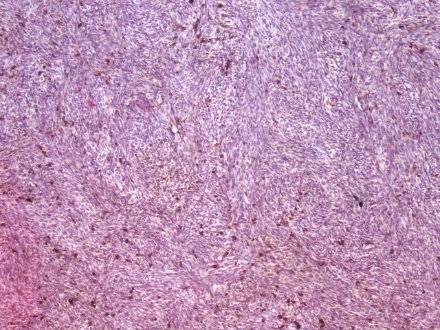
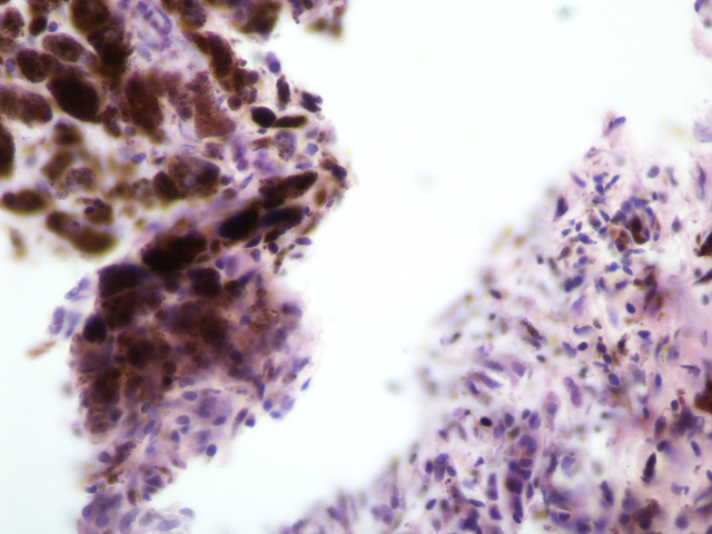
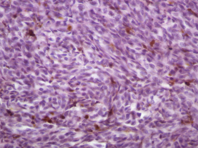
Ocular melanoma - site unspecified
Cytology images
Images hosted on other servers:

FNA of parotid metastasis
Differential diagnosis
- Extension of extraocular melanoma
- Metastatic melanoma: clinical history of melanoma, more circumscribed, no intraepithelial tumor
- Spindle cell carcinoma
