
Esophagus
Esophagitis
Radiation esophagitis
Author: Elliot Weisenberg, M.D.

Last author update: 1 October 2012
Last staff update: 17 May 2022
Copyright: 2003-2025, PathologyOutlines.com, Inc.
PubMed Search: Radiation[TI] esophagitis[TI]


Table of Contents
Definition / general | Gross images | Microscopic (histologic) description | Microscopic (histologic) imagesCite this page: Weisenberg E. Radiation esophagitis. PathologyOutlines.com website. https://www.pathologyoutlines.com/topic/esophagusirradiation.html. Accessed April 2nd, 2025.
Definition / general
- Complication of treatment for cancer of lung, mediastinum or esophagus with doses of 30 gray or higher
- Radiation esophagitis is primary dose limiting acute toxicity for radiation therapy of thoracic neoplasms (Semin Radiat Oncol 2004;14:280)
- Use of cytotoxic chemotherapy has an additive effect (World J Gastroenterol 2005;11:2626)
- Acute damage: mucosal necrosis, submucosal edema
- Chronic injury: due to fractionated doses of 60 gray or more; submucosal fibrosis, capillary telangiectasia, thick walled arterial vessels, mucus glandular atrophy, atypical fibroblasts; grossly are ulcers, strictures or fistulas
Gross images
AFIP images
Esophageal squamous cell carcinoma
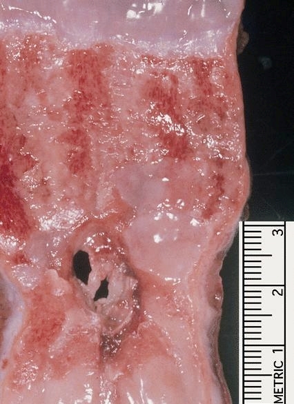
Radiation has destroyed tumor
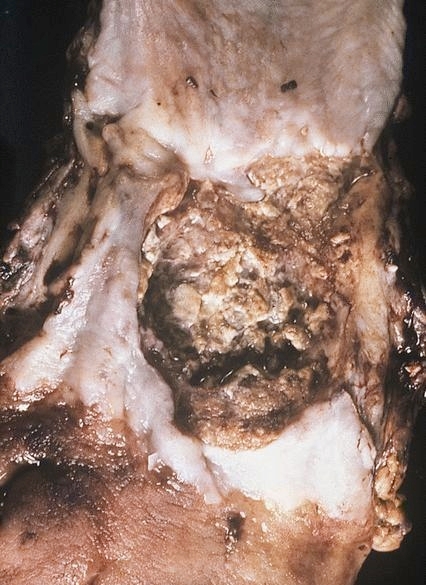
Deep ulcer with necrotic base
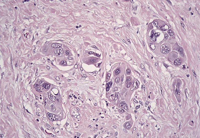
Microscopic (histologic) description
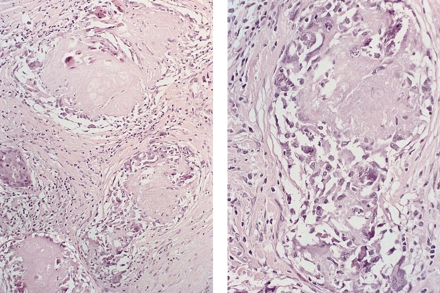
- Variably sized cells in all layers, often increased cytoplasm but usually normal nuclear to cytoplasmic ratio
- Nuclei enlarged with bizarre shapes, may be hyperchromatic or smudged
- May have large eosinophilic nucleoli; usually no mitotic figures
Microscopic (histologic) images
AFIP images
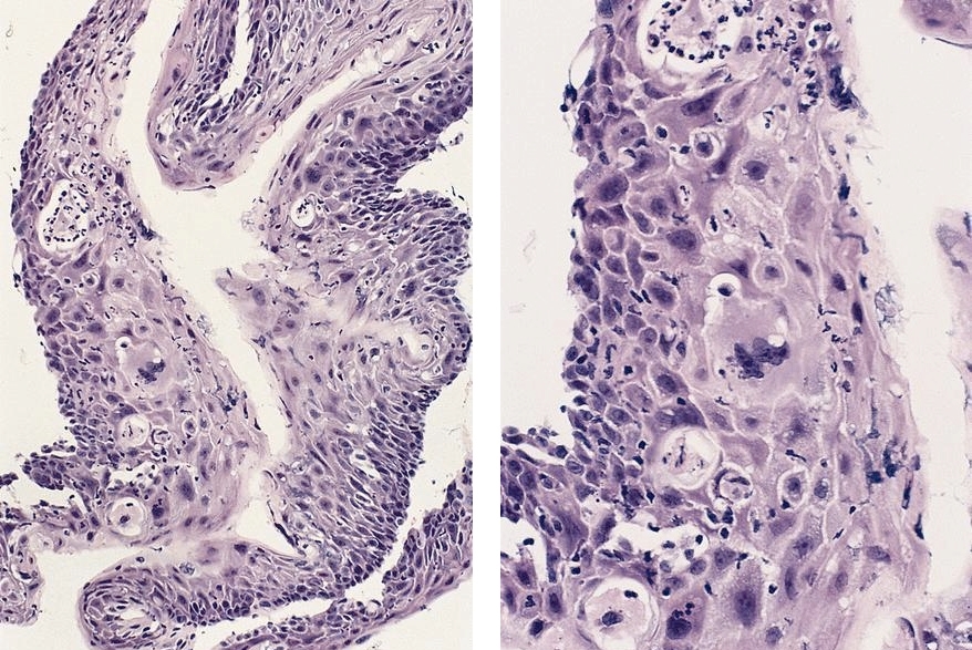
Normally oriented
epithelium is
thinner than
normal
Radiated esophageal squamous cell carcinoma
Irradiated squamous carcinoma
Images hosted on other servers:

Neovascularization (highlighted with CD31)
