
Esophagus
Other nonneoplastic
Esophagitis dissecans superficialis
Author: Hillary Zalaznick, M.D.
Editorial Board Member: Raul S. Gonzalez, M.D.

Last author update: 1 March 2017
Last staff update: 14 February 2024 (update in progress)
Copyright: 2017-2024, PathologyOutlines.com, Inc.
PubMed search: Esophagitis dissecans superficialis
Table of Contents
Definition / general | Essential features | Terminology | Epidemiology | Etiology | Clinical features | Diagnosis | Case reports | Treatment | Clinical images | Microscopic (histologic) description | Microscopic (histologic) images | Differential diagnosis | Additional references | Board review style question #1 | Board review style answer #1Cite this page: Zalaznick H. Esophagitis dissecans superficialis. PathologyOutlines.com website. https://www.pathologyoutlines.com/topic/esophaguseds.html. Accessed April 20th, 2024.
Definition / general
- Benign condition of uncertain etiology
- Superficial strips of squamous mucosa peel off into the esophageal lumen with normal underlying mucosa
- Tends to resolve without sequelae
Essential features
- Characteristic endoscopic finding of long, vertical white strips of peeling epithelium with normal underlying mucosa
- Constellation of nonspecific histologic findings
- Splitting of the squamous epithelium at different levels above the basal layer
- Variably sized cysts and bullae
- Thick layer of parakeratosis
- Basal cell hyperplasia
- Focal or minimal inflammation
Terminology
- Also called "sloughing esophagitis"
Epidemiology
- More common in older individuals (median age in the seventh decade)
Etiology
- Unknown
- Has been variably associated with certain medications (NSAIDs, bisphosphonates), polypharmacy, prior trauma, underlying motility disorders, heavy smoking, alcohol, hot beverages, immunosuppression, celiac disease, concurrent bullous skin conditions and impaired mobility
Clinical features
- Range from asymptomatic to dramatic
- Original reports described patients vomiting up casts of esophageal epithelium
- Esophageal symptoms such as dysphagia, odynophagia and reflux more common
- Can be an incidental finding
Diagnosis
- Characteristic endoscopic findings of peeling, white, vertical strips of epithelium > 2 cm long
- Normal underlying mucosa
- Lack of ulcerations or friability of adjacent mucosa
- Endoscopic appearance likened to an esophagus "filled with gift wrap paper" (Am J Surg Pathol 2009;33:1789)
- Biopsy may not be needed unless to rule out other conditions
Case reports
- 40 year old Hispanic woman with bullous systemic lupus erythematosus associated with esophagitis dissecans superficialis (Case Rep Rheumatol 2015;2015:930683)
- 67 year old man with unusual appearing esophagus on endoscopy (Case #420)
- 68 year old man with fungal esophagitis presenting with esophagitis dissecans superficialis (Gastroenterology Res 2016;9:108)
Treatment
- Typically proton pump inhibitor therapy and discontinuation of medications that may have triggered the condition
Clinical images
Images hosted on other servers:

Sloughing of large
esophageal mucosa
fragments

Endoscopy showing EDS

Vertical fissures in
distal esophagus with
sloughing of mucosa
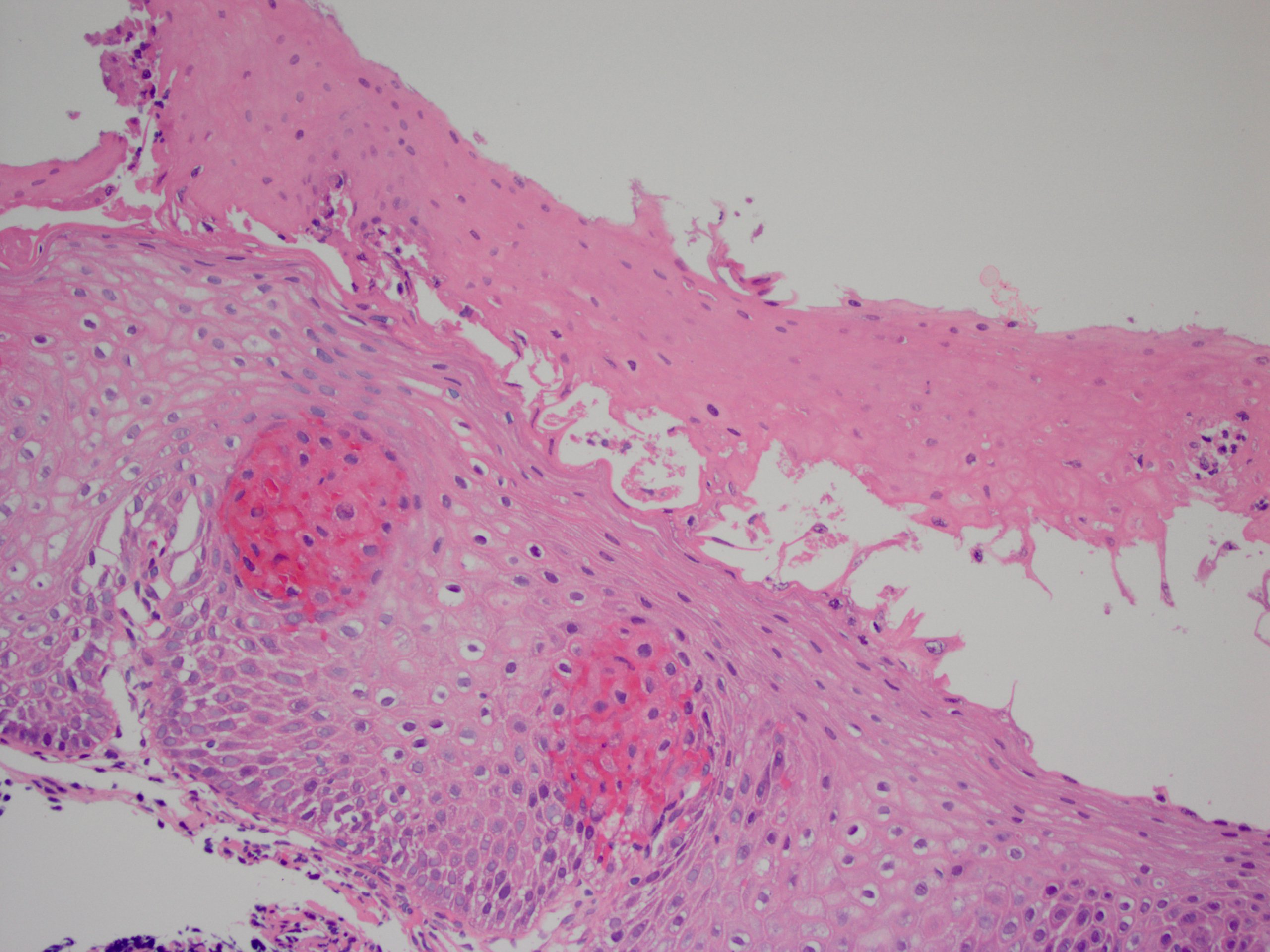
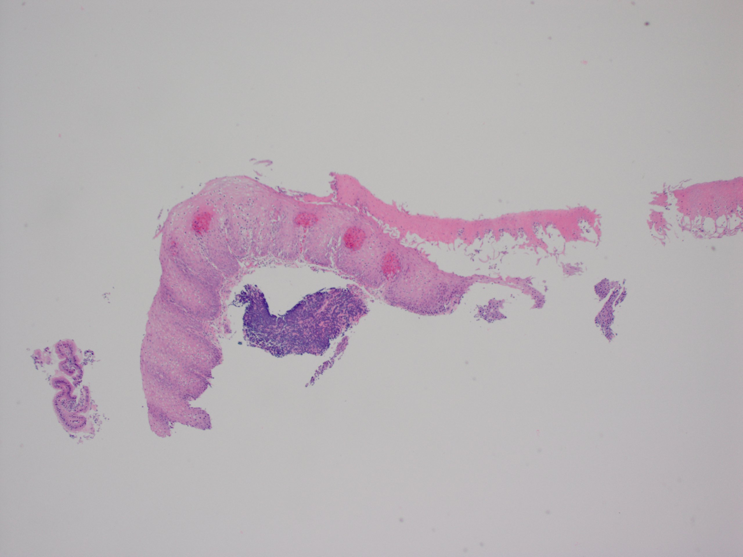
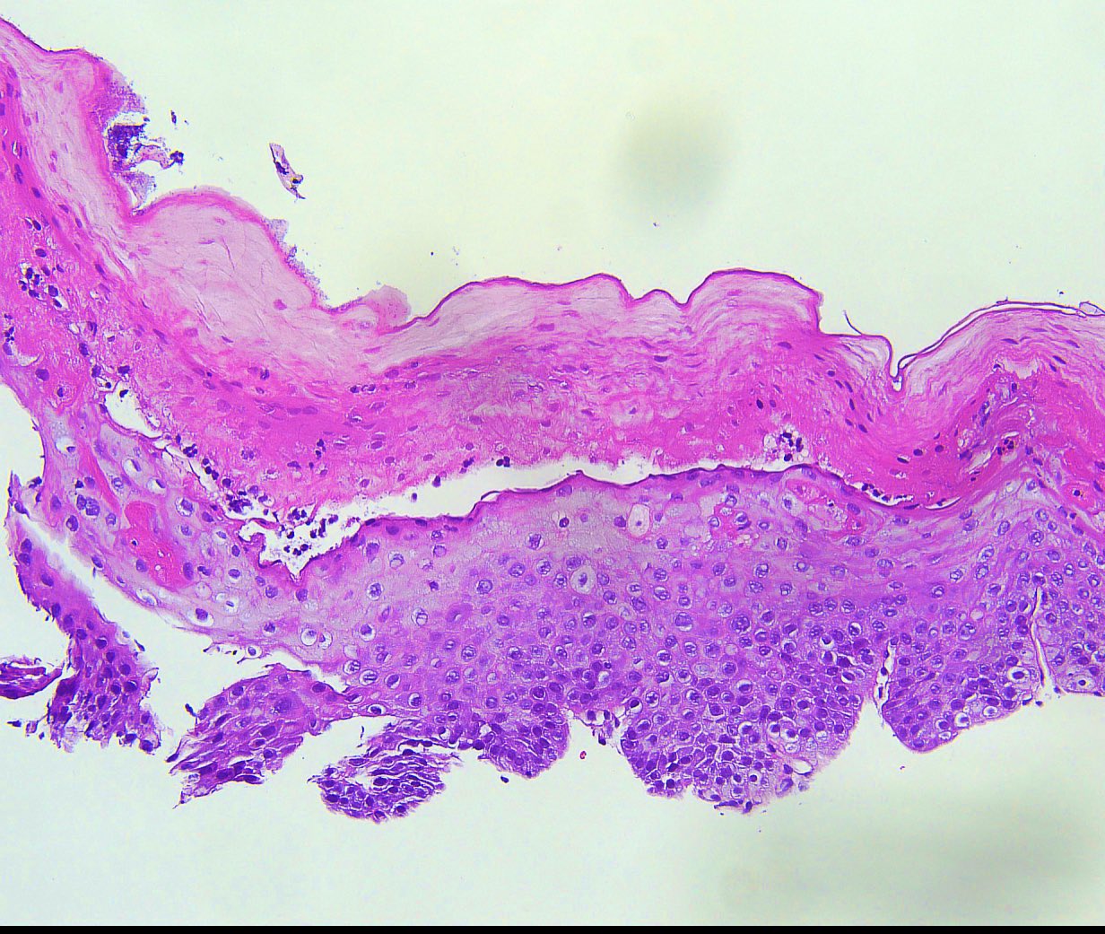
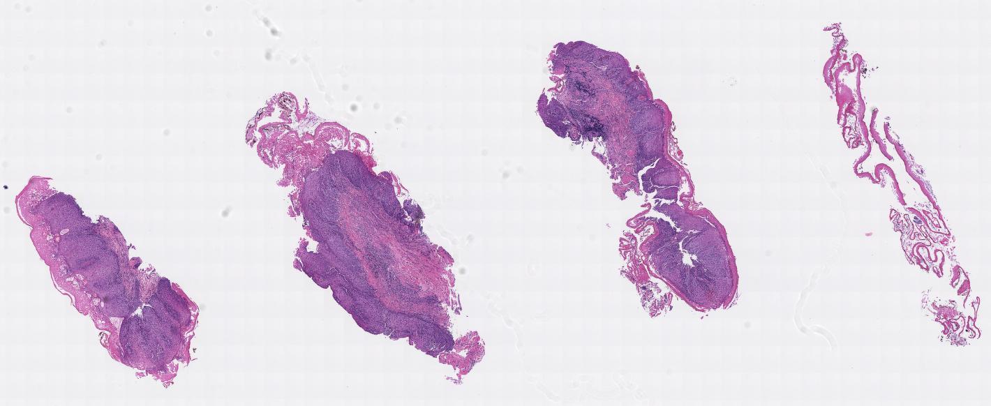
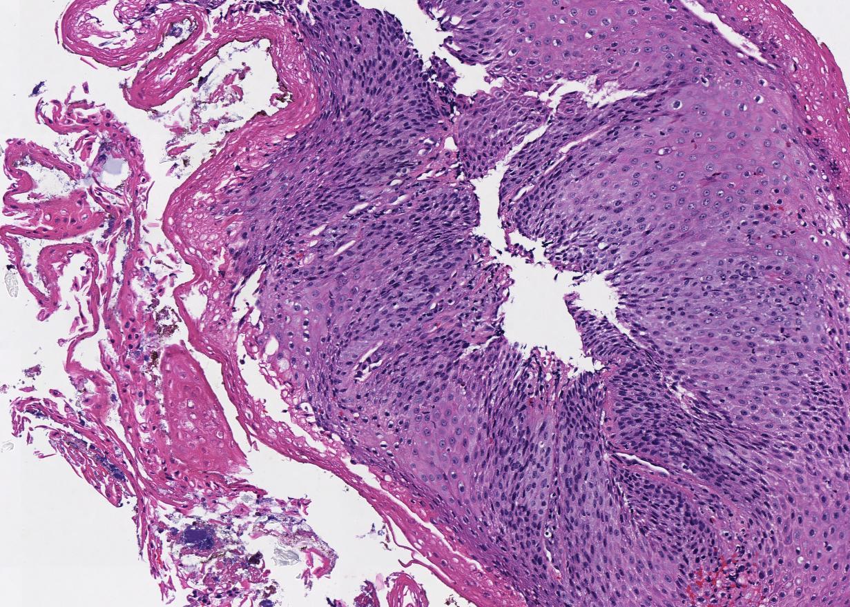
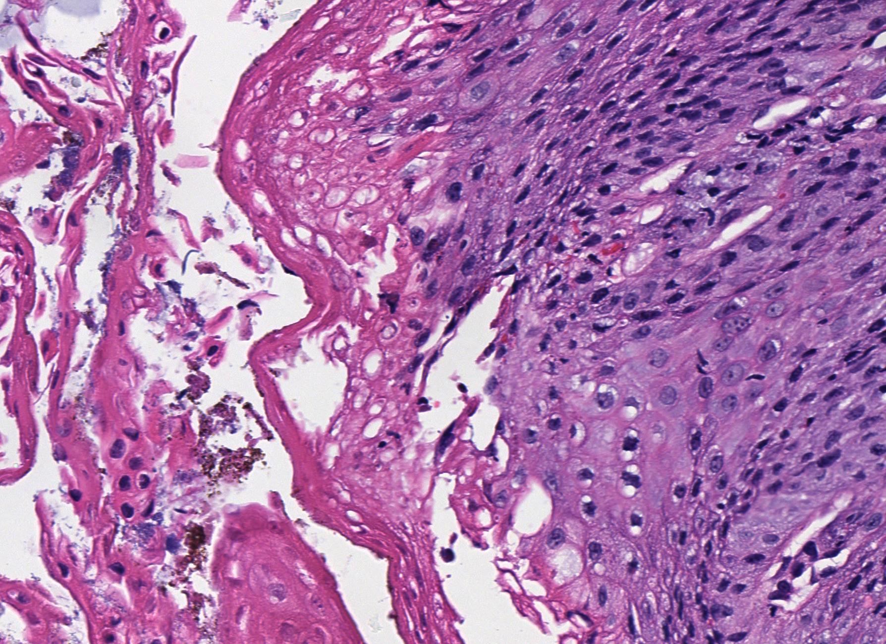
Microscopic (histologic) description
- Splitting of the squamous epithelium at different levels above the basal layer
- Variably sized cysts and bullae (termed "intraepithelial cystic degeneration" by Hart et al., Dig Dis Sci 2015;60:2049)
- Thick layer of parakeratosis that separates off into necrotic fragments
- "Two tone" appearance to epithelium
- Basal cell hyperplasia
- Fungal or bacterial colonies can be associated with the loose fragments
- Inflammation is typically minimal or focal (despite "esophagitis" being in the name)
Microscopic (histologic) images


Differential diagnosis
- Endoscopically:
- Candida esophagitis (white plaques)
- Iatrogenic trauma (e.g., prior endoscopy)
- Histologically:
- Artefactual splitting of epithelium due to biopsy or during grossing
- Bullous skin diseases such as pemphigus vulgaris
- Stricture (parakeratosis)
Additional references
Board review style question #1
Which histologic feature is not commonly seen in esophagitis dissecans superficialis?
- Basal cell hyperplasia
- More than 20 eosinophils per high powered field
- Splitting of the squamous epithelium at different levels above the basal layer
- Thick layer of parakeratosis
- Variably sized cysts and bullae
Board review style answer #1
B. More than 20 eosinophils per high powered field
Comment Here
Reference: Esophagitis dissecans superficialis
Comment Here
Reference: Esophagitis dissecans superficialis
