
Ear
Middle ear and inner ear tumors - malignant
Endolymphatic sac tumor
Author: Nat Pernick, M.D.

Last author update: 1 October 2013
Last staff update: 1 March 2021
Copyright: 2002-2024, PathologyOutlines.com, Inc.
PubMed Search: Endolymphatic sac tumor [pathology]
Table of Contents
Definition / general | Terminology | Radiology description | Case reports | Treatment | Microscopic (histologic) description | Microscopic (histologic) images | Cytology description | Positive stains | Negative stains | Electron microscopy description | Differential diagnosis | Additional referencesCite this page: Pernick N. Endolymphatic sac tumor. PathologyOutlines.com website. https://www.pathologyoutlines.com/topic/earendolymphaticsac.html. Accessed April 25th, 2024.
Definition / general
- Uncommon, associated with von Hippel-Lindau syndrome (11% have these tumors) and female adnexal tumor of presumed wolffian origin (Am J Surg Pathol 1994;18:1254); also somatic mutations of VHL gene in non VHL patients (Cancer Res 2000;60:5963)
- Most arise within intraosseous portion of the endolymphatic duct / sac, with precursor lesions present in VHL patients (Cancer Res 2005;65:10847)
- Median age 30s, range of 11 - 71 years
- Symptoms: early sensorineural hearing loss, tinnitus and episodic vertigo
Terminology
- The 2017 WHO now separates aggressive papillary tumor from endolymphatic sac tumor
- Also called papillary adenoma of endolymphatic sac / temporal bone, adenocarcinoma of temporal bone / mastoid, low grade adenocarcinoma of probable endolymphatic sac origin, Heffner tumor
Radiology description
- Tumor in posterior medial petrous ridge of temporal bone (site of endolymphatic sac)
Case reports
- 20 year old woman with endolymphatic sac tumor associated with von Hippel-Lindau disease (Mod Path 2001;14:727)
- 32 year old woman with papillary neoplasm of the endolymphatic sac associated with Hippel-Lindau disease (J Clin Pathol 1994;47:959)
- 42 year old woman with endolymphatic sac tumor associated with von Hippel-Lindau disease (Arch Pathol Lab Med 2003;127:1387)
- 77 year old man with aspirated cyst (Mod Pathol 2001;14:920)
- Patient with mutation of von Hippel-Lindau tumor suppressor gene in a sporadic endolymphatic sac tumor (Hum Pathol 2001;32:1272)
- Patients with low grade papillary adenomatous tumors of the temporal bone (Mod Pathol 1995;8:603)
Treatment
- Radical surgery including mastoidectomy and temporal bone resection with possible loss of cranial nerves
- Tumor grows slowly with only one reported metastasis (J Neurosurg Spine 2005;3:68) but may recur with inadequate excision
- Is infiltrative, destructive and may cause death
- Bleeds profusely at surgery
Microscopic (histologic) description
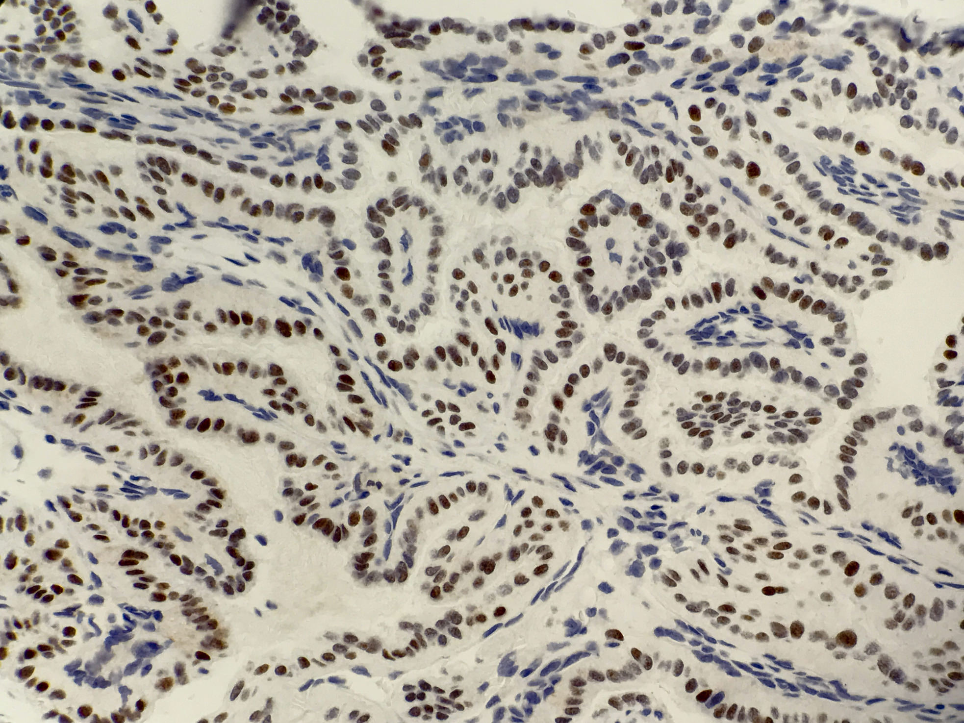
- Simple papillary structures composed of single layer of columnar to cuboidal epithelium, often with distinct cell boundaries
- May have apparent myoepithelial layer that actually is flattened stroma; epithelial cells have pale-clear cytoplasm, uniform central or luminal nuclei
- Often granulation tissue reaction with small vascular spaces and mixed inflammatory infiltrate is present next to tumor cells
- Occasional thyroid like hypercellular areas with cystic glandular spaces containing colloid like material
- May have areas of recent hemorrhage with cholesterol clefts
- Minimal pleomorphism
- No / rare mitotic figures or necrosis
- Resembles choroid plexus papilloma
Microscopic (histologic) images
Contributed by Kelly Magliocca D.D.S., M.P.H.
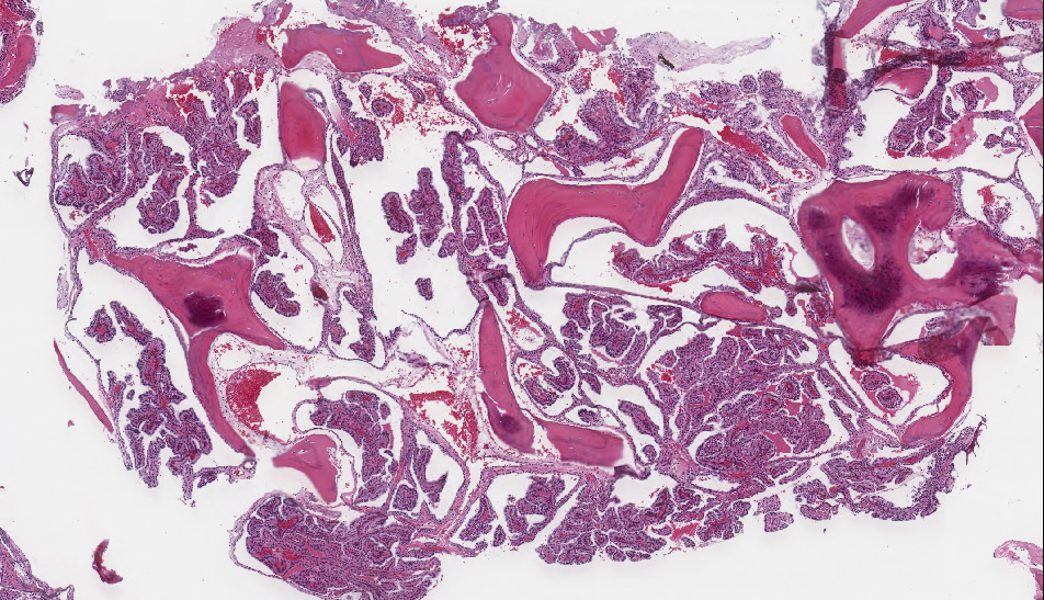
Endolymphatic sac tumor, 2x
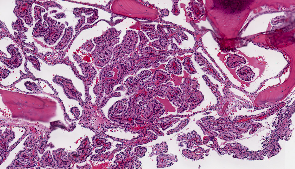
Endolymphatic sac tumor, 5x
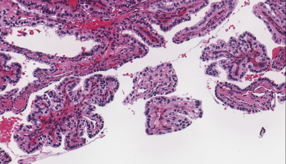
Endolymphatic sac tumor, 15x
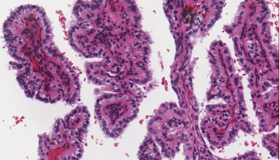
Endolymphatic sac tumor, 20x

Endolymphatic sac tumor, 40x

CAIX, 20x
PAX8, 40x

Negative RCC, 20x
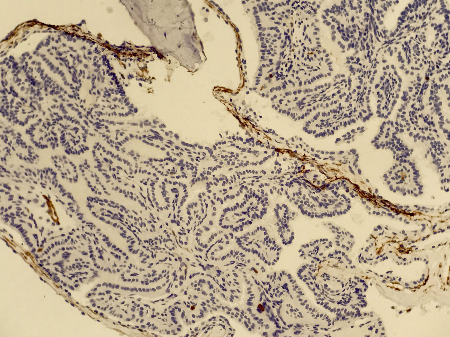
Negative CD10, 20x
Images hosted on other servers:

Various images

Papilla with hemorrhagic material

Papilla with vacuolated cells and colloid-like material
Cytology description
- Rare epithelial cell clusters, some with papillary features
- Foamy macrophages
- Epithelial cells have eosinophilic and focally vacuolated cytoplasm, some with pigmented granules resembling hemosiderin, well defined cell borders, bland nuclei
Positive stains
Negative stains
Electron microscopy description
- Intercellular junctional complexes, microvilli, basement membrane, rough ER, glycogen, secretory granules
Differential diagnosis
- Choroid plexus papilloma-carcinoma: originates within brain ventricles, S100+
- Jugulotympanic paraganglioma: vascular, but not papillary cystic, "zellballen", keratin negative
- Metastatic renal cell carcinoma: S100-, GFAP-, synaptophysin-, kidney tumor present on CT
- Metastatic thyroid carcinoma: thyroglobulin+, characteristic nuclear features
- Middle ear adenoma: not papillary, doesn't invade or destroy bone
Additional references
Back to top
