

CNS nontumor
Toxic and metabolic disorders
Alcoholic cerebellar degeneration
Author: Kymberly A. Gyure, M.D.

Last author update: 1 May 2016
Last staff update: 14 March 2024 (update in progress)
Copyright: 2002-2024, PathologyOutlines.com, Inc.
PubMed Search: Alcoholic cerebellar degeneration

Table of Contents
Definition / general | Essential features | Epidemiology | Sites | Pathophysiology | Clinical features | Laboratory | Radiology description | Radiology images | Prognostic factors | Case reports | Gross description | Gross images | Microscopic (histologic) description | Microscopic (histologic) images | Differential diagnosis | Additional referencesCite this page: Gyure K.A. Alcoholic cerebellar degeneration. PathologyOutlines.com website. https://www.pathologyoutlines.com/topic/cnsalcoholic.html. Accessed April 18th, 2024.
Definition / general
- Atrophy of the cerebellar vermis seen in the setting of chronic alcoholism
Essential features
- Characterized clinically by ataxia and gait disturbances in the setting of chronic alcoholism
- Pathologic features include cerebellar atrophy affecting the anterior / superior vermis with a loss of Purkinje cells and corresponding Bergmann gliosis
Epidemiology
- Occurs in approximately 10% of alcoholic patients
Sites
- Anterior / superior cerebellar vermis
Pathophysiology
- Cerebellar changes may be related in part to thiamine deficiency
- Alterations in GABA receptor dependent neurotransmission have also been proposed as a pathogenic mechanism
Clinical features
- Truncal ataxia
- Unsteady gait
- Nystagmus
- May be clinically asymptomatic in some individuals
Laboratory
- No specific laboratory abnormalities
Radiology description
- Atrophy of the anterior / superior vermis
Radiology images
Images hosted on other servers:
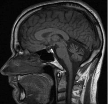
Marked diffuse cerebellar atrophy
Prognostic factors
- Cerebellar damage remains even after abstinence from ethanol
- Prevention of cerebellar damage by treatment of alcoholism is recommended
Case reports
- 79 year old man with progressive gait disturbance and repeated falls (J Korean Med Sci 2015;30:1539)
Gross description
- Atrophy of the anterior / superior cererellar vermis with widened sulci
Gross images
Contributed by Kymberly A. Gyure, M.D.
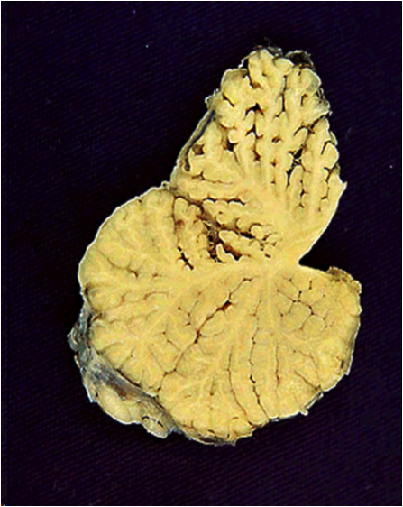
Alcoholic cerebellar degeneration
Microscopic (histologic) description
- Loss of cerebellar Purkinje cells with corresponding Bergmann gliosis
- Narrowing of the molecular layer and a reduced number of granular cells may also be seen
Microscopic (histologic) images
Contributed by Kymberly A. Gyure, M.D.

Alcoholic cerebellar degeneration
Differential diagnosis
- Age related cerebellar atrophy (generally milder)
- Other causes of cerebellar vermal atrophy: phenytoin use, heavy metal poisoning or a subset of the spinocerebellar ataxias
Additional references
