

Cervix
General
Histology
Authors: Kyle Devins, M.D., Lauren Schwartz, M.D.
Editorial Board Member: Carlos Parra-Herran, M.D.
Editor-in-Chief: Debra L. Zynger, M.D.


Last author update: 19 May 2020
Last staff update: 7 May 2021
Copyright: 2003-2024, PathologyOutlines.com, Inc.
PubMed Search: Cervix[TI] histology[TI]

Table of Contents
Definition / general | Essential features | Physiology | Diagrams / tables | Clinical features | Gross description | Gross images | Microscopic (histologic) description | Microscopic (histologic) images | Virtual slides | Cytology description | Positive stains | Negative stains | Additional references | Board review style question #1 | Board review style answer #1 | Board review style question #2 | Board review style answer #2Cite this page: Devins K, Schwartz L. Histology. PathologyOutlines.com website. https://www.pathologyoutlines.com/topic/cervixnormalhistology.html. Accessed April 25th, 2024.
Definition / general
- Lower portion of the uterus which connects the uterine corpus to the vaginal canal
Essential features
- Exocervix is lined by stratified squamous epithelium
- Endocervix is lined by columnar mucinous epithelium
- Transformation zone is the hormonally responsive zone of metaplasia between the exocervix and endocervix
- Squamocolumnar junction is the precise histologic transition between squamous and glandular epithelium
- Transformation zone is usual site of persistent infection with high risk subtypes of human papillomavirus (HPV), the most common cause of cervical carcinoma
Physiology
- 2 anatomic portions:
- Portio vaginalis: anatomic portion of cervix inferior to vaginal reflection and within vaginal canal
- Portio supravaginalis: anatomic portion of cervix superior to vaginal reflection
- Ectocervix (exocervix): external mucosal surface of portio vaginalis
- Normally lined by nonkeratinizing stratified squamous epithelium
- Endocervix (endocervical canal): mucosa lined cylindrical canal leading from ectocervix to uterine cavity
- Normally lined by single layer of mucinous columnar epithelium
- External os: anatomic opening of the endocervical canal onto the ectocervix
- Represents the inferior limit of the endocervix
- Internal os: anatomic widening of the endocervical canal as it opens and gradually transitions into the uterine cavity
- Represents the superior limit of the endocervix and distal aspect of the lower uterine segment
- Squamocolumnar junction: histologic junction between squamous epithelium of ectocervix and columnar glandular epithelium of endocervix; location changes throughout life in response to hormonal status and age
- Squamocolumnar junction is located on outer surface of portio vaginalis at birth due to effect of maternal hormones; rapidly recedes into endocervical canal until menarche (J Reprod Med 1976;16:221)
- In early adolescence, squamocolumnar junction migrates distally from the external os onto the exocervix in response to pubertal hormones, forming the ectropion
- Ectropion: physiologic ectopic columnar epithelium present on exocervix beginning at adolescence
- Ectropion is gradually replaced by squamous metaplasia throughout reproductive years and the functional squamocolumnar junction slowly recedes towards the external os
- Transformation zone: the physiologic area of metaplastic squamous epithelium between the exocervix and the endocervix; it begins distally at the original squamocolumnar junction present at adolescence and extends proximally to the current functional squamocolumnar junction
- Squamocolumnar junction often recedes within external os following menopause
Diagrams / tables
Contributed by Kyle Devins, M.D.

Schematic of exocervix and transformation zone
Images hosted on other servers:

Location of glandular and squamous epithelium
Clinical features
- Transformation zone is the usual site of persistent infection with high risk subtypes of human papillomavirus (HPV), the most common cause of cervical carcinoma (J Clin Pathol 2002;55:244)
Gross description
- Cylindrical structure forming lower ~ third of uterus, connecting uterine corpus to vaginal canal
- Anatomic position within the pelvic cavity between the urinary bladder (anterior) and rectum (posterior)
- Anterior portion can be identified by a higher peritoneal reflection, due to location of the bladder in vivo (J Clin Pathol 1993;46:388)
Gross images
Contributed by Kyle Devins, M.D.
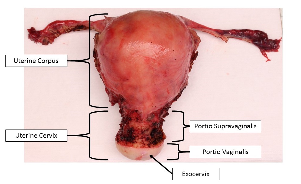
Uterus and cervix
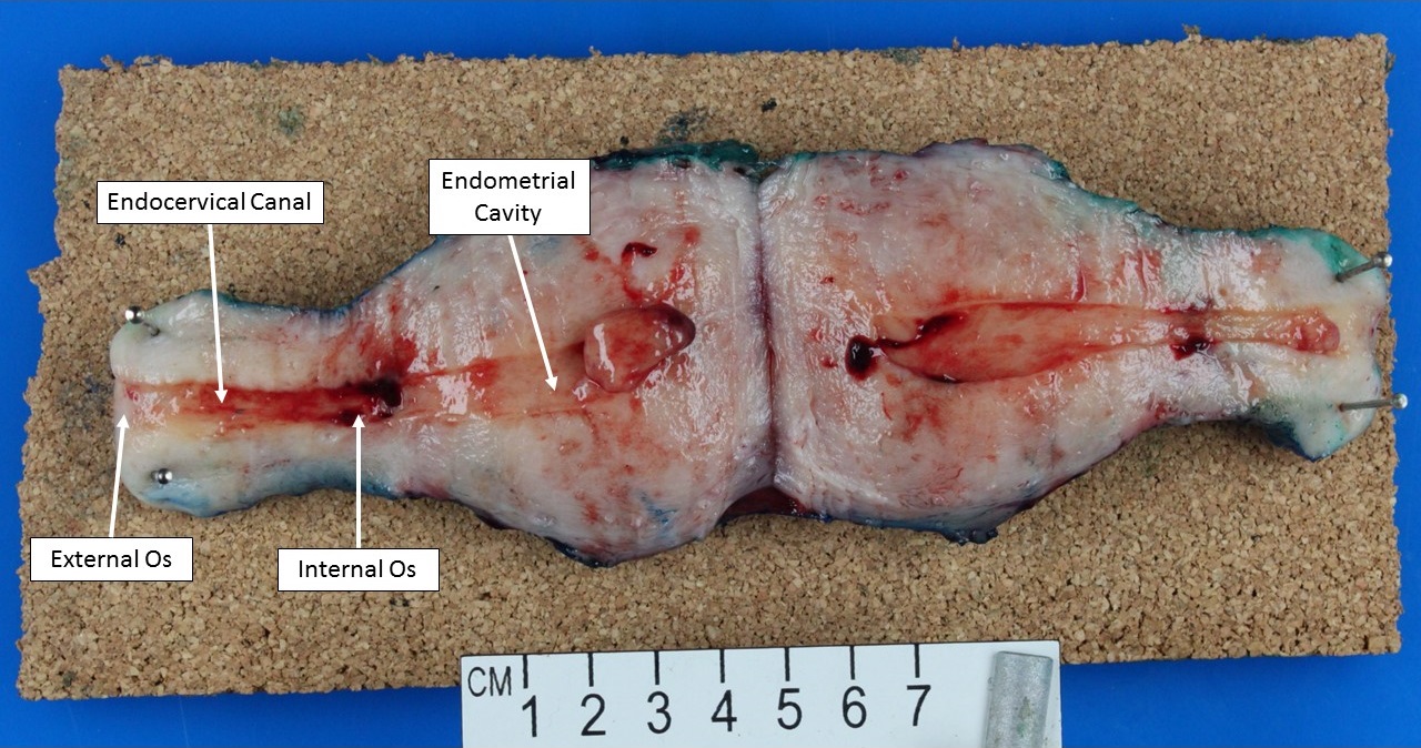
Bivalved uterus and cervix
Microscopic (histologic) description
- Ectocervix
- Stratified nonkeratinizing squamous epithelium
- Basal cells: deepest layer; dense nuclear chromatin, uniform oval nuclei oriented perpendicular to basement membrane, scant cytoplasm
- Parabasal cells: located just above the basal cell layer; slightly more cytoplasm than basal cells; may be multiple cell layers thick
- Intermediate cells: abundant cytoplasm which may be pink or clear due to glycogen accumulation
- Superficial cells: small, round nuclei; abundant pink or clear cytoplasm; cells flatten and are oriented parallel to basement membrane
- Hormone responsive
- Superficial cells predominate in early cycle due to estrogen
- Intermediate cells predominate in late cycle due to progestins
- Loss of intermediate and superficial cells (atrophy) occurs postmenopause
- Rare melanocytes, Langerhans cells and endocrine cells have been identified (Br J Obstet Gynaecol 1983;90:400, Virchows Arch A Pathol Anat Histopathol 1987;411:293)
- Stratified nonkeratinizing squamous epithelium
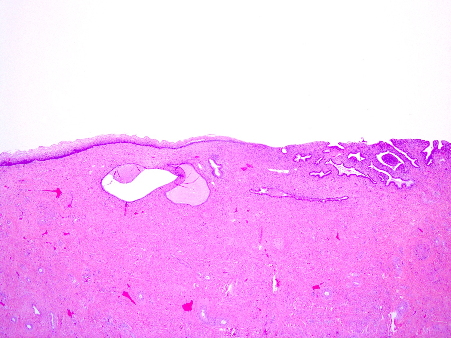
- Endocervix
- Single layer of mucinous columnar cells with dense, uniform, oval, basally oriented nuclei and apical mucin
- Mucin has a pale bluish appearance in H&E preparations; with PAS-Alcian blue stain, apical mucin stains intense blue / purple (due to the presence of acid type mucin)
- Ciliated cells can be found (usually in the context of tuboendometrioid metaplasia)
- Inconspicuous underlying reserve cell layer
- Forms infoldings, clefts and glands of variable shape
- Transformation zone
- Metaplastic cells: formed by endocervical reserve cells differentiating toward squamous lineage
- Located at transition between glandular and squamous epithelia
- Similar appearance to parabasal cells with relatively scant cytoplasm and dense nuclei
- Endocervical epithelium may overlie metaplastic cells
- Variable nonspecific inflammatory infiltrate consisting of lymphocytes, plasma cells and even neutrophils is common and is not necessarily associated with infection
- Metaplastic cells: formed by endocervical reserve cells differentiating toward squamous lineage
- Cervical stroma
- Mostly fibrous tissue with some haphazard smooth muscle fibers
- Blood vessels often numerous and prominent
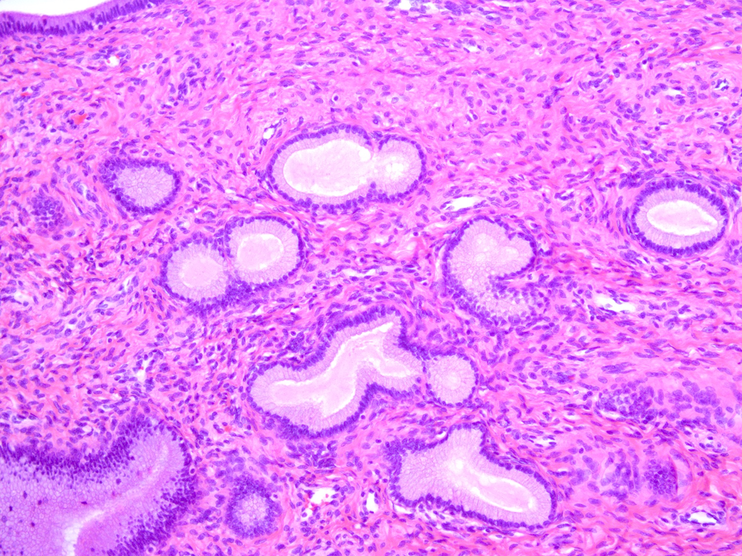
- Mesonephric rests / remnants
- Remnants of involuted embryologic mesonephric (Wolffian) ducts
- Present in lateral cervical wall in ~33% of women
- Microscopic clusters of tubules lined by single layer of cuboidal cells with eosinophilic luminal secretions (Am J Surg Pathol 1990;14:1100)
Microscopic (histologic) images
Contributed by Kyle Devins, M.D.
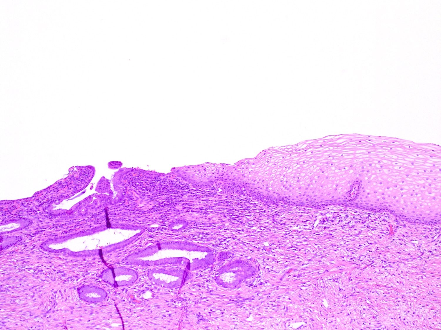
Squamocolumnar junction

Cervical squamous epithelium
Benign endocervical glands
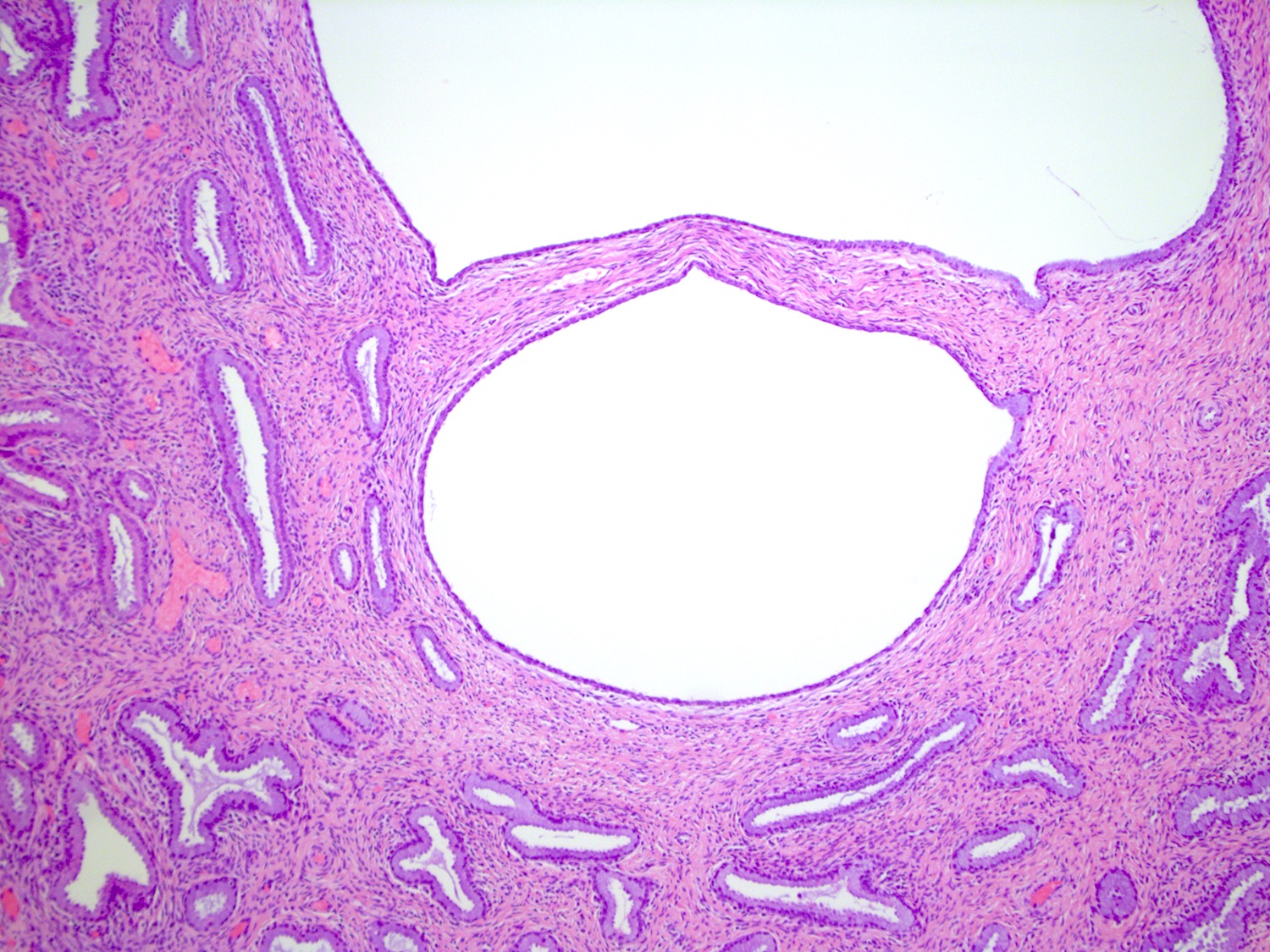
Dilated endocervical glands

Normal cervical squamous p16 IHC
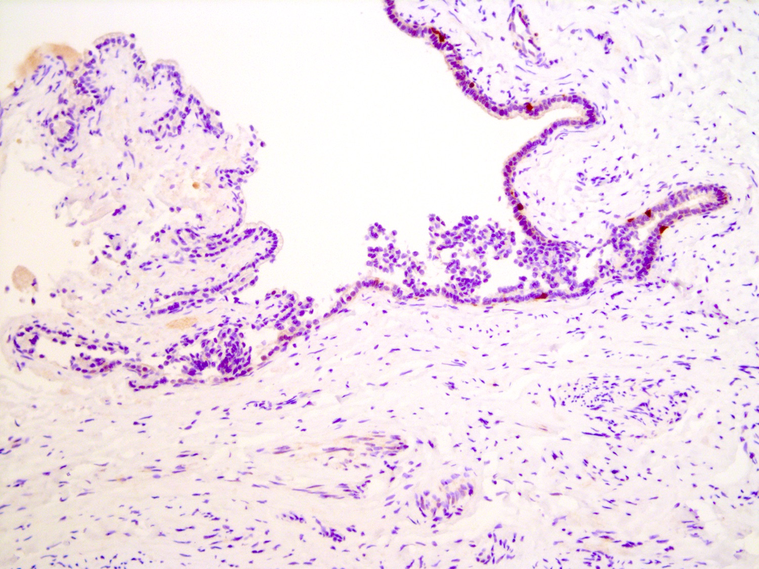
Normal endocervical p16 IHC
Virtual slides
Images hosted on other servers:

Cervix, normal transformation zone
Cytology description
Positive stains
Negative stains
- p16: negative or patchy (Adv Anat Pathol 2006;13:8)
Additional references
Board review style question #1
Which of the following is true about the squamocolumnar junction of the cervix, shown in the image?
- Relatively static and does not undergo significant histologic changes after birth
- Inflammatory infiltrate at the squamocolumnar junction is abnormal and indicates infection
- Migrates distally from the external os during menopause
- Migrates distally from the external os in response to estrogens during adolescence
Board review style answer #1
D. The squamocolumnar junction and transformation zone are hormonally responsive. The squamocolumnar junction actively migrates distally onto the exocervix in response to estrogens at puberty.
Comment Here
Reference: Cervix - histology
Comment Here
Reference: Cervix - histology
Board review style question #2
Which of the following best describes the appearance of normal endocervical epithelium?
- Columnar cells with basally located nuclei, apical mucin and occasional cilia
- Tall pseudostratified nuclei with conspicuous apoptotic bodies and apical mitotic figures
- Stratified cuboidal cells with scattered mucocytes
- Stratified nonkeratinizing squamous epithelium
Board review style answer #2
A. Normal endocervical epithelium consists of columnar cells with round, basally oriented nuclei and pink apical cytoplasm. Ciliated cells can be found.
Comment Here
Reference: Cervix - histology
Comment Here
Reference: Cervix - histology
