Table of Contents
Definition / general | Case reports | Gross description | Microscopic (histologic) description | Microscopic (histologic) images | Positive stains | Negative stains | Electron microscopy description | Differential diagnosis | Additional referencesCite this page: Perunovic B. Blue nevus. PathologyOutlines.com website. https://www.pathologyoutlines.com/topic/cervixbluenevus.html. Accessed January 17th, 2025.
Definition / general
- Present in up to 2% of cervices; may be more common in Japanese women, particularly if step sections are obtained (Acta Pathol Jpn 1991;41:751)
- 20% are multiple
- Usually an incidental finding
Case reports
- 32 year old woman with incidental finding (Appl Immunohistochem Mol Morphol 2004;12:79)
- 2 patients with endocervical location (Ceska Gynekol 2004;69:411)
Gross description
- Blue / black, flat up to 3 cm
- Usually ill defined in lower endocervix
Microscopic (histologic) description
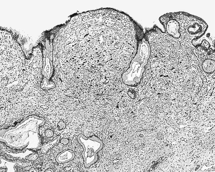
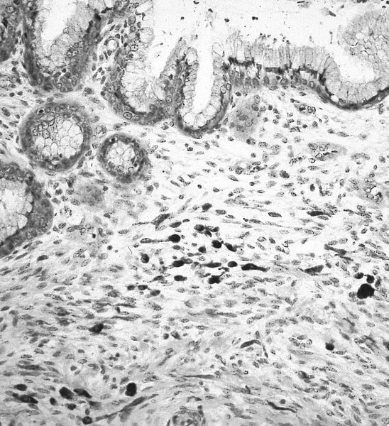
- Elongated, wavy dendritic cells in clusters or individually, below endocervical epithelium
- Cytoplasm has brown melanin
- Stromal macrophages
Positive stains
- Fontana-Masson (melanin turns black). S100, HMB45
Negative stains
- Iron stains
Electron microscopy description
- Dendritic cytoplasmic processes, electron dense membrane bound melanin granules, premelanosomes (Arch Pathol Lab Med 1983;107:87)
Differential diagnosis
Additional references


