
CD Markers
CD44


Copyright: 2003-2024, PathologyOutlines.com, Inc.
PubMed Search: CD44[TI] pathology review[PT] full text[SB]
CD44
Editorial Board Member: Christian M. Schürch, M.D., Ph.D.
Deputy Editor-in-Chief: Maria Tretiakova, M.D., Ph.D.
Last author update: 27 January 2022
Last staff update: 27 January 2022
Copyright: 2003-2024, PathologyOutlines.com, Inc.
PubMed Search: CD44[TI] pathology review[PT] full text[SB]

Table of Contents
Definition / general | Essential features | Terminology | Pathophysiology | Diagrams / tables | Clinical features | Interpretation | Uses by pathologists | Prognostic factors | Microscopic (histologic) description | Microscopic (histologic) images | Positive staining - normal | Positive staining - disease | Negative staining | Molecular / cytogenetics description | Molecular / cytogenetics images | Sample pathology report | CD44R | Board review style question #1 | Board review style answer #1Cite this page: Ismail S, Elshimali JYI. CD44. PathologyOutlines.com website. https://www.pathologyoutlines.com/topic/cdmarkerscd44.html. Accessed April 20th, 2024.
Definition / general
- Complex, transmembrane, multistructural adhesion glycoprotein
- Surface marker of cancer stem cells (Exp Hematol Oncol 2020;9:36, Methods Mol Biol 2018;1692:31)
Essential features
- Complex transmembrane adhesion glycoprotein
- Surface marker of cancer stem cells
- Plays a crucial role in cell adhesion, differentiation, migration and signaling (Exp Hematol Oncol 2020;9:36)
- CD44 might represent a therapeutic target in various tumor types
Terminology
- Cluster of differentiation CD44
- Nonkinase, transmembrane glycoprotein (J Hematol Oncol 2018;11:64)
- Also called Hermes antigen, HCAM or lymphocyte homing receptor (J Oral Maxillofac Pathol 2019;23:267)
- Also known as phagocytic glycoprotein 1 (Blood 2006;107:4149)
Pathophysiology
- Contributes to cellular adhesion through binding to hyaluronic acid (HA) and through inter CD44 binding by attached glycosylation moieties (J Cell Biol 1995;131:1623, J Oral Maxillofac Pathol 2019;23:267)
- Binding to HA induces the expression of inflammatory genes in macrophages, including Osteopontin, leading to aggregation of macrophages, lymphocytes and fibroblasts (Science 1996;271:509)
- Member of the cartilage link protein family
- Also binds to chondroitin, collagen, laminin and fibronectin (J Cell Biol 1992;116:817)
- Cytoplasmic tail initiates cell signaling pathways through binding to adaptor protein (Nat Rev Mol Cell Biol 2003;4:33)
- Splice variants provide proliferation initiating signals for lymphopoiesis, myelopoiesis and angiogenesis (J Clin Invest 1996;97:596, J Immunol 1997;159:2549)
- Also, expression is induced through the binding between CD44 promoter region and transcriptional factors Sp1, Egr1, P53, NFκB and ETS1 (J Oral Maxillofac Pathol 2019;23:267)
- Contributes to cancer cell division, proliferation and invasion through mediating protein kinases, intracellular pathways and proteinases (J Hematol Oncol 2018;11:64)
- Contributes to the adaptive plasticity of cancer cells (J Hematol Oncol 2018;11:64)
Diagrams / tables
Images hosted on other servers:

CD44 molecule structure with ligands
Clinical features
- Might represent a therapeutic target in various tumor types, including ovarian cancer (Front Oncol 2020;10:589601)
- High expression of CD44 is associated with recurrence and metastases in several cancer types, including gastric, colorectal and bladder cancer (Oncol Rep 2016;36:2852)
- Treatment is based on suppression of CD44 expression by peptide mimetics, aptamers and natural compounds (J Hematol Oncol 2018;11:64)
- CD44 deletion enhances the fibrogenic response, increases leukocyte infiltration and delays the accumulation of fibrillar collagen (Matrix Biol 2019;75-76:314)
Interpretation
- Membranous
Uses by pathologists
- Biomarker for cancer stem cells (Methods Mol Biol 2018;1692:31, Stem Cells Int 2016;2016:2087204)
- Prognostic marker in various cancer subtypes and a marker for rapid progression and metastasizing (Stem Cells Int 2016;2016:2087204, Front Oncol 2019;9:39)
- Differentiate reactive urothelium from urothelial carcinoma in situ (Med J Islam Repub Iran 2016;30:400, Am J Surg Pathol 2001;25:1074, Appl Immunohistochem Mol Morphol 2018;26:180)
Prognostic factors
- Increased expression is associated with invasiveness and increased chemotherapy resistance
- Might represent a therapeutic target in various tumor types
- CD44v high expression correlates to progression and poor outcome in various cancers, including non-Hodgkin lymphoma, hepatocellular carcinoma, breast, bone, ovarian, pancreatic, colorectal, bladder, gastric, head and neck squamous cell carcinomas, leukemias and lymphomas (Front Oncol 2020;10:589601)
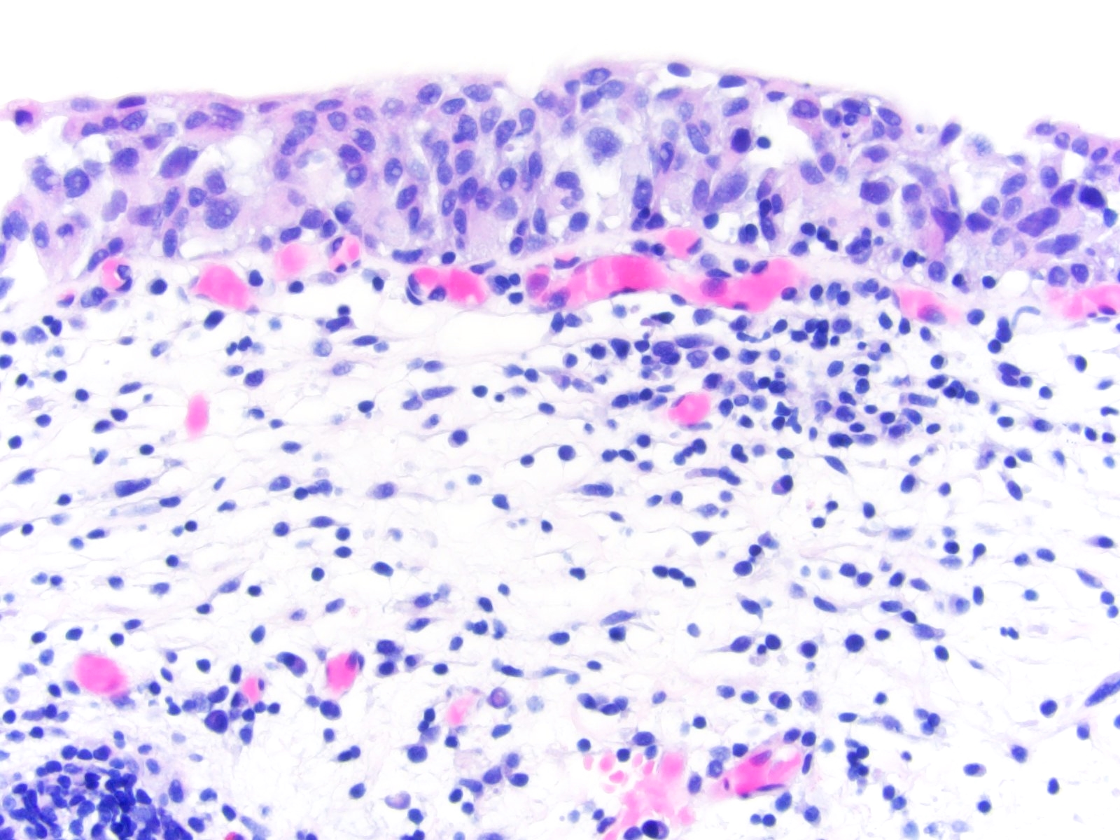
Microscopic (histologic) description
- Positive cells should demonstrate a strong plasma membrane staining pattern
- Cells that lack membranous staining or demonstrate only cytoplasmic staining are considered negative
- Reference: PLoS One 2016;11:e0165253
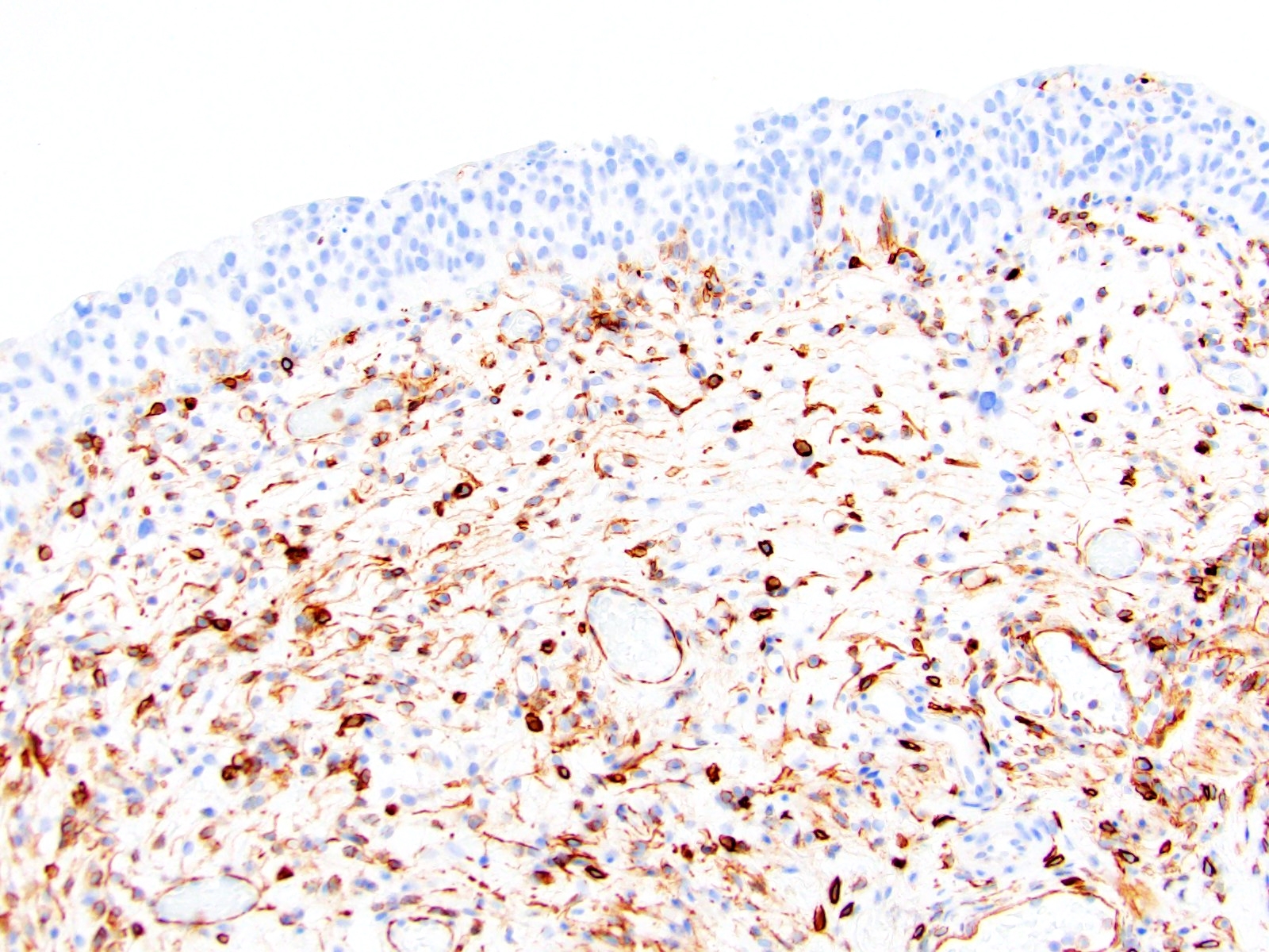
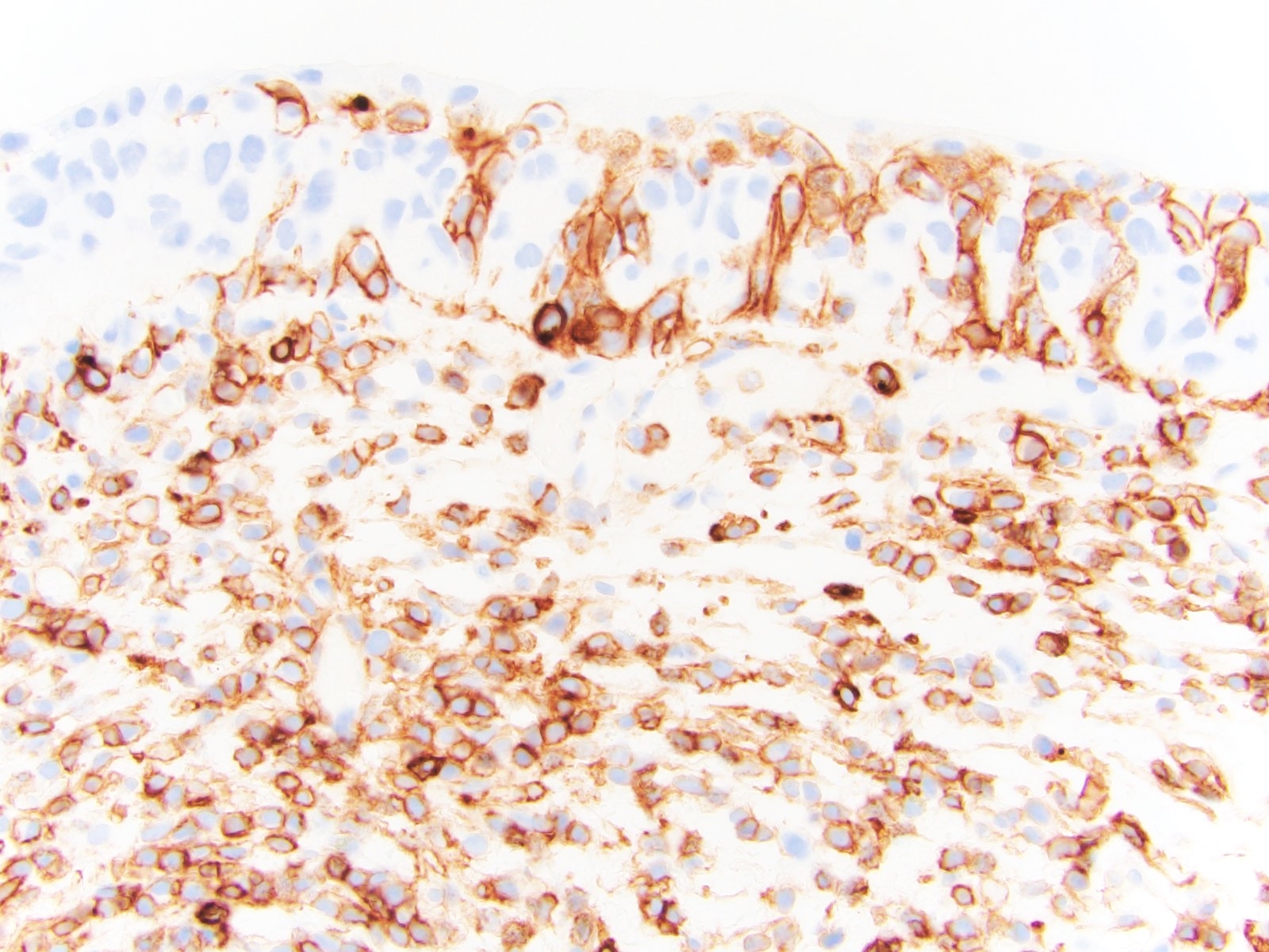
Microscopic (histologic) images
Contributed by John Yahya I. Elshimali, M.D. and Maria Tretiakova, M.D., Ph.D.
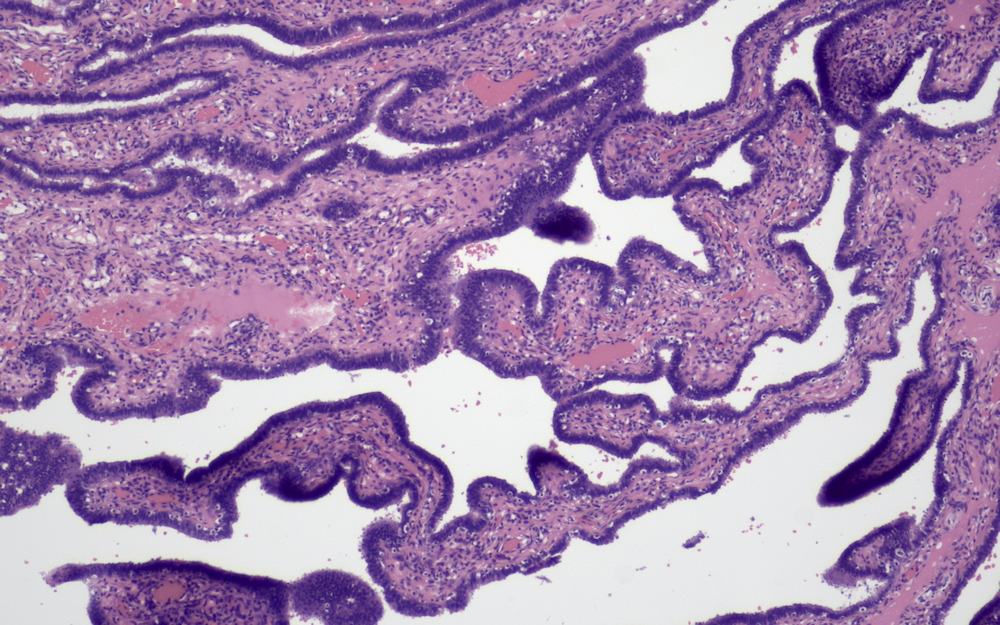
Epithelium of fallopian tube

Fallopian stromal cells
CD44 positive epithelial cells in fallopian tube
CD44 positive epithelial cells in fallopian tube
Positive CD44 expression in fallopian tube epithelium
Decidual and stromal cells in fallopian pregnancy positive for CD44
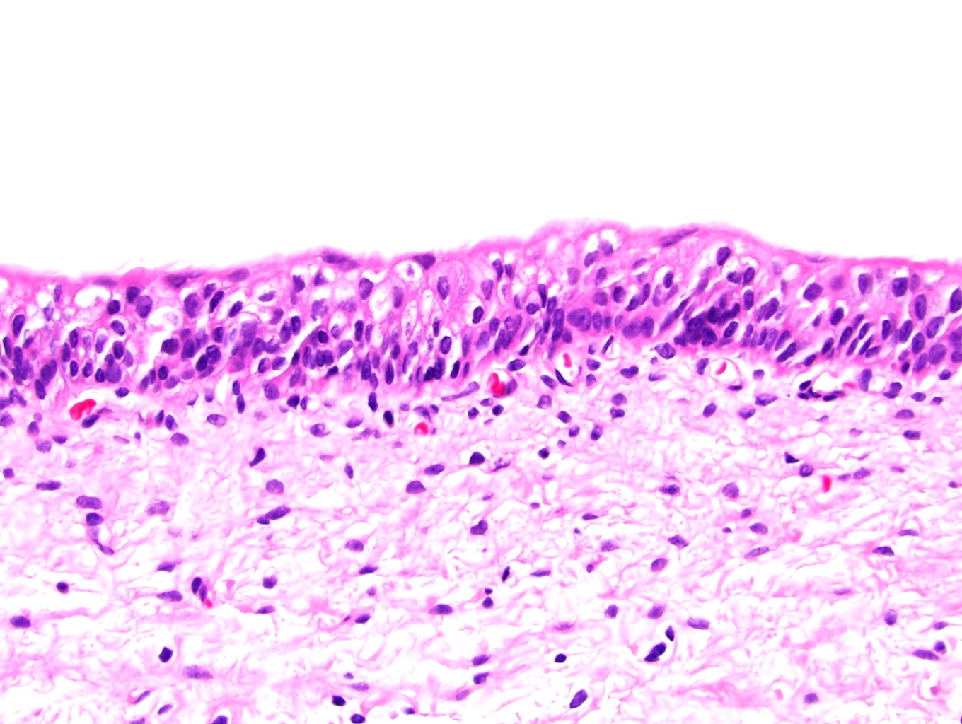
Normal bladder mucosa
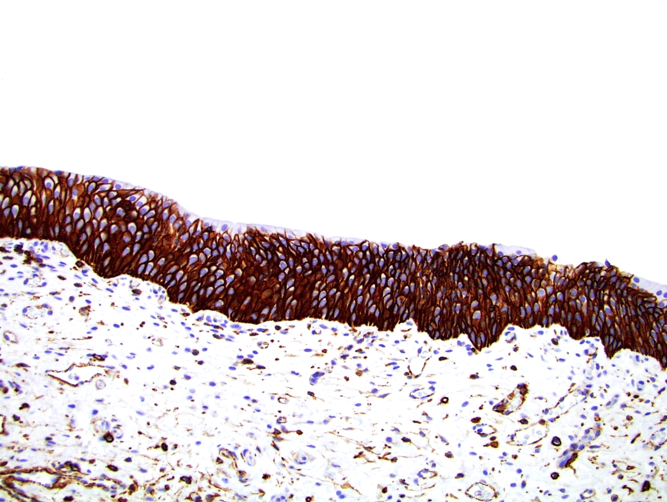
CD44 - full thickness in normal bladder mucosa
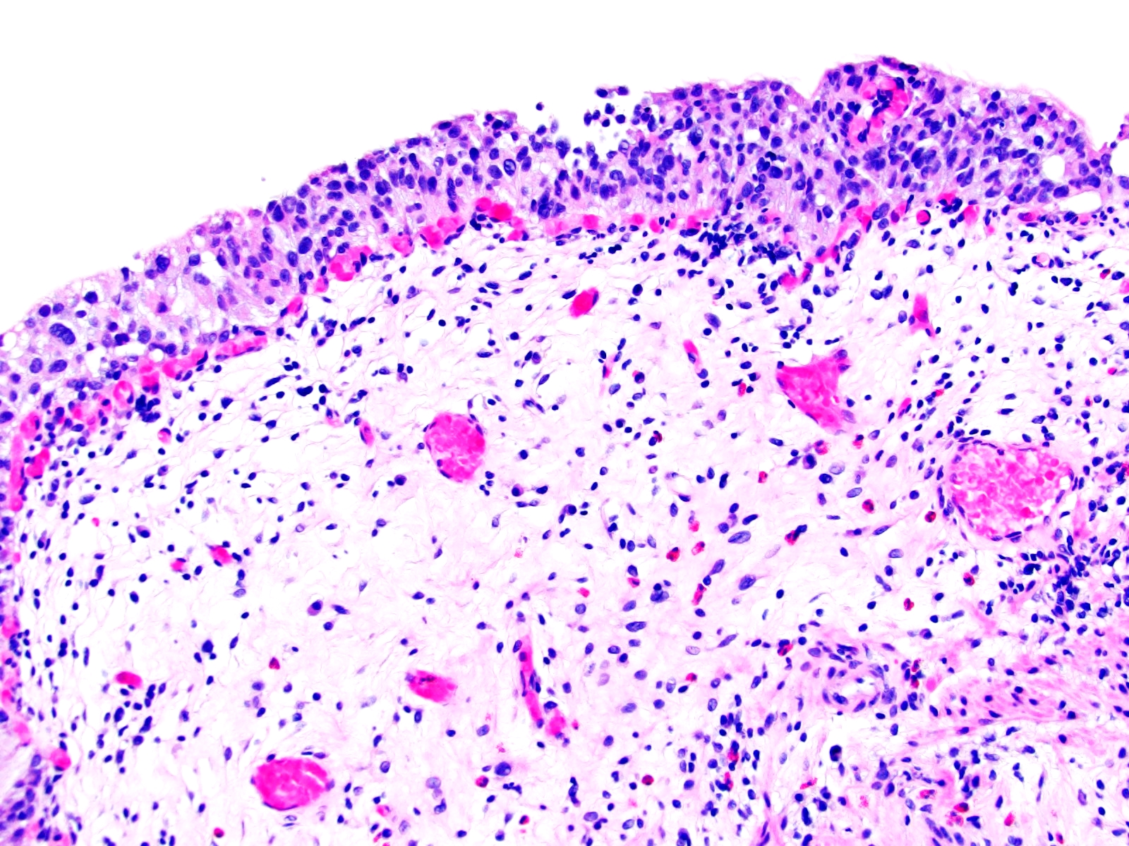
Bladder CIS
CD44 - loss of CD44 expression
Pagetoid CIS vs reactive inflammation
CD44 - patchy loss of expression in pagetoid CIS
Positive staining - normal
- Fallopian tube epithelium (Stem Cells 2012;30:2487)
- Myeloid cells in bone marrow biopsies, memory T cells, activated lymphocytes and other hematopoietic cells (Commun Integr Biol 2010;3:508)
- Also, high expression in white matter cells of the brain, epithelial cells in the stomach, colon, bladder, liver and cervix (Brain 2013;136:209, J Biol Chem 2013;288:16085, Stem Cells 2013;31:2024, Verh Dtsch Ges Pathol 1995;79:144, Clin Cancer Res 1995;1:1125)
- Proliferating cells and smooth muscle cells express high amounts of CD44 (J Oral Maxillofac Pathol 2019;23:267)
- CD44 is expressed in most cells, CD44v is expressed only in subpopulations of epithelial and hematopoietic cells, particularly during embryogenesis and hematopoiesis, on leukocytes during activation and frequently on cancer initiating cells (CIC) (Front Cell Dev Biol 2018;6:97)
- CD44v6: pancreatic ductal cells (Front Cell Dev Biol 2018;6:97)
Positive staining - disease
- Tubal intraepithelial carcinomas, serous tumors of the ovary, lung squamous cell carcinoma, breast cancer, soft tissue sarcomas (Biomolecules 2015;5:3051, PLoS One 2010;5:e14062, Breast Dis 2020;39:1, Virchows Arch 2000;436:574)
- Reactive urothelium (Med J Islam Repub Iran 2016;30:400, Am J Surg Pathol 2001;25:1074)
- Colorectal adenocarcinoma (CD44v) (Zhonghua Zhong Liu Za Zhi 1996;18:347)
- Head and neck squamous cell carcinoma (CD44v3-CD44v6-CD44v10) (Laryngoscope 2009;119:1518)
- B cell chronic lymphocytic leukemia CD44s (Front Immunol 2015;6:177)
- Pancreatic cancer (CD44v6) (J Oncol 2019;2019:3516973)
- Thyroid cancer (CD44v6) (Diagn Mol Pathol 2011;20:233)
- Prostate adenocarcinoma (CD44v6-CD44v9-CD44v10) (Lab Invest 2004;84:894)
- Leukemia and hematopoietic malignancies (CD44v8-10) (J Hematol Oncol 2018;11:64)
- Liposarcoma and clear cell sarcoma have lower expression of CD44 (J Oral Maxillofac Pathol 2019;23:267)
- Triple negative breast cancer: high expression (PLoS One 2013;8:e78259)
Negative staining
- Neuroblastoma (Mol Cell Biol 1991;11:5446)
- Basal and squamous cell carcinoma of the skin (Br J Dermatol 1996;134:465, Br J Dermatol 2009;160:1251)
- Bladder carcinoma in situ and normal urothelium (Med J Islam Repub Iran 2016;30:400, Am J Surg Pathol 2001;25:1074, Anal Quant Cytopathol Histpathol 2015;37:29)
Molecular / cytogenetics description
- Molecular mass of 85 - 200 kDa (Front Cell Dev Biol 2017;5:18)
- Single chain glycoprotein encoded by CD44 gene located on chromosome 11, (11p13) (Front Cell Dev Biol 2017;5:18)
- Known as the major hyaluronan (HA) receptor (Front Cell Dev Biol 2018;6:97)
- Has 2 isoforms: the standard isoform (CD44s) and the variant isoform (CD44v) (Exp Hematol Oncol 2020;9:36)
- Composed of an extracellular domain, a membrane proximal region, a transmembrane domain and a cytoplasmic tail
- Lacks TATA and CCAAT in upstream regulatory region (J Hematol Oncol 2018;11:64)
- CD44s is composed of 363 amino acids (aa) with a molecular mass of 37 kDa (J Oral Maxillofac Pathol 2019;23:267)
- CD44 is encoded by 19 exons, CD44s is encoded by 10 constant exons and CD44v is encoded by splicing and combination of the 10 exons and the other 9 variable exons (J Hematol Oncol 2018;11:64)
- Main specific ligand is hyaluronic acid (HA)
- Activation of CD44 in breast cancer by HA promotes cell proliferation through the expression of BCL2 and P glycoprotein (J Hematol Oncol 2018;11:64)
Molecular / cytogenetics images
Images hosted on other servers:

CD44 transmembrane receptor function
Sample pathology report
- Breast, right mastectomy:
- Invasive triple negative breast carcinoma (see comment)
- CD44 IHC (clone 156-3C11, Thermo Fisher Scientific)
- Comment: CD44 is a marker for cancer stem cell and a biomarker for embryonic stem cells. Surgically resected specimen was fixed in 10% buffered formalin and embedded in paraffin. Positive cells were defined as those demonstrating a strong plasma membrane staining pattern. Cells that lacked membranous staining were considered negative. Positive rates were semiquantitatively assessed. The tumor was considered positive since more than 5% of cancer cells revealed a CD44 positive staining pattern on the cell membrane.
CD44R
- Definition / general
- Variant isoform of CD44
- Also known as CD44v9 (Front Cell Dev Biol 2020;8:417)
- Potential marker for tumor initiating cells (Cancer Sci 2016;107:609)
- Pathophysiology
- Variant isoform of CD44 that is generated by alternative splicing of CD44 gene
- CD44v9 is associated with recurrence and poor response to chemotherapy due to reactive oxygen species suppression through stabilizing the glutamate-cystine transporter on the cytoplasmic membrane (Cancer Sci 2018;109:2801)
- Uses by pathologists
- Predicts prognosis and resistance to chemotherapy in hepatocellular carcinoma and invasive bladder cancer (Cancer Sci 2018;109:2801, Cancer Sci 2019;110:1431)
- High expression of CD44v9 is associated with rapid progression and cancer specific death in metastatic invasive bladder cancer (Front Cell Dev Biol 2020;8:417)
- Positive staining – disease
- Colonic adenocarcinoma, cholangiocarcinoma (Mediators Inflamm 2018;2018:4867234)
- High expression is associated with poor outcomes in esophageal squamous cell carcinoma, head and neck squamous cell carcinoma, gastric cancer, triple negative breast cancer, hepatocellular carcinoma, bladder cancer (Cancer Med 2018;7:6258, PLoS One 2015;10:e0116596, Br J Cancer 2013;109:379, Breast Cancer 2019;26:47, Cancer Sci 2018;109:2801, Urol Oncol 2016;34:337.e19)
Board review style question #1
A CD44 immunostain is shown above. Which of the following statements regarding CD44 is correct?
- CD44 deletion enhances the fibrogenic response, increases the kinetics of leukocyte infiltration and delays the accumulation of FAP+ fibroblasts
- CD44 is encoded by 20 exons, CD44v is encoded by 10 constant exons
- CD44v9 is associated with increased free survival in metastatic invasive bladder cancer
- The main ligand for CD44 is collagen
Board review style answer #1
A. CD44 deletion enhances the fibrogenic response, increases the kinetics of leukocyte infiltration and delays the accumulation of FAP+ fibroblasts. The main ligand for CD44 is hyaluronic acid (HA). CD44 is encoded by 19 exons; CD44s is encoded by 10 constant exons. CD44v9 is associated with poor prognosis in metastatic invasive bladder cancer.
Comment Here
Reference: CD44
Comment Here
Reference: CD44

