
Stains & CD markers
CD34

Copyright: 2002-2024, PathologyOutlines.com, Inc.
PubMed Search: CD34
CD34
Author: Wiebke Solass, M.D.
Editorial Board Members: Brandon Umphress, M.D., Christian M. Schürch, M.D., Ph.D.
Last author update: 8 September 2023
Last staff update: 8 September 2023
Copyright: 2002-2024, PathologyOutlines.com, Inc.
PubMed Search: CD34

Table of Contents
Definition / general | Essential features | Terminology | Pathophysiology | Interpretation | Uses by pathologists | Prognostic factors | Microscopic (histologic) description | Microscopic (histologic) images | Positive staining - normal | Positive staining - disease | Negative staining | Sample pathology report | Board review style question #1 | Board review style answer #1Cite this page: Solass W. CD34. PathologyOutlines.com website. https://www.pathologyoutlines.com/topic/cdmarkerscd34.html. Accessed May 2nd, 2024.
Definition / general
- 110kDa glycosylated transmembrane protein
- Marker of vascular endothelial cells
- Sensitive but not specific marker for neoangiogenesis
- May mediate attachment of hematopoietic stem cells to bone marrow extracellular matrix or directly to stromal cells, although specific function is unknown
Essential features
- Good marker for neoangiogenesis
- Helpful to distinguish liver lesions (focal nodular hyperplasia [CD34-] versus hepatocellular carcinoma [CD34+ in sinusoidal spaces])
- Screening marker to differentiate between soft tissue neoplasms
- Marker for hematopoietic progenitor cells - blasts
Terminology
- Hematopoietic progenitor cell antigen CD34
- CD34+ stromal cells are called dendritic interstitial cells
Pathophysiology
- Intercellular adhesion protein and cell surface glycoprotein; ligand is CD62L (L selectin)
- May mediate attachment of hematopoietic stem cells to bone marrow extracellular matrix or directly to stromal cells, although specific function is unknown
- CD34 staining defines adult hematopoietic stem cells but CD34+ cells can also differentiate into neural cells (Exp Neurol 2006;197:399)
Interpretation
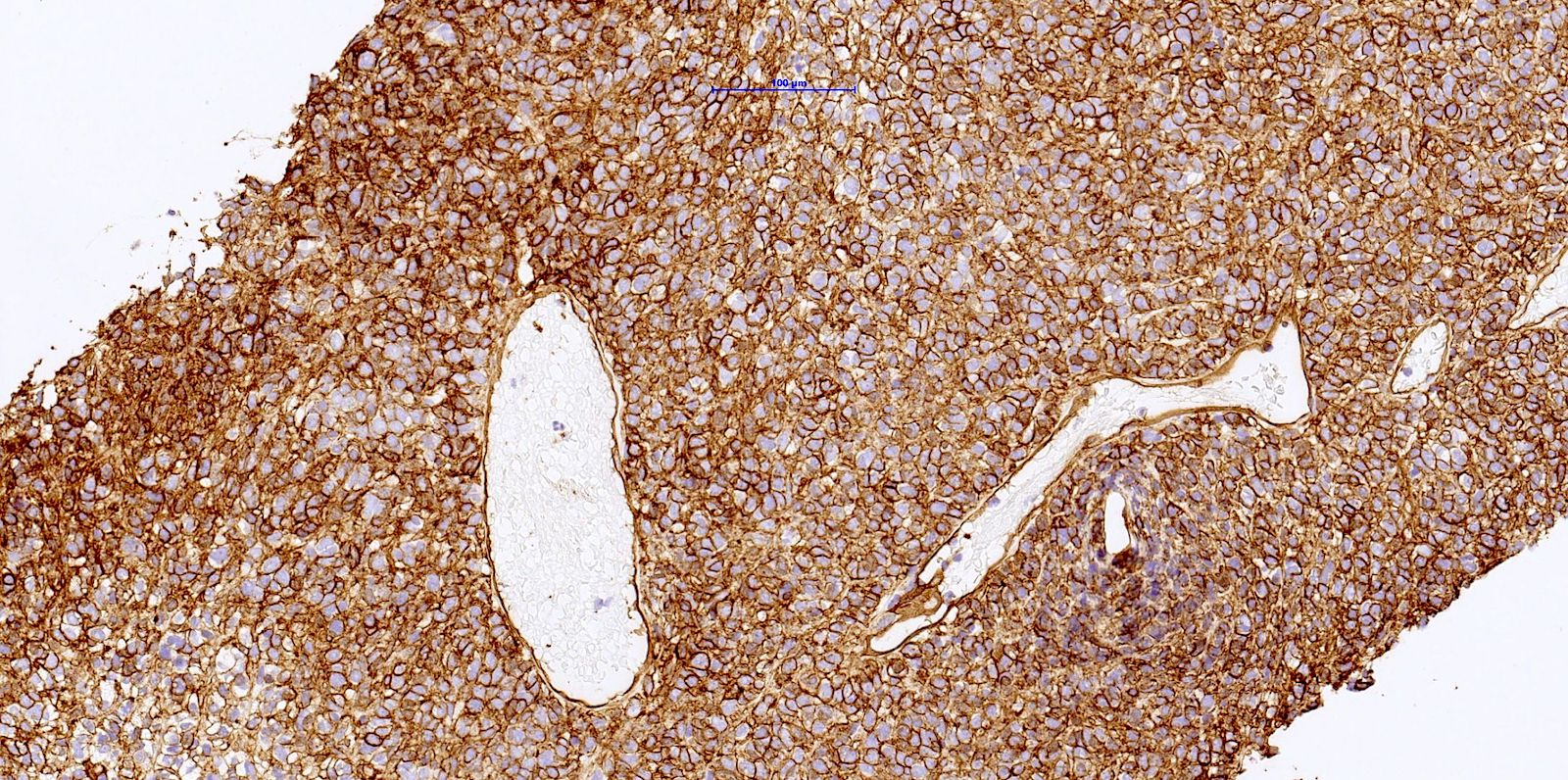
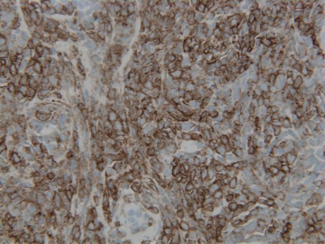
- Membranous stain
- Endothelium acts as a positive internal control
- Staining represents presence of protein, not cross reactivity, despite wide range of tissues that are CD34+ (Am J Pathol 2000;156:21)
Uses by pathologists
- Dermatopathology and soft tissue
- Distinguish CD34+ dermal neoplasms, such as Kaposi sarcoma (CD34+), dermatofibrosarcoma protuberans (DSFP) (CD34+) and epithelioid sarcoma (often CD34+) from dermatofibroma (CD34-)
- Distinguish solitary fibrous tumor (CD34+) from desmoplastic mesothelioma (CD34-) (Hum Pathol 1995;26:428)
- Distinguish hemangiopericytoma (CD34+) from endometrial stromal sarcoma (CD34-) (Mod Pathol 2005;18:40)
- Classify other soft tissue tumors
- Hematopathology
- Distinguish lymphocyte rich thymoma (CD34+ cells) from T cell acute lymphoblastic leukemia (T ALL, CD34-) (Am J Clin Pathol 2004;121:268)
- Quantify and purify lymphohematopoietic stem cell / progenitor cells for research and for clinical bone marrow transplantation (Folia Histochem Cytobiol 2006;44:53, Breast Cancer 2005;12:178)
- Identify blasts in general and in hypoplastic marrows (Am J Clin Pathol 2010;134:749)
- Distinguish acute leukemia and myelodysplastic syndrome (CD34+ blasts present) from aplastic anemia (no blasts / low marrow CD34+ cells) (Leukemia 2006;20:458)
- Identify hematogones and megakaryocytes (CD34+), although not all myeloblasts are CD34+ (Arch Pathol Lab Med 2002;126:823)
- May support the diagnosis of essential thrombocythemia
- Poor prognostic factor in newly diagnosed acute myeloid leukemia (AML) (Neoplasma 2005;52:402)
- Other
- Determine vascularization of tumors or disease processes (Saudi Med J 2006;27:154)
- Distinguish vascular invasion (CD34+) from displaced epithelium (CD34-) (J Clin Pathol 2002;55:780)
- Confirm diagnosis of gastrointestinal stromal tumor (GIST) (Arch Pathol Lab Med 2002;126:1189)
- May confirm diagnosis of chronic intestinal pseudo-obstruction (Am J Surg Pathol 2003;27:228)
- Use with other stains to distinguish meningeal solitary fibrous tumor hemangiopericytoma from meningioma (Am J Surg Pathol 2015;39:1377)
- Determine microvessel density; high microvessel density (using CD34) is associated with
- Poorer prognosis in esophageal squamous cell carcinoma (J Clin Pathol 2001;54:940)
- Poor survival in nonsmall cell lung cancer (J Clin Pathol 2004;57:591)
- Prostate specific antigen (PSA) recurrence of prostate cancer (Am J Clin Pathol 2000;113:555)
Prognostic factors
- Poor prognostic factor in newly diagnosed acute myeloid leukemia (Neoplasma 2005;52:402)
- Determine microvessel density; high microvessel density (using CD34) is associated with
- Poorer prognosis in esophageal squamous cell carcinoma (J Clin Pathol 2001;54:940)
- Poor survival in nonsmall cell lung cancer (J Clin Pathol 2004;57:591)
- PSA recurrence of prostate cancer (Am J Clin Pathol 2000;113:555)
Microscopic (histologic) description
- Membranous
Microscopic (histologic) images


Positive staining - normal
- Endothelium of blood vessels (including alveolar wall capillaries and glomeruli but not hepatic sinusoids or splenic sinusoids), endothelium of some lymphatics (J Histochem Cytochem 2006;54:385)
- Hematopoietic progenitor cells / hematogones (less mature) (Am J Clin Pathol 2009;132:573)
- Also dendritic interstitial cells, dermal dendrocytes, endometrial stroma, endoneurium, fibroblasts, fibrocytes, follicle cells, interstitial cells of Cajal (20%), mast cells, megakaryocytes, neural stem cells in CNS, osteoblasts, perivascular stroma, umbilical cord blood, vascular adventitial fibroblastic cells in stomach (Mod Pathol 2003;16:293, Am J Pathol 2000;156:1157, Exp Neurol 2006;197:399, J Clin Pathol 2001;54:428)
Positive staining - disease
- Acral pseudolymphomatous angiokeratoma of children (APACHE) (J Cutan Pathol 2015;42:50)
- Acute lymphoblastic leukemia, pre-B (Am J Clin Pathol 2009;132:940, Am J Clin Pathol 2004;121:512)
- Alveolar soft part sarcoma (Indian J Ophthalmol 2017;65:889)
- Acute myeloid leukemia (40 - 60%) (Arch Pathol Lab Med 2003;127:42)
- Angioblastoma / tufted angioma (J Clin Pathol 2005;58:214)
- Angiofibroma, cellular (50%) (Mod Pathol 2011;24:82)
- Angiomyofibroblastoma (variable) (Arch Pathol Lab Med 2000;124:1679)
- Angiosarcoma (70%) (Am J Surg Pathol 1998;22:683)
- Benign epithelioid peripheral nerve sheath tumor (fibroblasts are CD34+) (Am J Surg Pathol 2005;29:39)
- Benign stromal spindle cell tumors (Cureus 2023;15:e41961)
- Blood vessels in tumors (Blood 1990;75:2417)
- Calcifying fibrous pseudotumor (Int J Surg Pathol 2002;10:189)
- Cellular blue nevus (J Cutan Pathol 2001;28:145)
- Cellular digital fibroma (J Cutan Pathol 2005;32:413)
- Chordoid glioma (focal) (Am J Surg Pathol 2002;26:1330)
- Chronic myelomonocytic leukemia (Blood Cancer Discov 2022;3:536)
- Congenital / infantile fibrosarcoma (Semin Diagn Pathol 2023;40:295)
- Cystic panfolliculoma surrounding fibrotic stroma is CD34+ (Arch Pathol Lab Med 2006;130:389)
- Dendritic fibromyxolipoma (Mol Clin Oncol 2021;14:7)
- Dendritic interstitial cells at margin of salivary gland tumors (Arch Pathol Lab Med 2001;125:232)
- Dermal dendrocytoma (Am J Dermatopathol 2002;24:50)
- Dermatofibrosarcoma protuberans (Br J Dermatol 1992;127:79, Am J Surg Pathol 2003;27:27)
- Elastofibroma (spindle cells) (Anticancer Res 2021;41:2211)
- Epithelioid hemangioendothelioma (Am J Clin Pathol 1991;96:25)
- Epithelioid sarcoma (50%) (Hum Pathol 1999;30:934, Am J Surg Pathol 1997;21:130)
- Fibroadenoma (breast) (Hum Pathol 2023;139:17)
- Fibroblastic connective tissue nevus (Am J Surg Pathol 2012;36:1509)
- Fibromas with solitary fibrous tumor-like features (Semin Diagn Pathol 2023;40:295)
- Fibrous hamartoma of infancy (Am J Surg Pathol 2014;38:394)
- Ganglioglioma (Acta Neuropathol Commun 2023;11:50)
- Gardner type fibroma of soft tissue (Semin Diagn Pathol 2023;40:295)
- Glioneural hamartoma (J Neuropathol Exp Neurol 2002;61:575)
- Gastric carcinoma, diffuse type (vascular adventitial fibroblastic cells) (J Clin Pathol 2004;57:970)
- Gastrointestinal stromal tumor (50 - 80%) (Am J Surg Pathol 2005;29:52)
- Giant cell angiofibroma (Am J Surg Pathol 2000;24:971)
- Giant cell fibroblastoma (Am J Surg Pathol 2003;27:27)
- Glomus tumor / glomangioma (variable) (J Cutan Pathol 1993;20:15, Tohoku J Exp Med 1997;182:241)
- Hamartomatous tumors of chronic drug resistant epilepsy
- Hemangioblastoma (J Int Med Res 2021;49:3000605211027774)
- Hemangioendothelioma (Medicine (Baltimore) 2023;102:e32914)
- Hemangioma (variable) (Am J Surg Pathol 1999;23:97)
- Hemosiderotic fibrohistiocytic lipomatous lesion (Mod Pathol 2000;13:1192)
- Hepatic sinusoids in conditions associated with altered vascular flow and neoplasms (Res Pract 2023;249:154741)
- Hepatocellular carcinoma (staining of almost all sinusoidal spaces) (Am J Surg Pathol 2008;32:433)
- Inflammatory fibrous polyps of stomach / GI tract (Hum Pathol 2002;33:307, Am J Surg Pathol 2004;28:107)
- Juxtaglomerular cell tumor (may be focal) (Am J Clin Pathol 2001;116:854, Am J Surg Pathol 2004;28:1098)
- Kaposiform hemangioendothelioma (Am J Surg Pathol 2004;28:559)
- Kaposi sarcoma (90%) (strong and diffuse in spindle cells) (J Clin Pathol 2002;55:619)
- Kaposi sarcoma-like pyogenic granuloma (J Clin Pathol 2002;55:619)
- Lipomatous hemangiopericytoma (Am J Surg Pathol 1997;21:195)
- Lipomatous tumors (benign and malignant) (Am J Surg Pathol 1997;21:195)
- Liposarcoma (myxoid, pleomorphic, well differentiated) (Am J Surg Pathol 2002;26:601)
- Lymphangioma (50%) (Hum Pathol 2005;36:426)
- Lymphangioendothelioma (Autops Case Rep 2023;13:e2023435)
- Lymphatic endothelial cells in tumors (Am J Pathol 2006;168:1045)
- Malignant fibrous histiocytoma (Pathol Res Pract 1997;193:51)
- Malignant peripheral nerve sheath tumors (low grade stronger / more frequent than high grade) (Am J Surg Pathol 2003;27:1337)
- Meningioma (some) (Am J Surg Pathol 2002;26:125)
- Metanephric adenofibroma (often) (Int J Clin Exp Pathol 2015;8:3250)
- Metanephric adenosarcoma of kidney (stroma only) (Am J Surg Pathol 2001;25:1451)
- Metanephric stromal tumor of kidney (patchy) (Am J Surg Pathol 2000;24:917)
- Microcystic adnexal carcinoma (Am J Surg Pathol 2001;25:464)
- Mucosal epithelioid nerve sheath tumors (Am J Surg Pathol 2005;29:1310)
- Myelodysplastic syndrome (myeloid blasts)
- Myofibroblastoma (breast, cervicovaginal, soft tissue) (Am J Surg Pathol 2001;25:1022)
- Myxoma of heart (Cardiovasc Pathol 2023;63:107511)
- Myxoma of soft tissue (50%) (In Vivo 2023;37:503)
- Neurofibroma (may be focal) (Am J Clin Pathol 2000;114:123)
- Nuchal fibrocartilaginous pseudotumor of soft tissue (Histol Histopathol 2014;29:831)
- Nuchal type fibroma (Histol Histopathol 2014;29:831)
- Onychomatricoma (Am J Dermatopathol 2010;32:1)
- Papillary thyroid carcinoma (BMC Med Imaging 2021;21:159)
- Paratesticular fibrous pseudotumor (Am J Surg Pathol 2010;34:569)
- Periductal stromal sarcoma of breast (Am J Surg Pathol 2003;27:343)
- Perineurioma (variable) (Am J Surg Pathol 2005;29:859)
- Phyllodes tumor of breast (more common if benign) (J Surg Res 2000;94:84)
- Pleomorphic fibroma of skin (Semin Diagn Pathol 2023;40:295)
- Pleomorphic hyalinizing angiectatic tumor (Arch Pathol Lab Med 2000;124:423)
- Primary renal myxofibrosarcoma (Pathol Res Pract 2015;211:619)
- Pseudoangiomatous stromal hyperplasia of breast (J Clin Pathol 1998;51:204, Am J Surg Pathol 1995;19:270)
- Pseudocyst of adrenal gland (Ann Diagn Pathol 2022;57:151888)
- Reactive angioendotheliomatosis of skin (Am J Dermatopathol 2015;37:581)
- Schwannoma (Antoni B areas) (Am J Clin Pathol 2000;114:123)
- Sclerosing angiomatoid nodular transformation of spleen (capillaries) (Am J Surg Pathol 2004;28:1268)
- Sclerosing hemangioma of lung (Sci Rep 2014;4:6102)
- Sclerosing liposarcoma (spindle and atypical cells) (Int J Surg Case Rep 2023;109:108513)
- Solitary fibrous tumor; expression may be lost in high grade areas (Am J Surg Pathol 1994;18:992, Hum Pathol 1995;26:440, Am J Surg Pathol 2009;33:1314)
- Solitary sclerotic fibroma of skin (J Cutan Pathol 2003;30:631)
- Spindle cell epithelioma of vagina (Am J Dermatopathol 2022;44:764)
- Spindle cell lipoma / pleomorphic lipoma (Am J Dermatopathol 2022;44:764)
- Storiform collagenoma (J Cutan Pathol 2020;47:291)
- Stromal sarcoma of prostate (Diagn Pathol 2012;7:173)
- Subconjunctival herniated orbital fat (Am J Surg Pathol 2007;31:193)
- Superficial acral fibromyxoma (Hum Pathol 2001;32:704)
- Superficial cervicovaginal myofibroblastoma (Pathology 2005;37:144)
- Superficial CD34 positive fibroblastic tumor (Pathology 2015;47:479)
- Thymoma (ectopic hamartomatous variant; otherwise weak) (Am J Surg Pathol 2005;29:1208)
- Transient myeloproliferative disorder (staining of blasts) (Am J Clin Pathol 2001;116:204)
- Vaginal mixed tumor / spindle cell epithelioma (Mod Pathol 2004;17:1243)
- Vulvar fibroma, prepubertal (Am J Surg Pathol 2004;28:1601)
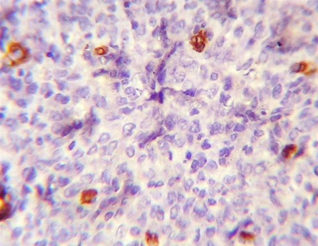
Negative staining
- Normal hepatic sinusoids, bone marrow mast cells
- Adenomatoid tumor (Am J Surg Pathol 2003;27:969)
- Angiomyxofibromatous tumor (Am J Surg Pathol 2006;30:545)
- Anaplastic sarcoma of kidney (Mod Pathol 2018;31:169)
- Angiomyolipoma (Histopathology 2004;44:462)
- Atypical fibrous histiocytoma of skin (focal in 10%) (Am J Surg Pathol 2002;26:35)
- Benign fibrous histiocytoma (but deep tumors are often CD34+)
- Benign vascular proliferations in irradiated skin (Am J Surg Pathol 2002;26:328)
- Calcifying aponeurotic fibroma of soft tissue (Histopathology 2004;44:462)
- Carcinoma (Adv Anat Pathol 2020;27:114)
- Chondromyxoid fibroma (Ann Diagn Pathol 2023;66:152174)
- Clear cell sarcoma of kidney (Histopathology 2004;44:462)
- Desmoplastic fibroblastoma (Oral Surg Oral Med Oral Pathol Oral Radiol 2019;128:e92)
- Epithelioid sarcoma-like hemangioendothelioma (Am J Surg Pathol 2003;27:48)
- Ewing sarcoma / primitive neuroectodermal tumor (PNET) (Ann Diagn Pathol 2023;65:152152)
- Fibroblastic polyp of colon (may be weak / focal) (Am J Surg Pathol 2004;28:374)
- Fibroepithelial polyp of renal pelvis (Urology 2009;73:929.e9)
- Fibromatosis (J Clin Pathol 2004;57:1119, Am J Surg Pathol 2002;26:1296)
- Follicular dendritic cell sarcoma
- Giant cell tumor of tendon sheath
- Gliosarcoma (Arch Pathol Lab Med 1997;121:129)
- Granulocytic sarcoma (47%) (Arch Pathol Lab Med 2001;125:1448)
- Hibernoma (but spindle cell subtype is CD34+) (Cancer Diagn Progn 2023;3:282)
- Histiocytic sarcoma (Am J Surg Pathol 2004;28:1133)
- Inflammatory myofibroblastic tumor (Pathol Res Pract 2023;242:154335)
- Interdigitating dendritic cell sarcoma (Am J Surg Pathol 2002;26:530)
- Leiomyoma (usually) and vascular leiomyoma (Am J Surg Pathol 2001;25:1355, J Clin Pathol 2002;55:395)
- Leiomyosarcoma (usually) (Arch Pathol Lab Med 2017;141:1072)
- Leukemia cutis (Am J Clin Pathol 2009;132:101)
- Littoral cell angioma of spleen (Am J Surg Pathol 1997;21:827)
- Low grade fibromyxoid sarcoma (Arch Pathol Lab Med 2017;141:1072)
- Mammary NOS type sarcoma (Am J Surg Pathol 2006;30:450)
- Melanoma (rarely CD34+) (J Cutan Pathol 2005;32:685)
- Mesothelial cyst (Am J Surg Pathol 1997;21:334)
- Mesoplastic nephroma (Histopathology 2004;44:462)
- Metaplastic carcinoma of breast, spindle cell type (J Clin Pathol 2023 Aug 16 [Epub ahead of print])
- Multicentric reticulohistiocytosis (J Rheumatol 2014;41:780)
- Myelolipoma (Am J Surg Pathol 2006;30:838)
- Myoepithelioma of breast (Cancers (Basel) 2022;14:476)
- Myofibrosarcoma of breast (Cancers (Basel) 2022;14:476)
- Myoid gonadal stromal tumor (Am J Clin Pathol 2014;142:675)
- Myointimoma (Int J Impot Res 2021;33:583)
- Myxofibrosarcoma (38%) (J Clin Pathol 2003;56:666)
- Nodular fasciitis (Radiol Case Rep 2023;18:1564)
- Ossifying fibromyxoid tumor (Diagn Cytopathol 2015;43:646)
- Paratesticular leiomyosarcoma (30%) (Integr Cancer Ther 2020;19:1534735419900554)
- PEComa (Am J Surg Pathol 2005;29:1558)
- Pigmented villonodular synovitis (Clin Podiatr Med Surg 2023;40:381)
- Plexiform fibrohistiocytic tumor (Clin Podiatr Med Surg 2023;40:381)
- Plexiform fibromyxoma (Am J Surg Pathol 2009;33:1624)
- Plexiform xanthomatous tumor (Am J Surg Pathol 2002;26:1302)
- Pseudomycogenic hemangioendothelioma (Am J Surg Pathol 2011;35:190)
- Reactive fibroblastic and myofibroblastic proliferation of vulva (Am J Surg Pathol 2011;35:110)
- Schwannoma (Int J Clin Oncol 2021;26:284)
- Sclerosing epithelioid fibrosarcoma of the kidney (Diagn Pathol 2015;10:186)
- Sclerosing epithelioid fibrosarcoma of soft tissue (Skeletal Radiol 2023 Jul 31 [Epub ahead of print])
- Sclerosing hemangioma of lung (Hum Pathol 2004;35:503)
- Sclerosing paraganglioma (Head Neck Pathol 2015;9:300)
- Sclerosing perineurioma (J Gen Intern Med 2022;37:3188)
- Sinonasal hemangiopericytoma (Arch Pathol Lab Med 2001;125:686)
- Synovial sarcoma (focal in 6% of monophasic tumors) (Am J Surg Pathol 2002;26:1434)
- Thymoma (BMC Cancer 2023;23:161)
Sample pathology report
- Left thoracic tumor, resection:
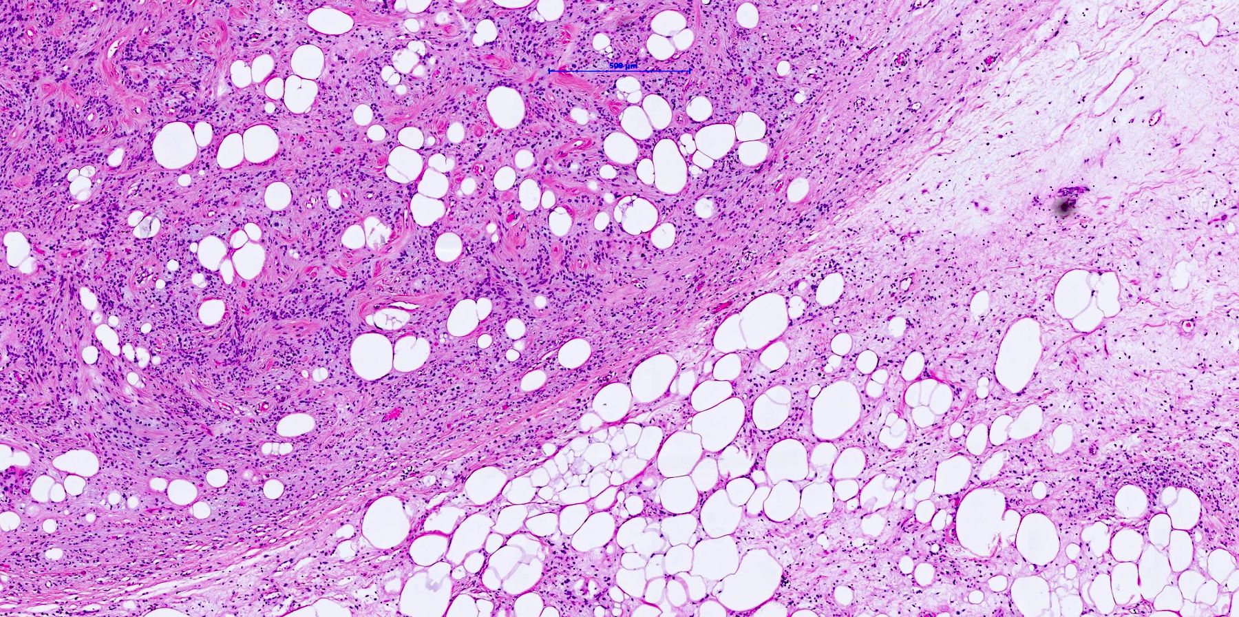
- Spindle cell lipoma (maximum 11.5 cm) (see comment)
- MDM2 FISH: negative (no gene amplification)
- Comment: In combination with the additional molecular examinations, the findings fit primarily with a spindle cell lipoma (MDM2 negative and CD34 positive). There is no evidence for liposarcoma / atypical lipomatous tumor and the criteria for the diagnosis of an atypical spindle cell lipomatous tumor are not fulfilled.
Board review style question #1


If positive, which marker can support the diagnosis of a gastrointestinal stromal tumor (GIST)?
- CD34
- Desmin
- EMA
- S100
- SMA
Board review style answer #1
A. CD34. Under the given answers CD34 is the distinguishing marker for soft tissue neoplasms and in the majority of GIST positive. Other markers supporting the diagnosis of GIST are DOG1 and c-KIT (CD117). Answers B and E are incorrect because desmin and SMA are usually negative in GIST but positive in leiomyoma. Answer D is incorrect because S100 is a strong marker for neuronal differentiation and should be negative in GIST (Curr Opin Gastroenterol 2019;35:555). Answer C is incorrect because EMA is highly expressed in carcinomas and not in GIST.
Comment Here
Reference: CD34
Comment Here
Reference: CD34

