Table of Contents
Definition / general | Essential features | Pathophysiology | Clinical features | Uses by pathologists | Case reports | Microscopic (histologic) images | Positive staining - normal | Positive staining - disease | Negative staining | Flow cytometry description | Sample pathology report | Board review style question #1 | Board review style answer #1 | Board review style question #2 | Board review style answer #2Cite this page: Frauenfeld L, Schürch CM. CD20. PathologyOutlines.com website. https://www.pathologyoutlines.com/topic/cdmarkerscd20.html. Accessed January 15th, 2025.
Definition / general
Essential features
- Widely used B cell marker (also CD19, CD79a, PAX5)
- Retained on mature B cells until plasma cell differentiation
- Anti-CD20 therapy (e.g., rituximab) is available for treatment of non-Hodgkin B cell lymphoma and autoimmune diseases
Pathophysiology
- 33kd phosphoprotein with 3 hydrophobic regions that traverse the cell membrane, creating a structure similar to an ion channel that allows for the influx of calcium required for cell activation
- Initially expressed on B cells after CD19 / CD10 expression and before CD21 / CD22 and surface immunoglobulin expression; retained on mature B cells until plasma cell differentiation
- Delivers early signal in B cell activation
- FMC7 detects a conformational epitope on the CD20 molecule with probable cholesterol dependency (Cytometry 2001;46:98, Leukemia 2003;17:1384)
Clinical features
- Rituximab is a chimeric murine / human anti-CD20 antibody used to treat B cell lymphomas; treatment may cause selection of CD20 negative (but CD79a positive) tumor subclones (Am J Surg Pathol 2005;29:1399, Ann Hematol 2020;99:2141)
- Changes in morphology and CD20 expression after rituximab therapy vary widely (Am J Surg Pathol 2013;37:563, Ann Hematol 2020;99:2141)
- Rituximab is also used to treat autoimmune disorders, thrombotic thrombocytopenic purpura / hemolytic uremia syndrome (TTP / HUS), ABO incompatible transplantation and transplant rejection (Clin Immunol 2005;117:207, J Clin Aesthet Dermatol 2013;6:45, Ther Adv Neurol Disord 2018;11:1756286418773025, Neurology 2018;90:e1805, Clin Transplant 2005;19:423, Blood Transfus 2010;8:203, Animal Model Exp Med 2019;2:76, Transplant Proc 2005;37:1205, Int Rev Immunol 2019;38:118, Clin Transplant 2005;19:137, Clin J Am Soc Nephrol 2020;15:430, Am J Transplant 2018;18:927)
- Radioimmunotherapy with rituximab may be effective for follicular lymphoma and as myeloablative therapy followed by autologous stem cell support for relapsed / refractory B cell lymphoma (Clin Dev Immunol 2013;2013:875343, Oncotarget 2013;4:899)
- Dim CD20 expression on IgG4 plasma cells in IgG4 related lymphadenopathy may explain efficacy of rituximab in IgG4 related disease (Arch Pathol Lab Med 2013;137:1282, Eur J Intern Med 2020;74:92)
- Further anti-CD20 antibodies are ocrelizumab (mainly used in therapy of multiple sclerosis), ofatumumab (in use for relapsed CLL and multiple sclerosis) and obinutuzumab (in use for relapsed or refractory follicular lymphoma and advanced CLL) (Ther Adv Neurol Disord 2018;11:1756286418773025, N Engl J Med 2014;371:213, Neurology 2018;90:e1805, N Engl J Med 2017;377:1331, N Engl J Med 2019;380:2225)
- CD20 antigen may be expressed by reactive or lymphomatous cells of transformed mycosis fungoides as well as in other T cell lymphomas (see Positive staining - disease) (Am J Surg Pathol 2013;37:1845, Am J Dermatopathol 2013;35:833)
- CD20 mutation rarely causes common variable immunodeficiency 5 (OMIM: Membrane-Spanning 4 Domains, Subfamily A, Member 1; MS4A1 [Accessed 20 September 2021])
- Double hit lymphomas show reduced CD20 expression by flow cytometry (Am J Clin Pathol 2010;134:258)
Uses by pathologists
- Commonly used B cell marker used in the workup of benign and malignant processes
Case reports
- 21 year old man with nodular lymphocyte predominant Hodgkin lymphoma with large atypical cells (Case #284)
Microscopic (histologic) images
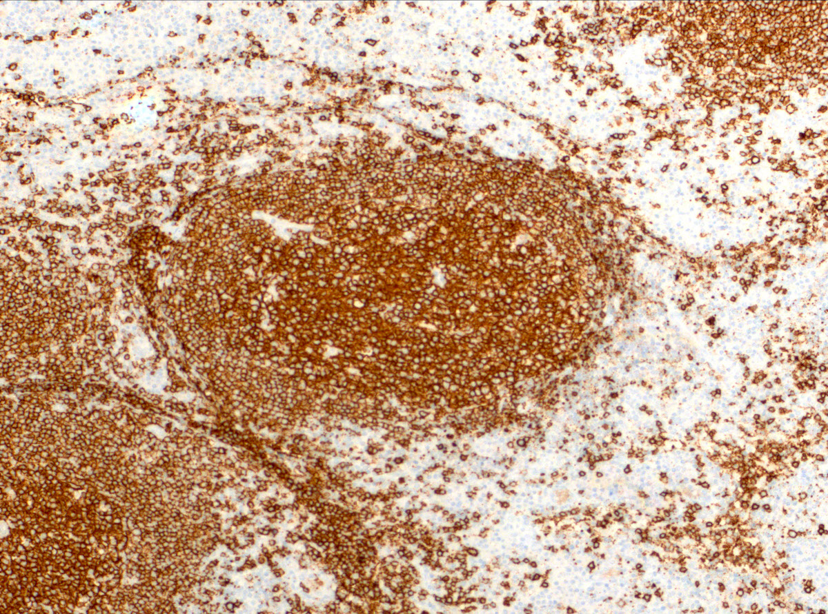
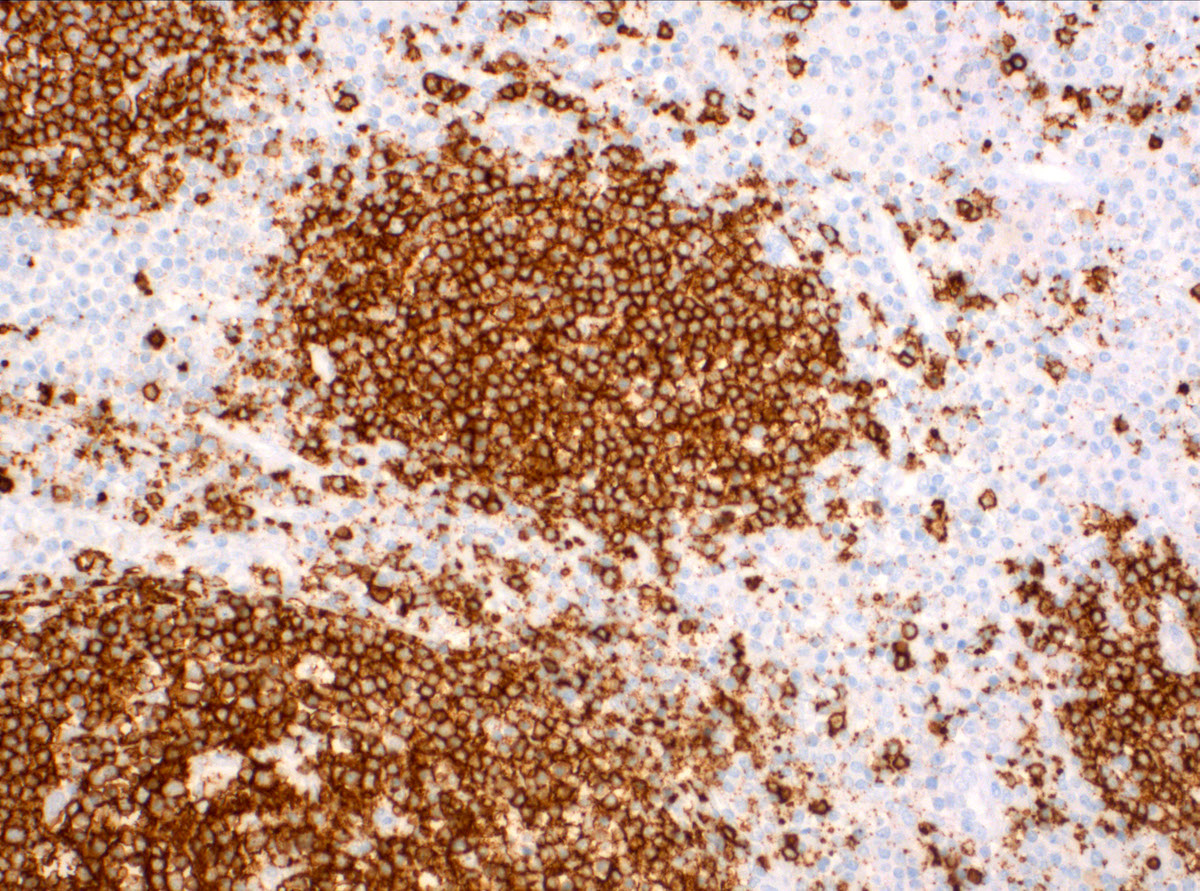
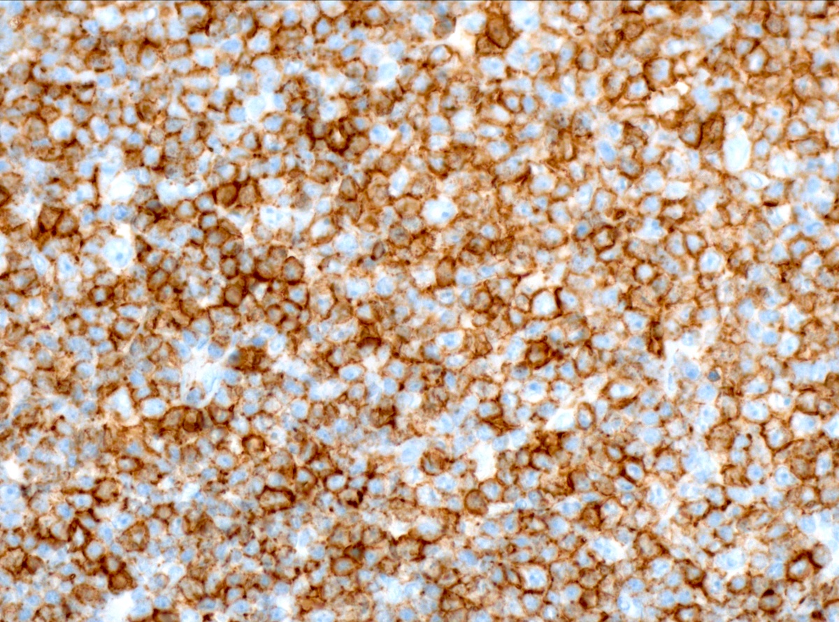
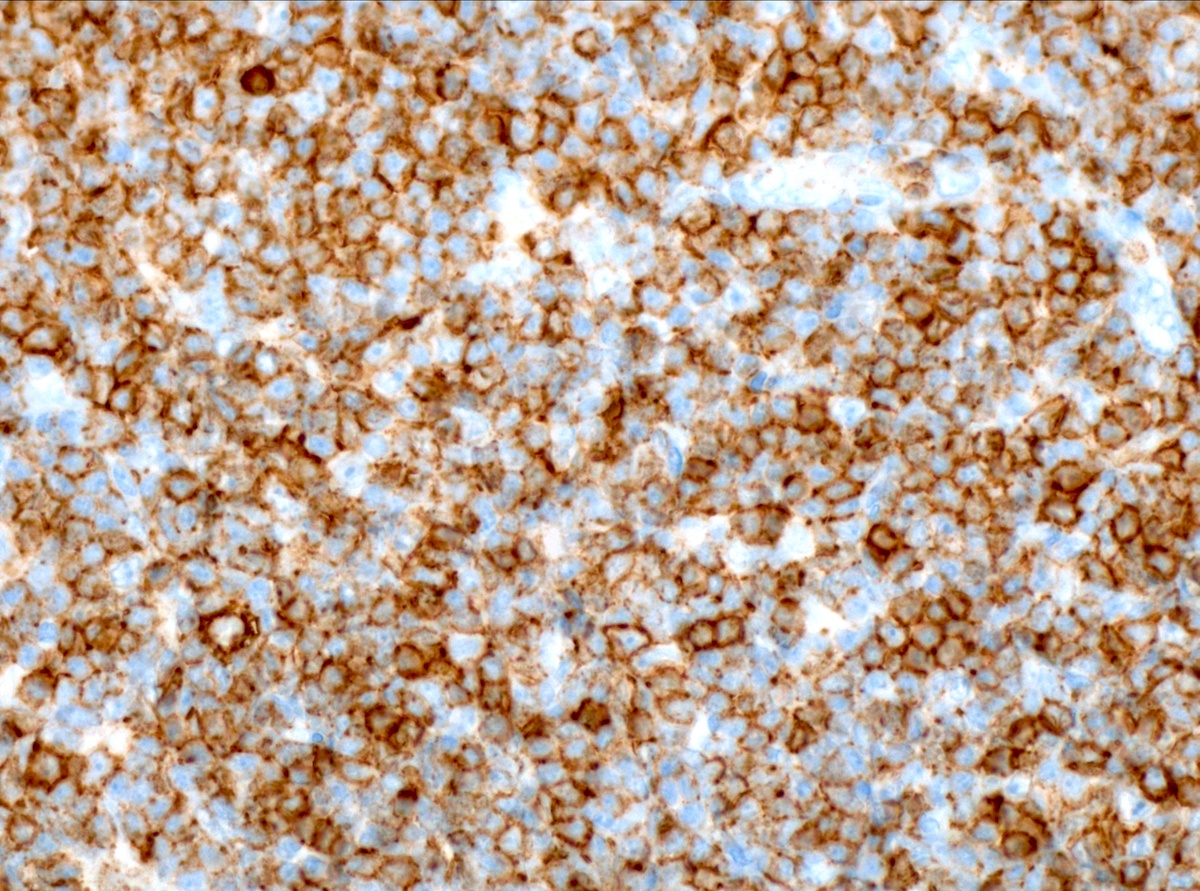
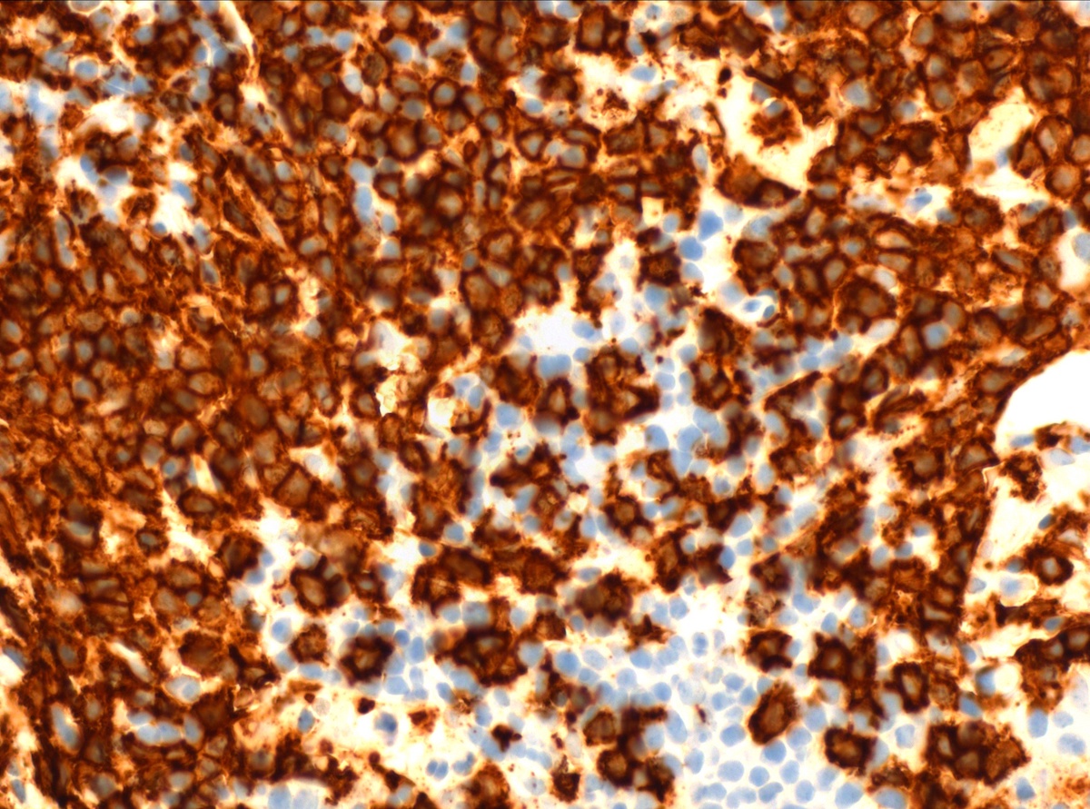
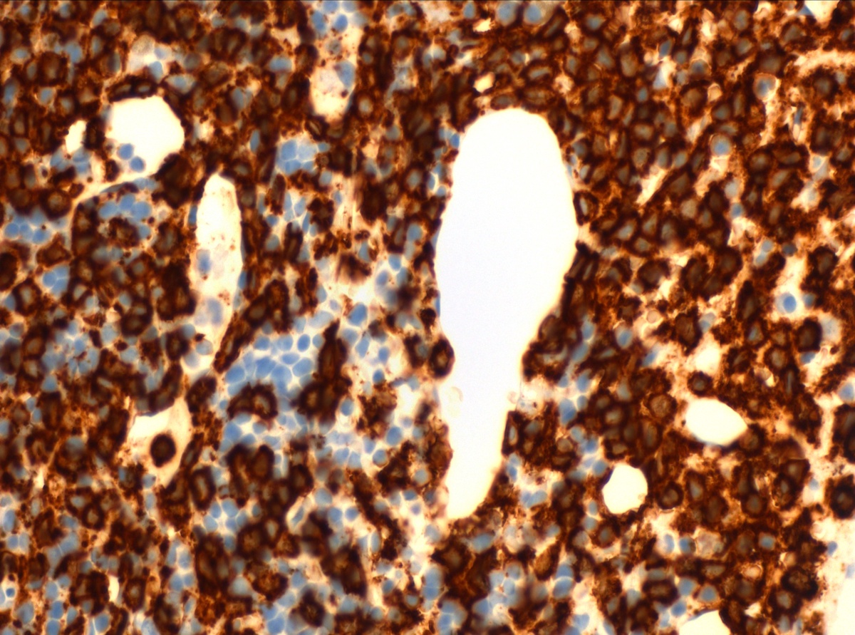
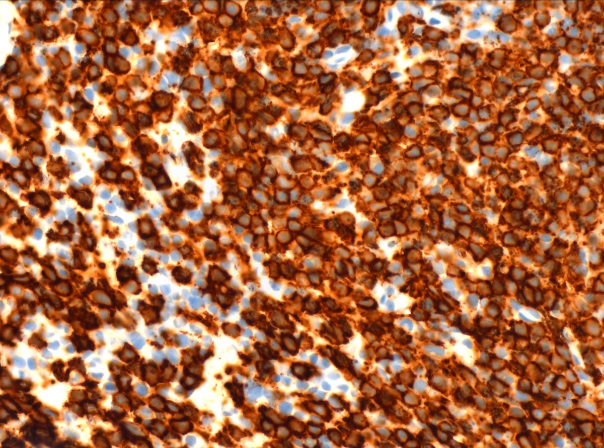
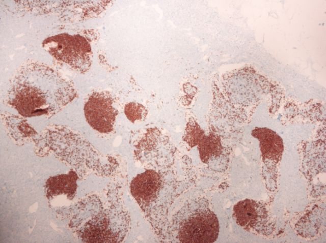
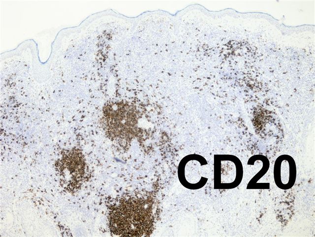
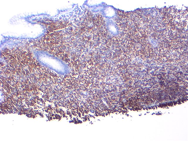
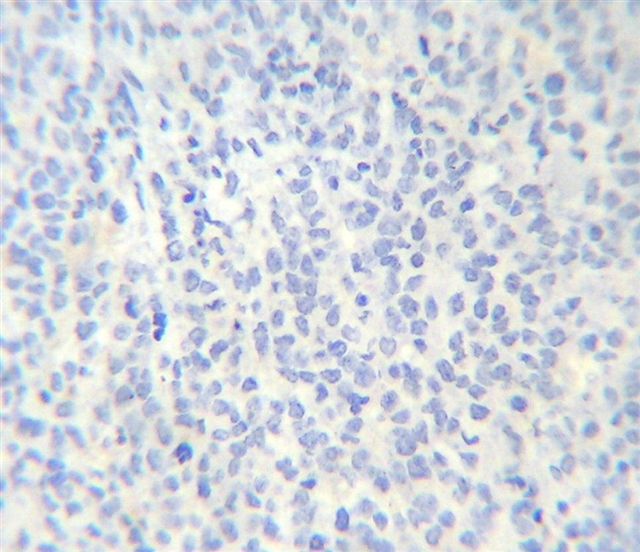
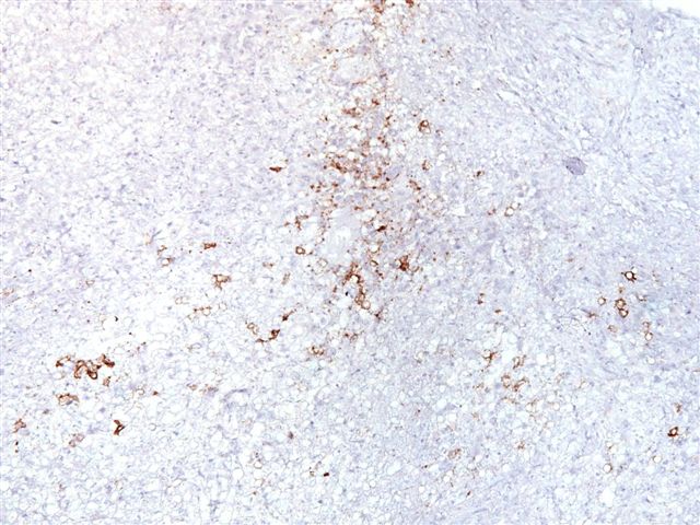
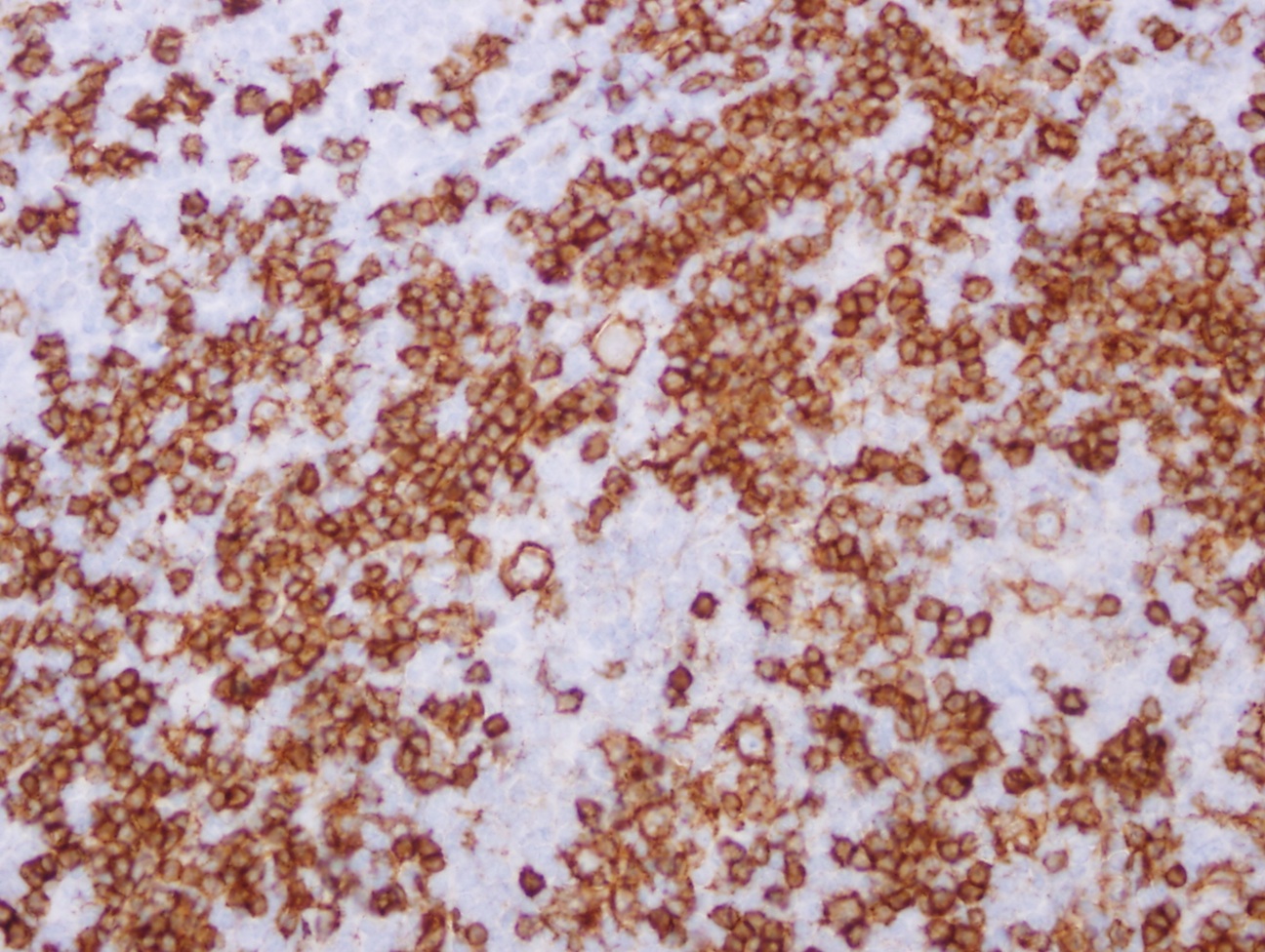
Contributed by Leonie Frauenfeld, M.D., Andrey Bychkov, M.D., Ph.D. and Kaveh Naemi, D.O.
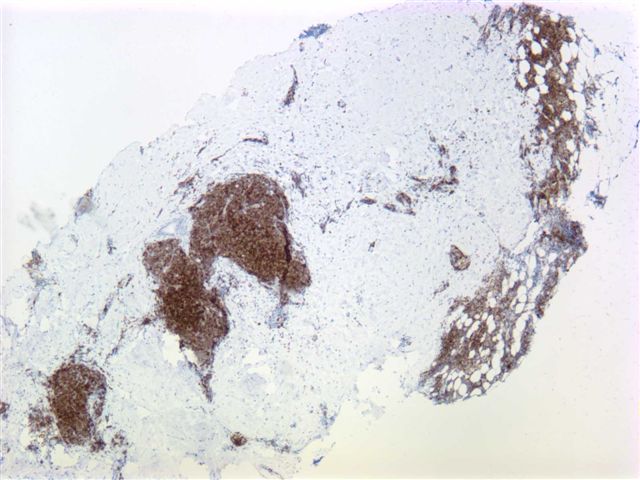
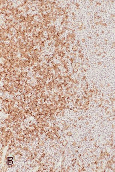
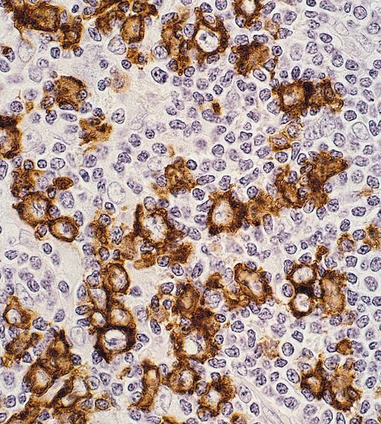
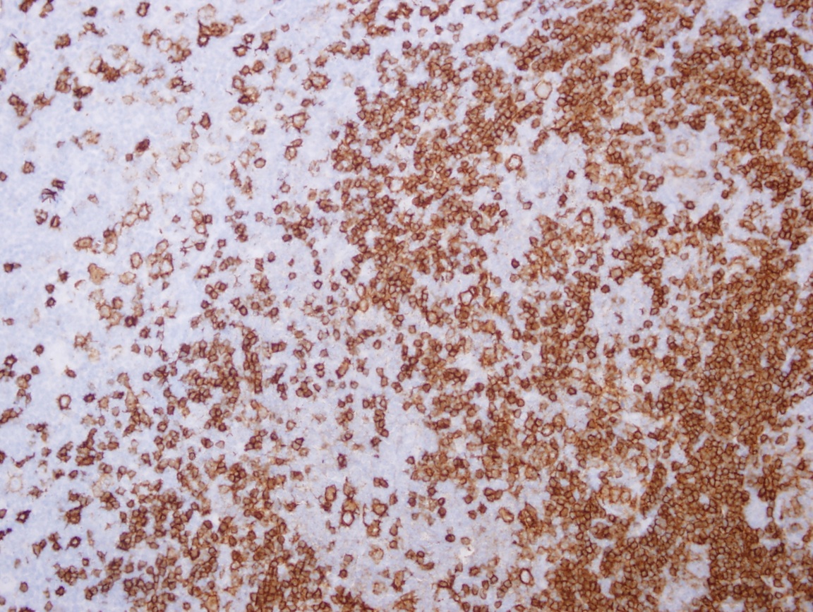
Cases #101, 118, 127, 130, 284 and AFIP images
Positive staining - normal
- Most B cells (considered a pan-B cell antigen), also follicular dendritic cells
- Hematogones can be CD20+ (acquire CD20 during maturation) and a panel is useful in distinguishing them from leukemia cells (Am J Clin Pathol 2000;114:66, Blood 2001;98:2498)
Positive staining - disease
- 90% of B cell lymphomas, although B cell lymphomas are CD20 negative after rituximab and other B cell markers (CD19, CD79a, PAX5) should be used (Am J Clin Pathol 2006;126:534, Biomark Res 2017;5:5, Ann Hematol 2020;99:2141)
- 40% of pre-B acute lymphoblastic leukemia / lymphoblastic lymphoma (ALL / LBL) (Hematology Am Soc Hematol Educ Program 2018;2018:9)
- Also spindle cell thymomas (Am J Surg Pathol 1992;16:988, Diagn Pathol 2007;2:13)
- 80% of nodular lymphocyte predominant Hodgkin lymphoma, 20% of classic Hodgkin lymphoma (may be an adverse prognostic factor) (Br J Haematol 2004;125:701, PLoS One 2009;4:e6341, Br J Haematol 2019;184:45)
- Dimly expressed in T cells (benign and neoplastic, particularly in bone marrow), some myelomas (especially multiple myeloma with lymphoplasmacytoid morphology) and occasionally acute myelogenous leukemia (Am J Surg Pathol 2008;32:1593, Am J Clin Pathol 1996;106:78, Am J Clin Pathol 1994;102:483, Mod Pathol 2004;17:1217, Blood 2003;102:1070, Leuk Lymphoma 2016;57:335, Am J Clin Pathol 2013;140:519, Virchows Arch 2020;476:337)
- Certain T cell lymphomas such as MEITL have a 20% positivity rate for CD20 (Pathology 2020;52:128)
Negative staining
- Nonhematopoietic cells, most T cells, basophils, plasma cells and mast cells
- Note: staining does not work well with Bouin fixative
Flow cytometry description
- CD20 can be evaluated by flow cytometric immunophenotyping as well as immunohistochemical stains
- Dim or bright CD20 expression can provide clues to diagnoses
- Brighter expression in follicular lymphomas than normal B cells (Am J Clin Pathol 2005;124:576)
- In chronic lymphocytic leukemia / small lymphocytic lymphoma (CLL / SLL), CD20 expression may be dim to negative
Sample pathology report
- Lymph node, excision:
- Diffuse large B cell lymphoma (DLBCL)
- Comment: Lymph node presents with effaced architecture and demonstrates a dense, sheet-like proliferation of immunoblastic cells with strong, homogeneous CD20 positivity. Additionally, the cells mark positive for CD10 and BCL6 but are negative for MUM1, according to a germinal center type DLBCL (Hans algorithm). The cells are positive for BCL2 and weakly positive for MYC (20%).
Board review style question #1
Board review style answer #1
Board review style question #2
CD20 is the target of monoclonal antibodies used in the therapy against
- Anaplastic large cell lymphoma
- Mycosis fungoides
- Rheumatoid arthritis
- T cell large granular lymphocytic leukemia (T-LGL)
Board review style answer #2


