
Breast
Other nonneoplastic
Subareolar sclerosing duct hyperplasia
Author: Cansu Karakas, M.D.

Last author update: 1 February 2016
Last staff update: 1 August 2023
Copyright: 2002-2024, PathologyOutlines.com, Inc.
PubMed Search: Subareolar sclerosing hyperplasia
Table of Contents
Definition / general | Clinical features | Treatment | Gross description | Microscopic (histologic) description | Microscopic (histologic) images | Differential diagnosisCite this page: Karakas C. Subareolar sclerosing duct hyperplasia. PathologyOutlines.com website. https://www.pathologyoutlines.com/topic/breastsubareolarsclerosing.html. Accessed April 16th, 2024.
Definition / general
- A sclerosing papillary variant of radial sclerosing lesion in the subareolar region, first described in 1987 (Cancer 1987;59:1927)
- Beneath areola, without involvement of surface of nipple (called nipple adenoma if nipple is involved)
- Note: this is the only article describing this lesion in the literature
Clinical features
- 26 to 73 years, mean 46 years
- A mass located beneath the nipple or areola in the breast
- Left and right breast affected equally
- Erosion or ulceration of the nipple are absent
- Nipple retraction may occur
- Several patients had blood discharge
- Nonspecific mammographic findings, may mimic carcinoma
Treatment
- Excision through circumareolar incision, sparing the nipple
- Recurrence may occur after incomplete excision
- Benign, no evidence that this condition is premalignant
Gross description
- Firm to hard, round to oval tumor with indistinct margins
- Mean 1.2 cm, range 0.6 to 2.0 cm
- Yellow streaks may be noted
Microscopic (histologic) description
- Prominent central elastosis and sclerosis in the center of the tumor, duct hyperplasia is more prominent in the periphery, causing distortion of the ductal pattern
- Cartilaginous metaplasia may occur in the sclerotic core
- Varying amounts of papillary ductal proliferation
- Papillary epithelial hyperplasia within ducts may exhibit considerable atypia
- Generally no cystic change, no papilary apocrine change, no squamous metaplasia
Microscopic (histologic) images
AFIP images
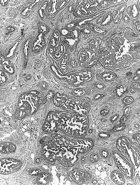
Papillary duct proliferation
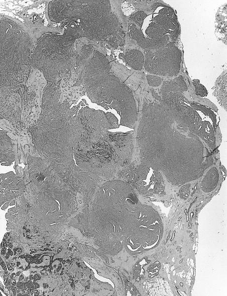
Solid foci in ducts

Papillary duct hyperplasia
Differential diagnosis
- Nipple adenoma / florid papillomatosis of the nipple: typically more nodular than satellite; apocrine metaplasia, squamous cyst, squamous metaplasia may occur
