
Breast
Other invasive carcinoma subtypes, WHO classified
Papillary
Authors: Zena Jameel, M.D., Marilin Rosa, M.D.
Editorial Board Members: Gary Tozbikian, M.D., Julie M. Jorns, M.D.


Last author update: 15 April 2021
Last staff update: 10 March 2023
Copyright: 2002-2024, PathologyOutlines.com, Inc.
PubMed search: Invasive papillary carcinoma[TIAB] breast
Table of Contents
Definition / general | Essential features | Terminology | ICD coding | Epidemiology | Sites | Etiology | Clinical features | Diagnosis | Radiology description | Prognostic factors | Case reports | Treatment | Gross description | Microscopic (histologic) description | Microscopic (histologic) images | Positive stains | Negative stains | Sample pathology report | Differential diagnosis | Additional references | Board review style question #1 | Board review style answer #1Cite this page: Jameel Z, Rosa M. Papillary. PathologyOutlines.com website. https://www.pathologyoutlines.com/topic/breastmalignantpapillary.html. Accessed April 24th, 2024.
Definition / general
- A rare subtype of invasive ductal carcinoma (IDC) (J Surg Res 2021;261:105)
- Comprises about 0.5 - 0.7% of all invasive breast cancers (J Surg Res 2021;261:105, J Cancer Res Clin Oncol 2013;139:77)
Essential features
- A rare subtype of invasive ductal carcinoma with infiltrative papillary growth
- Imperative to distinguish it from other invasive and noninvasive mammary lesions, as well as extramammary tumors metastasizing to the breast
Terminology
- Invasive papillary carcinoma (IPC)
ICD coding
Epidemiology
- Most commonly seen in non-Caucasian postmenopausal women in their sixth to eighth decade of life (J Cancer Res Clin Oncol 2013;139:77, J Surg Res 2021;261:105)
- More common in lower economic status and lower household income (J Surg Res 2021;261:105)
Sites
- No specific location in the breast
Etiology
- Insufficient clinicopathological data on this tumor (Mod Pathol 2021 Jan 18 [Epub ahead of print])
- Much of the published literature about IPC in fact describes variants of encapsulated or solid papillary carcinoma with invasion (Mod Pathol 2021 Jan 18 [Epub ahead of print])
Clinical features
- Due to the rarity of this tumor, specific clinical features have not been described
- Typically presents with bloody nipple discharge, an abnormal mass or radiographic abnormalities
- However, it is not clear if these features refer to true IPC or other types of papillary carcinomas misdiagnosed as IPC (J Surg Res 2021;261:105)
Diagnosis
- Mammogram
- Breast ultrasound (US)
- Breast MRI
Radiology description
- Solitary mass, solid, solid and cystic, round, oval, lobulated or irregular (Clin Radiol 1997;52:865, Curr Probl Diagn Radiol 2019;48:348)
- Hypoechoic solid, smooth walled abnormalities, with good sound transmission on ultrasound (Cureus 2020;12:e11026)
Prognostic factors
- Tumors should be graded according to the Nottingham grading system (Histopathology 1991;19:403)
- Tumors should be staged according to their pathological size and axillary lymph node status
- Poor prognostic factors include old age, advanced pathologic stage and patients without radiation (J Surg Res 2021;261:105)
- Conflicting data; some report favorable prognosis when compared with invasive ductal carcinoma (J Cancer Res Clin Oncol 2013;139:77)
- Others report similar prognosis (J Surg Res 2021;261:105)
Case reports
- 45 year old woman with a right breast lump (J Cancer Res Ther 2015;11:1029)
- 52 year old woman with a right nipple lump (BMJ Case Rep 2018;2018:bcr2017222817)
- 61 year old woman with a painless breast mass (Indian J Pathol Microbiol 2012;55:543)
- 79 year old woman with an irregular 9 mm right breast mass (Pathol Int 2019;69:183)
Treatment
- Surgical excision followed by adjuvant radiation and systemic therapy based on the predictive and prognostic factors (J Cancer Res Clin Oncol 2013;139:77)
Gross description
- Size ranging from less than 2 cm to more than 5 cm (Cureus 2020;12:e11026)
Microscopic (histologic) description
- Invasive carcinoma with > 90% papillary architecture (Mod Pathol 2021 Jan 18 [Epub ahead of print])
- Frankly invasive growth pattern without surrounding fibrous capsule (Mod Pathol 2021;34:78)
- Growth pattern is characterized by arborizing fibrovascular stalks lined by epithelial cells (J Cancer Res Clin Oncol 2013;139:77)
- Mitotic figures and necrosis are not common (Sci Rep 2016;6:24037)
- Carcinoma cells show cytologic atypia and stratification (J Cancer Res Clin Oncol 2013;139:77)
- Nuclear grade ranges from low to high (Sci Rep 2016;6:24037)
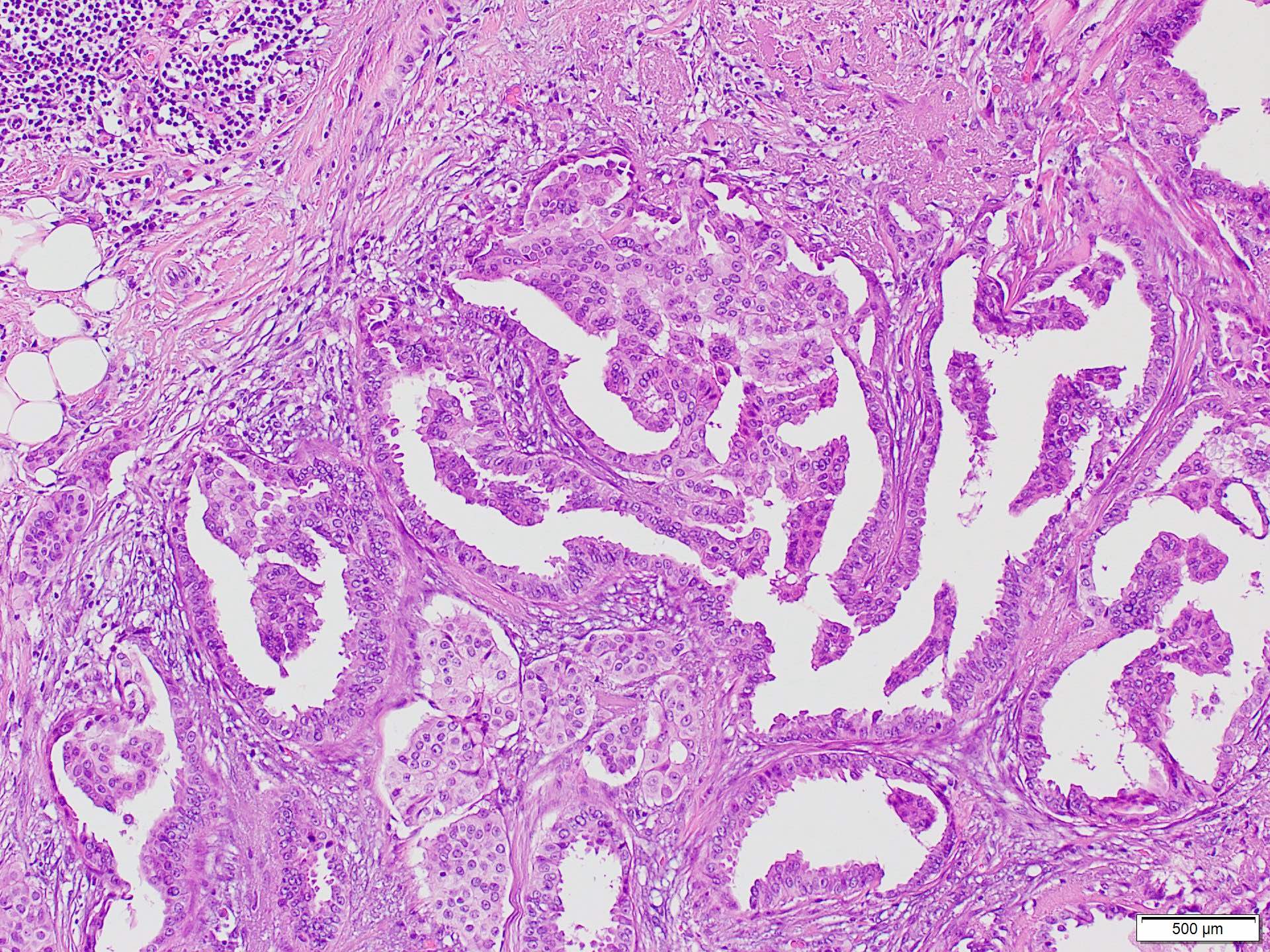
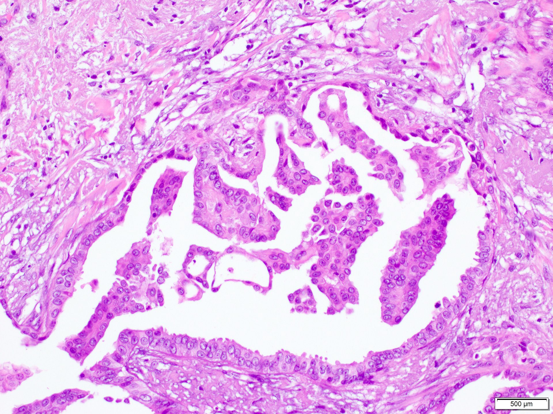
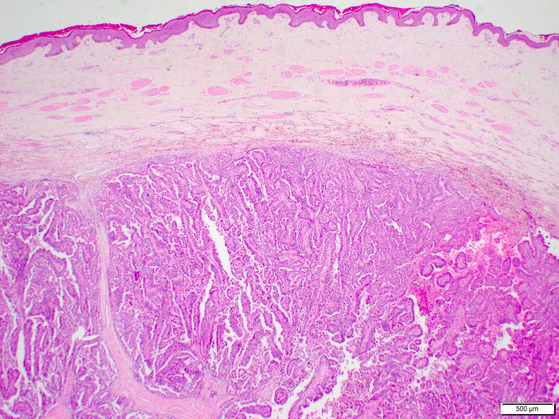
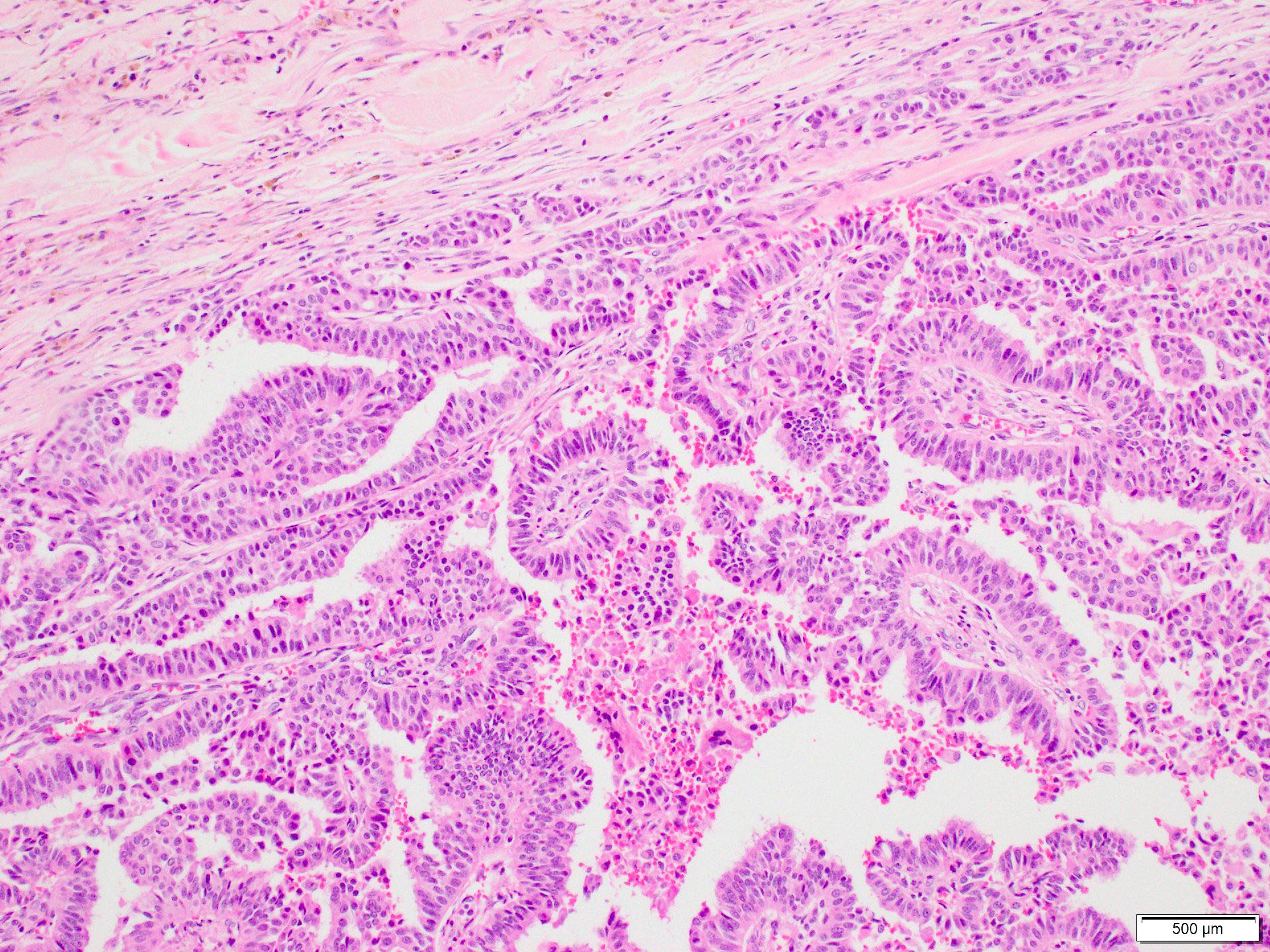
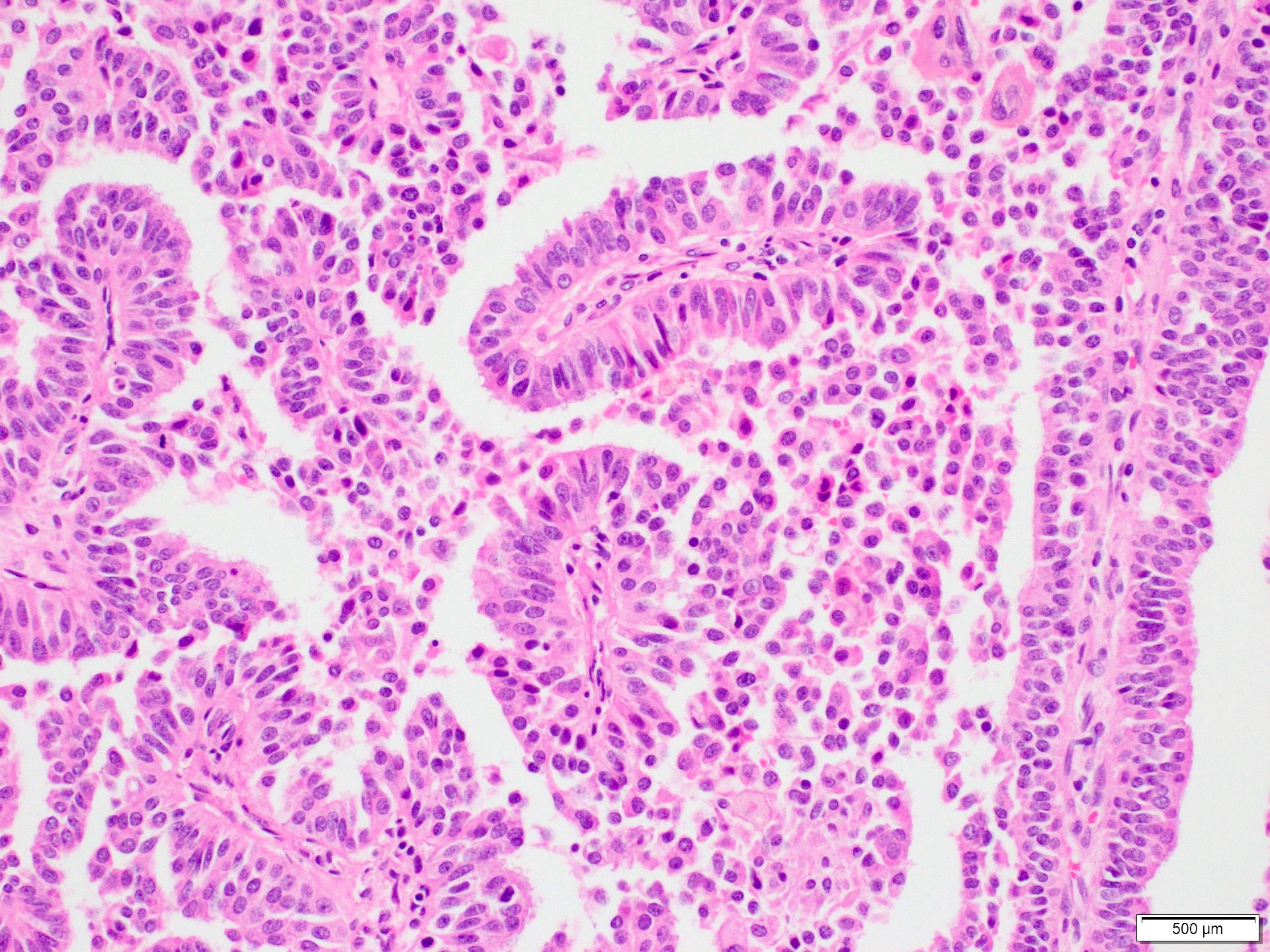
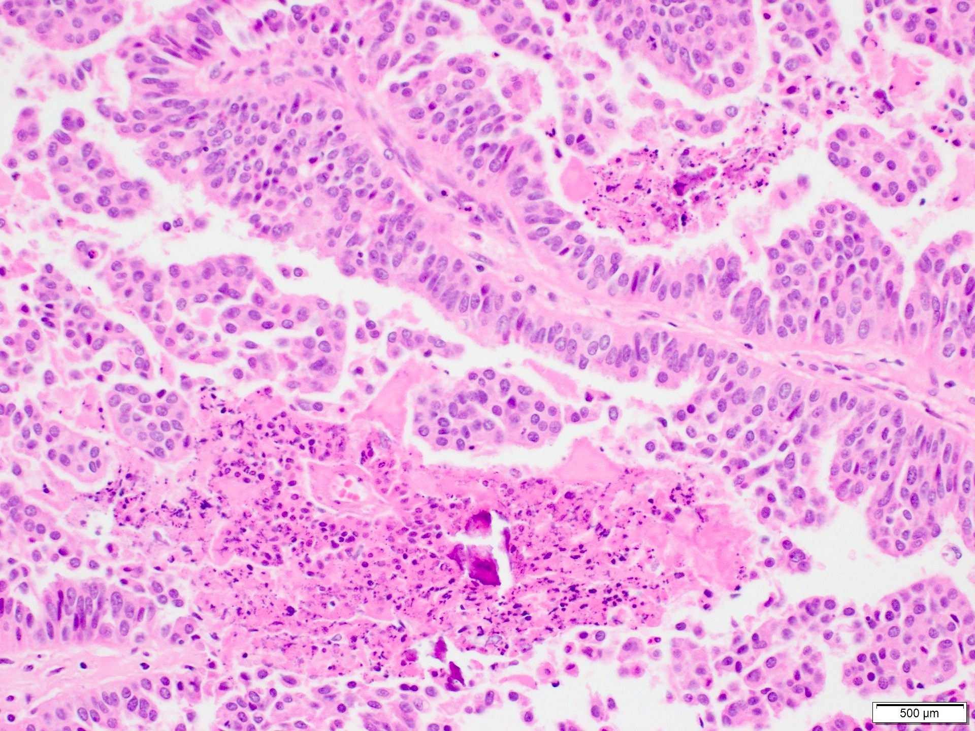
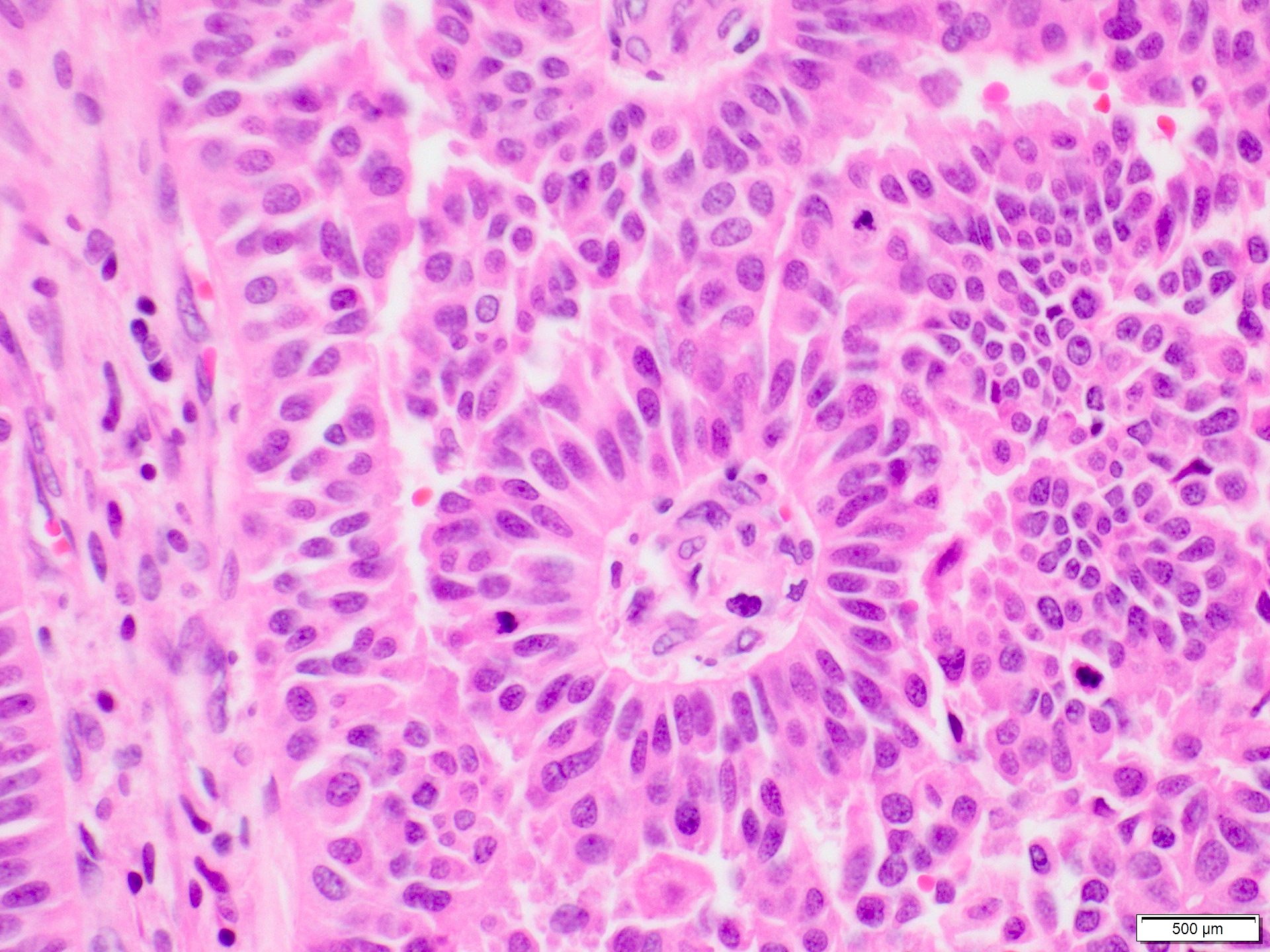
Microscopic (histologic) images
Contributed by Marilin Rosa, M.D.
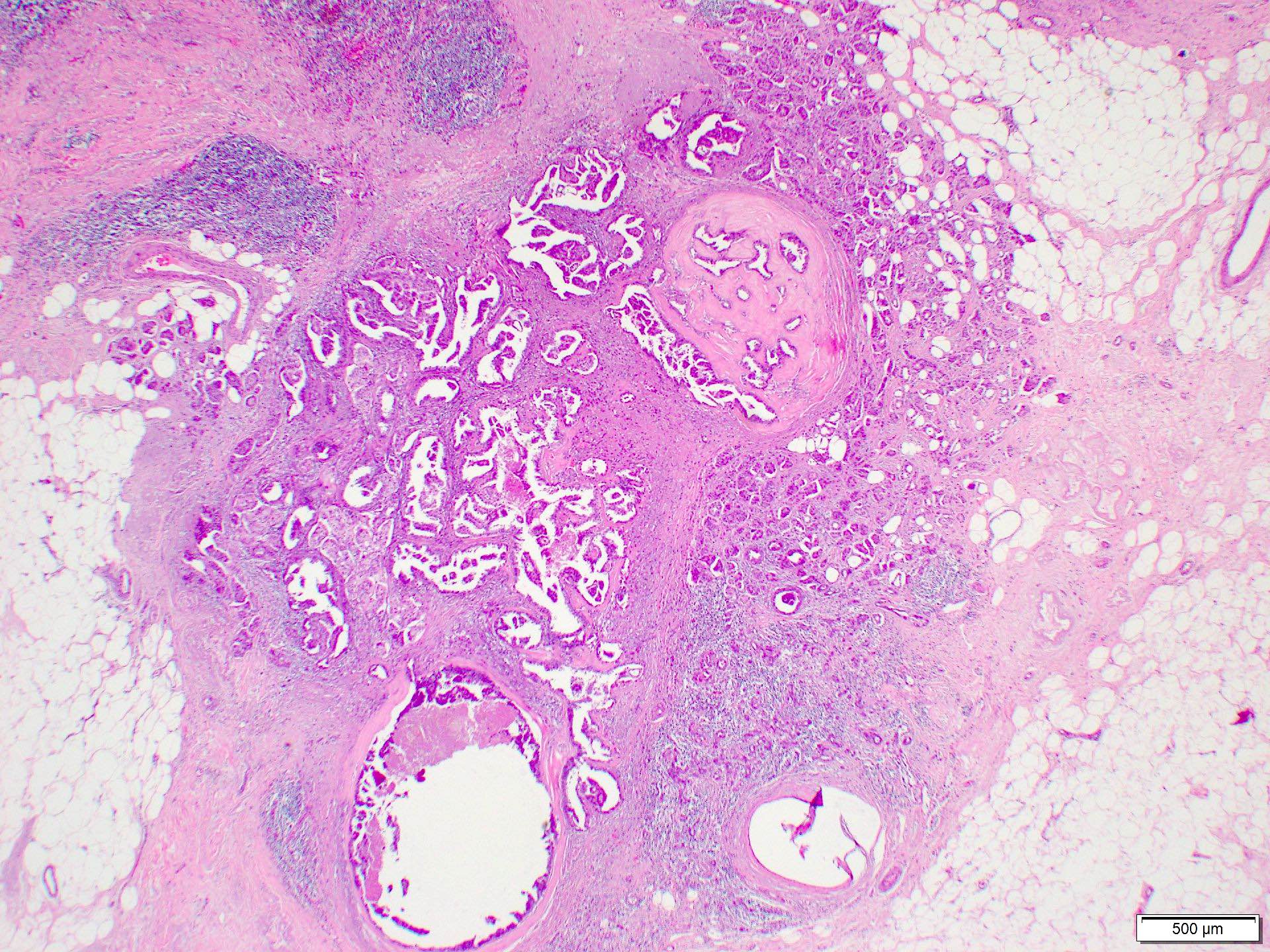
Invasive mixed papillary and micropapillary carcinoma
No surrounding fibrous capsule
Desmoplasia
Invasive papillary carcinoma
No surrounding fibrous capsule
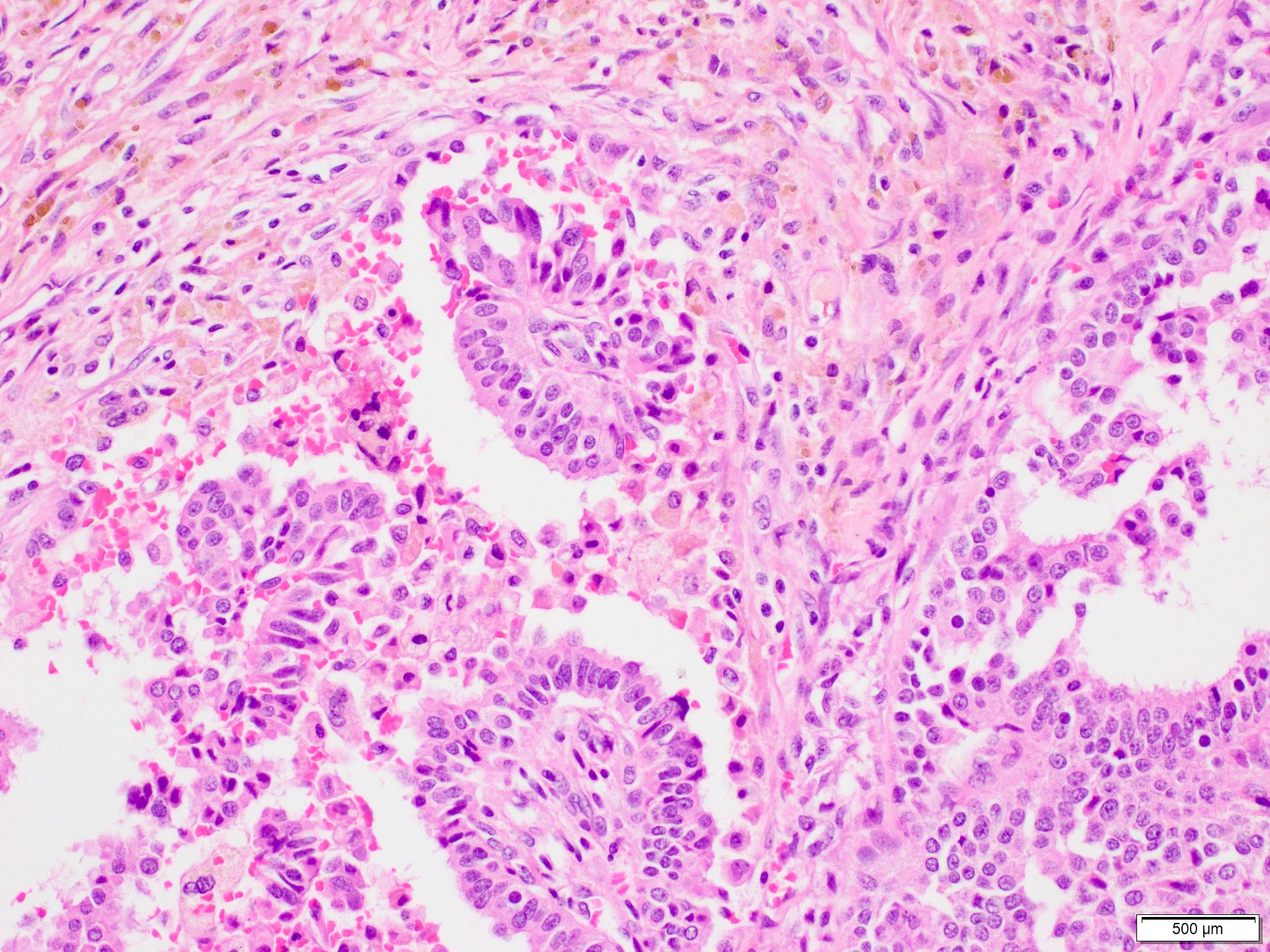
Invasive papillary fronds
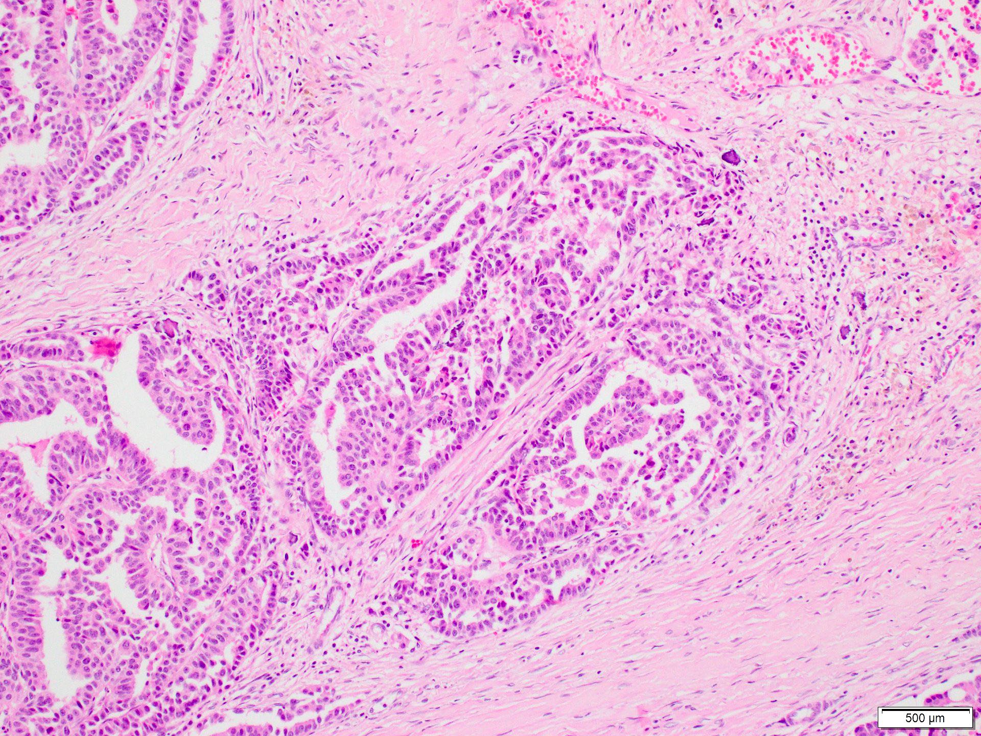
Invasive papillary carcinoma
Papillae lack myoepithelial cells
Necrosis and calcifications
Mitotic figures
Positive stains
Negative stains
- Myoepithelial cell stains, such as p63, calponin and smooth muscle myosin heavy chain (Mod Pathol 2021 Jan 18 [Epub ahead of print])
- HER2 (J Cancer Res Clin Oncol 2013;139:77)
- PAX8, WT1, TTF1, Napsin A and thyroglobulin (Mod Pathol 2021 Jan 18 [Epub ahead of print])
Sample pathology report
- Right / left breast, mastectomy:
- Invasive papillary carcinoma, Nottingham histologic grade (X), measuring (X) cm (please see detailed synoptic report)
- Associated ductal carcinoma in situ (DCIS), (X) nuclear grade, (X) patterns, comprising X% of tumor (if present).
- Lymphovascular invasion (is / is not) identified.
- Biopsy site changes are identified.
- Margins are (involved / uninvolved).
Differential diagnosis
- Encapsulated papillary carcinoma:
- Rounded pushing border
- Typically surrounded by a fibrous capsule
- Frank invasion may be present when tumor cells infiltrate beyond the fibrous capsule
- Solid papillary carcinoma, invasive and noninvasive:
- Expansile nodules with solid pattern and delicate fibrovascular cores
- Extracellular mucin may be present
- Invasive micropapillary carcinoma (an aggressive form of mammary carcinoma):
- More than 90% of tumor consists of morula-like epithelial clusters
- Tumor clusters are surrounded by empty spaces, devoid of fibrovascular cores and show reverse polarity
- Extramammary carcinoma metastatic to the breast, such as ovarian carcinoma, lung carcinoma with papillary features or thyroid papillary carcinoma (J Cancer Res Clin Oncol 2013;139:77, J Surg Res 2021;261:105):
- Tall cell carcinoma with reversed polarity (Am J Surg Pathol 2003;27:1114):
- Carcinoma cells arranged in solid, papillary and glandular structures, resembling thyroid follicles
- Carcinoma glands contain colloid-like material
- Tumor cells are columnar or cuboidal
- Many of the nuclei sit near the luminal membranes, hence the term reversed polarity
- Most tumors are negative for ER and PR
Additional references
Board review style question #1
Which of the following is a relevant feature of invasive papillary carcinoma of the breast?
- Commonly encountered breast tumor
- Graded according to nuclear size
- May mimic metastases from other organs
- Myoepithelial cells at the periphery
- Positive for Napsin A and PAX8
Board review style answer #1
C. It may mimic metastases from other organs (metastases to the breast from other organs such as ovary, lung and thyroid must be excluded)
Comment Here
Reference: Invasive papillary carcinoma
Comment Here
Reference: Invasive papillary carcinoma
