

Breast
Breast cancer
Inflammatory
Authors: Kiran Manjee, M.D., Julie M. Jorns, M.D.
Editorial Board Member: Kristen E. Muller, D.O.
Deputy Editor-in-Chief: Gary Tozbikian, M.D.


Last author update: 21 March 2023
Last staff update: 21 March 2023
Copyright: 2002-2025, PathologyOutlines.com, Inc.
PubMed Search: Inflammatory carcinoma breast


Table of Contents
Definition / general | Essential features | ICD coding | Epidemiology | Sites | Pathophysiology | Etiology | Clinical features | Diagnosis | Radiology description | Radiology images | Prognostic factors | Case reports | Treatment | Clinical images | Gross description | Microscopic (histologic) description | Microscopic (histologic) images | Virtual slides | Positive stains | Negative stains | Sample pathology report | Differential diagnosis | Additional references | Board review style question #1 | Board review style answer #1 | Board review style question #2 | Board review style answer #2Cite this page: Manjee K, Jorns JM. Inflammatory. PathologyOutlines.com website. https://www.pathologyoutlines.com/topic/breastmalignantinflamcar.html. Accessed January 3rd, 2025.
Definition / general
- Clinicopathologic entity characterized by rapid onset (≤ 6 months) and enlarged, erythematous and edematous (peau d’orange) breast due to dermal plugging of lymphatic vessels by tumor (Breast Cancer Res 2005;7:52)
- Involvement of dermal lymphatics alone without the classic clinical presentation is not considered inflammatory carcinoma
Essential features
- Clinical presentation of breast cancer with dermal lymphatic involvement
- Breast skin with erythema and edema occupying at least 33% of the breast is required to make the diagnosis
- Must be of rapid onset; duration of symptoms of ≤ 6 months
ICD coding
Epidemiology
- Accounts for 2 - 5% of all newly diagnosed breast cancer cases (J Natl Cancer Inst 2005;97:966)
- Contributes to 7% of breast cancer related mortality (J Natl Cancer Inst 2005;97:966)
- Incidence is higher in black women, intermediate in white women and lowest in Asian women (Ann Surg Oncol 2014;21:1267)
- Mean age is 56 years (but wide age range) (BMC Cancer 2021;21:138)
Sites
- Breast, breast skin (dermal lymphatics)
Pathophysiology
- Tumor microemboli in the lymphatic spaces causes obstruction that leads to the clinical presentation of peau d’orange appearance
- RhoC overexpression induces angiogenesis and facilitates rapid metastasis (Breast Cancer Res Treat 2004;84:3)
- E-cadherin accumulation / overexpression within tumor emboli (Anticancer Res 2010;30:3903)
Etiology
- No association with inherited genetic mutation
- No association with family history
Clinical features
- Rapid onset of erythema, edema, induration, warmth and tenderness of the breast skin (Ann Oncol 2011;22:515)
- Patients may have a history of being diagnosed with mastitis that does not respond to at least 1 week of antibiotics (Ann Oncol 2011;22:515)
- Erythema occupying at least 33% of the breast (Ann Oncol 2011;22:515)
- Duration of history < 6 months (Ann Oncol 2011;22:515)
- May or may not have a breast mass, typically not palpable due to rapid spread to lymphatics and distant sites but low volume of invasive carcinoma in breast
Diagnosis
- At least 2 skin punch biopsies between 2 - 8 mm of the most prominent area of skin discoloration (Ann Oncol 2011;22:515)
Radiology description
- Mammogram (Breast Cancer Res Treat 2008;109:417)
- Low sensitivity compared with other modalities (ultrasound, MRI) in identifying a breast tumor
- Skin thickening due to edema and dermal collagen deposition
- Ultrasound, MRI, PET / CT (J Med Imaging Radiat Oncol 2016;60:83, Radiol Med 2010;115:70, Acad Radiol 2022;29:637, Clin Imaging 1992;16:183, Breast Cancer Res Treat 2008;109:417)
- Breast skin thickening and edema are the most common findings
- Used to further evaluate for the presence of breast lesion(s), nodal disease and distant metastatic disease
Radiology images
Contributed by Kiran Manjee, M.D. and Julie M. Jorns, M.D.
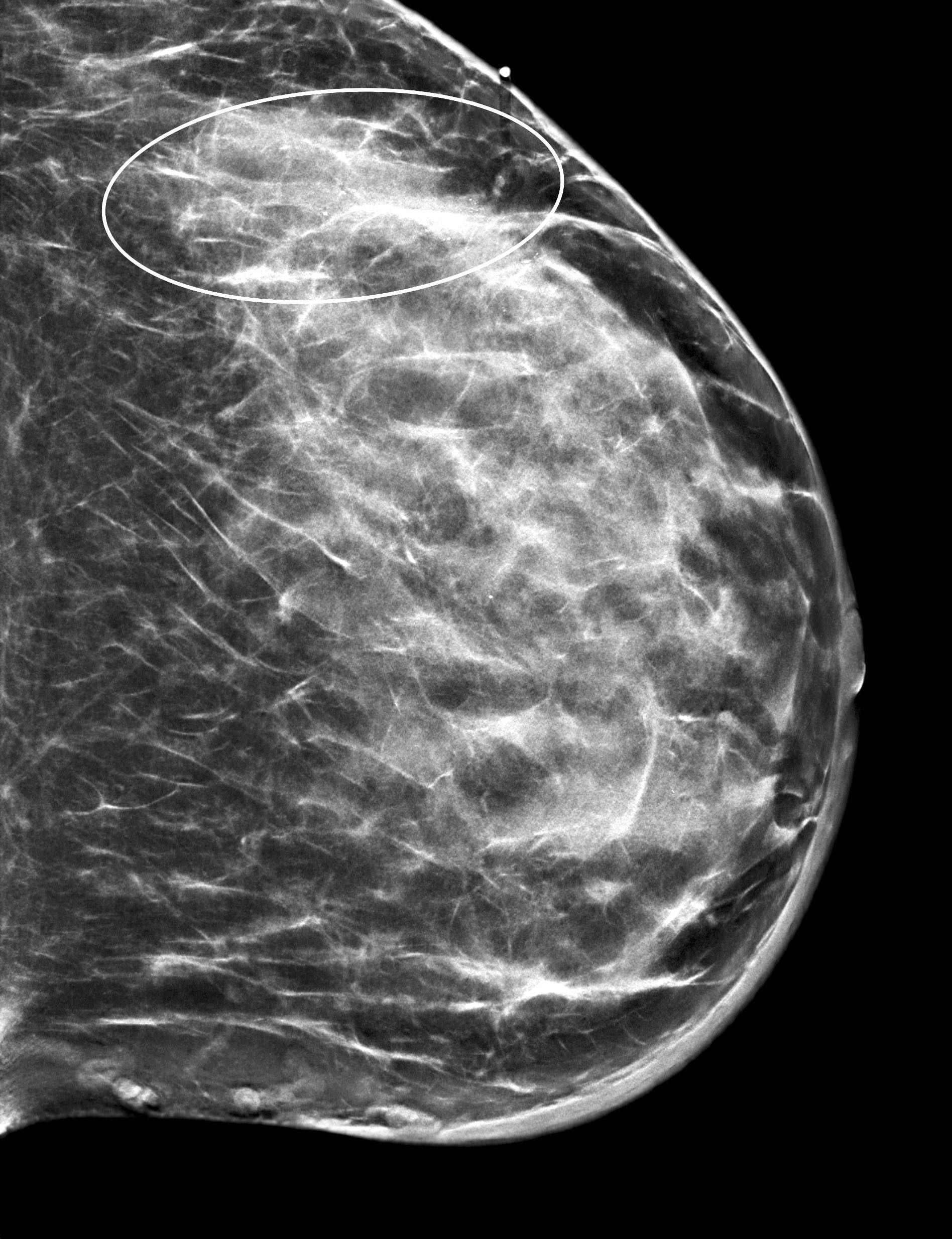
Skin thickening and mass
Prognostic factors
- Previous studies have reported poor survival rates; however, this may be due to advanced stage at presentation and delayed diagnosis
- 2010 - 2015 Surveillance, Epidemiology and End Results (SEER) database showed similar breast cancer specific survival in patients with inflammatory breast cancer and stage T4 noninflammatory breast cancer (BMC Cancer 2021;21:138)
- 2010 - 2011 SEER data showed worse outcomes in hormone receptor positive / HER2 negative and triple negative breast cancer compared with triple positive and hormone receptor negative / HER2 positive subtypes (J Cancer Res Clin Oncol 2017;143:161)
- Use of neoadjuvant therapy has improved survival rates
- Histologic subtype does not impact survival (PLoS One 2016;11:e0145534)
- 50% of patients with complete pathological response will subsequently recur
- Overall 5 year survival rate is 41% (Cancer.Net: Breast Cancer - Inflammatory: Statistics [Accessed 27 December 2022])
- 5 year survival with regional lymph node involvement is 56%
- 5 year survival with distant metastasis is 19%
- p53 status is not evaluated routinely but positive staining (≥ 10%) was associated with decreased response to chemotherapy (n = 59) (Clin Cancer Res 2004;10:6215)
Case reports
- 31 year old woman with inflammatory breast cancer that was misdiagnosed as plasma cell mastitis on first biopsy (Asian J Surg 2023;46:1153)
- 75 year old woman with invasive lobular carcinoma that presented clinically as inflammatory breast cancer (Cureus 2022;14:e27358)
- 78 year old man with inflammatory breast carcinoma (Int J Surg Case Rep 2021;80:105696)
Treatment
- Multidisciplinary team approach is critical
- Neoadjuvant chemotherapy, mastectomy with lymph node dissection, radiation therapy and adjuvant endocrine therapy if indicated (Ann Oncol 2011;22:515)
- If there is a good clinical response to neoadjuvant chemotherapy, some may be able to safely undergo breast conserving therapy instead of a mastectomy (Gland Surg 2018;7:520)
Clinical images
Contributed by Amanda L. Kong, M.D., M.S. and Mark R. Wick, M.D.
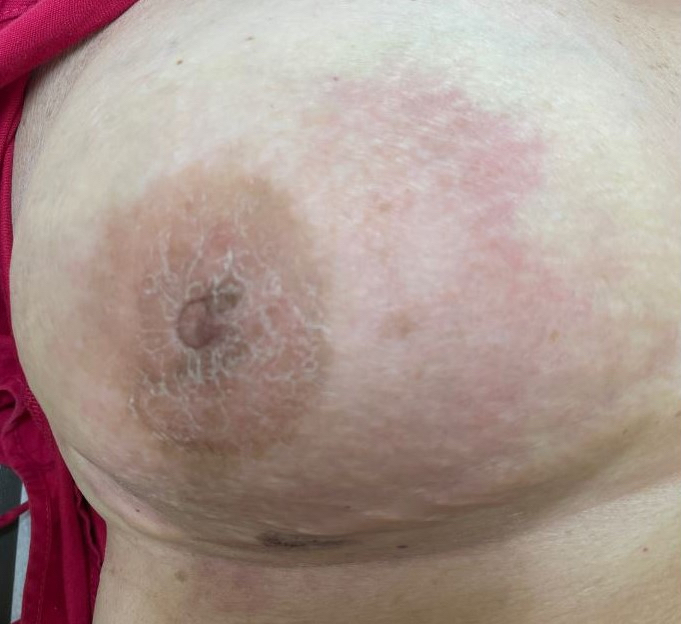
Erythema and edema
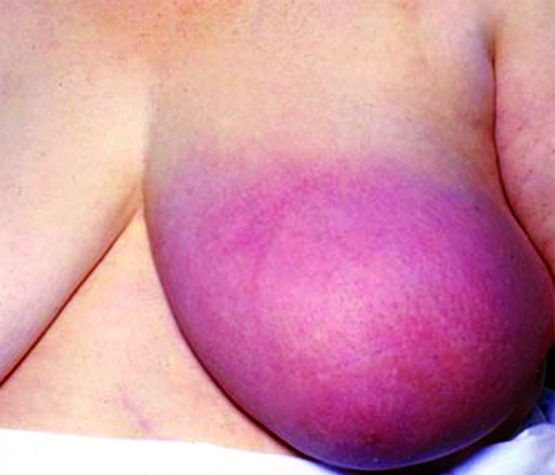
Discoloration
Gross description
- Skin findings and mass lesions are usually not present after neoadjuvant therapy
Microscopic (histologic) description
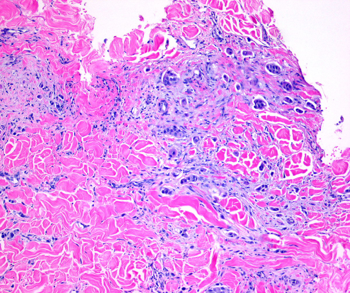
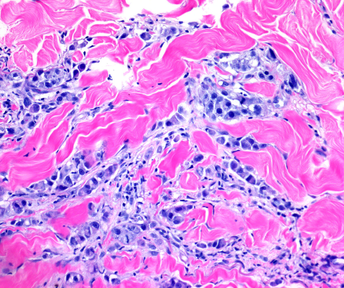
- Presence of tumor cells in dermal lymphovascular spaces
- Tumor cells or lymphatic spaces may be scant and therefore assessment at multiple levels is recommended
- It is not a specific histological subtype
- Most associated invasive carcinoma is high grade invasive ductal carcinoma, no special type; however, other types of breast cancers can present as inflammatory carcinoma (PLoS One 2016;11:e0145534)
- Ductal carcinoma in situ is often absent
- Classified as T4d in AJCC cancer staging
- T stage does not change following neoadjuvant therapy (ypT4d)
Microscopic (histologic) images
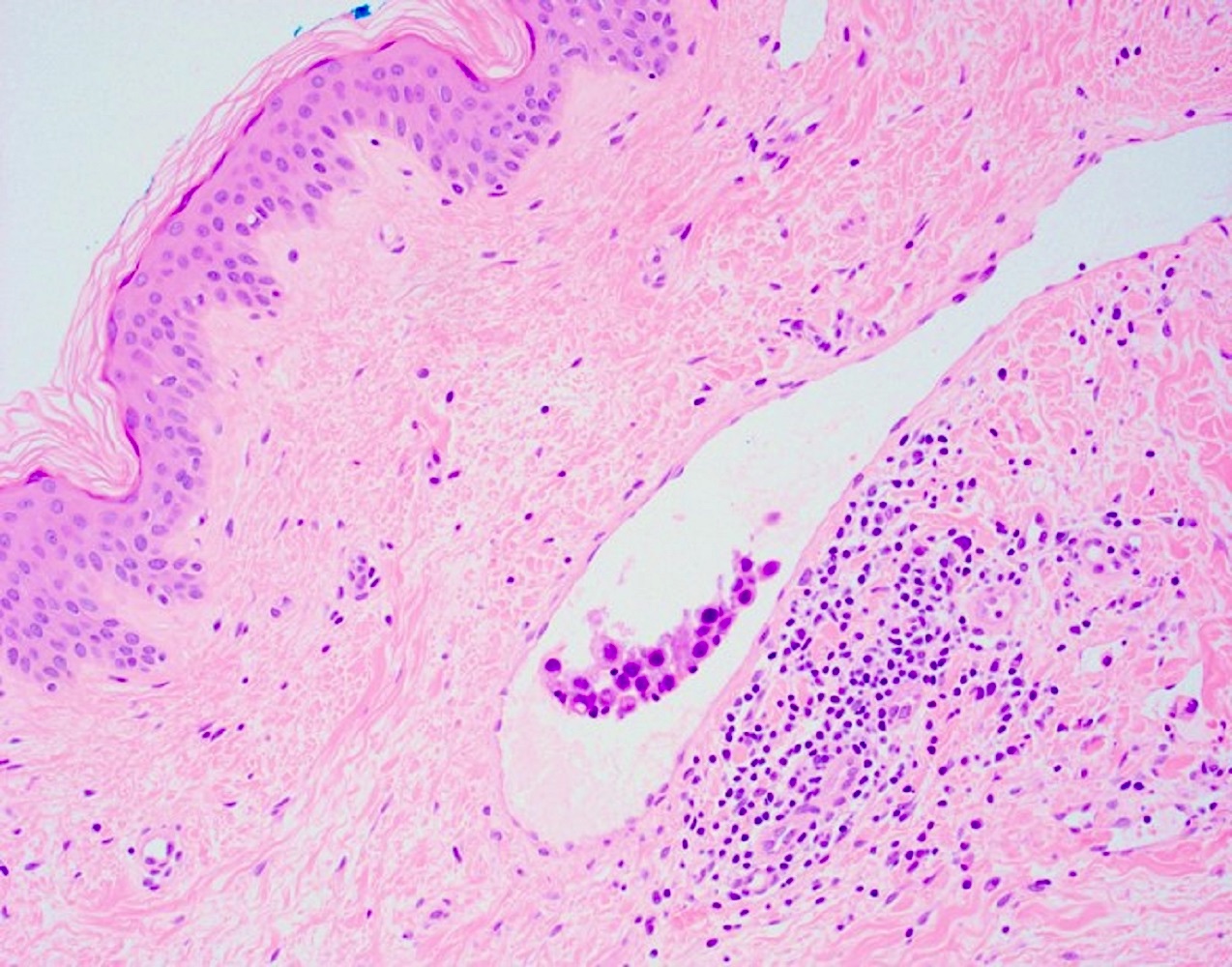
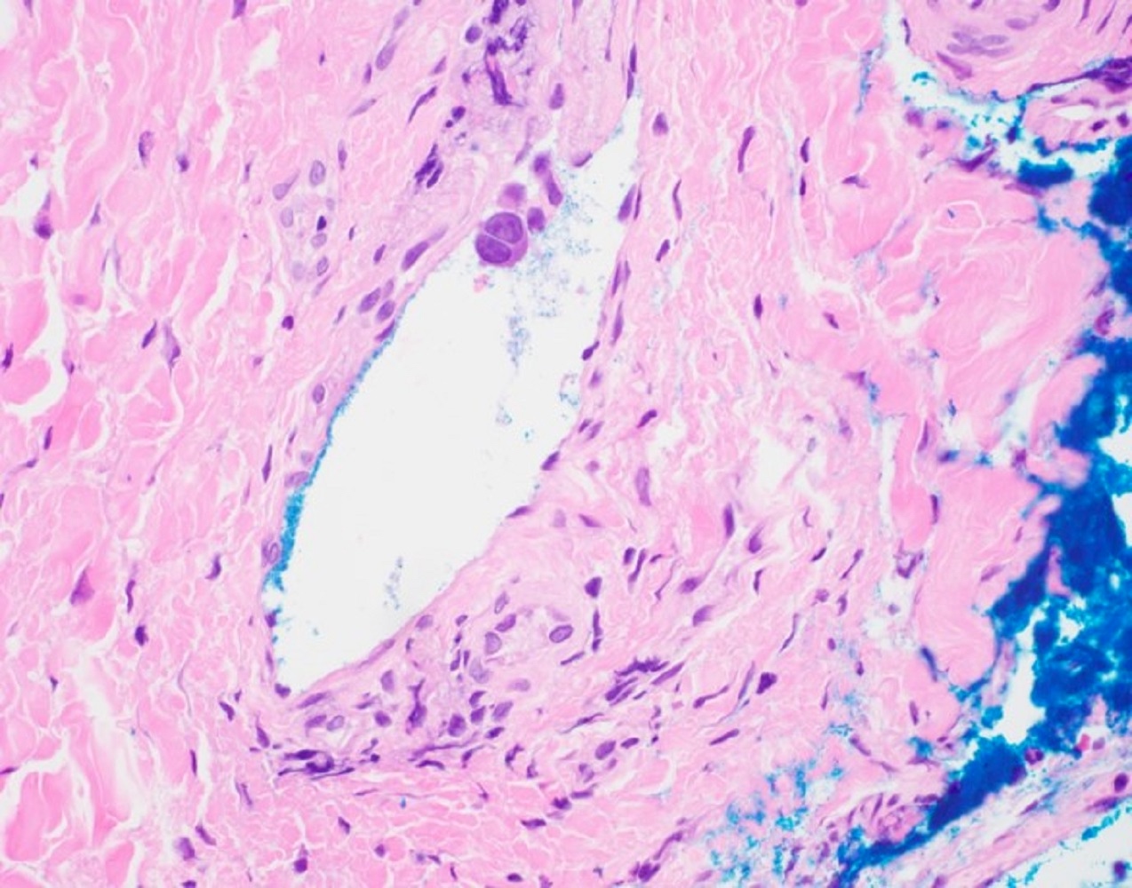
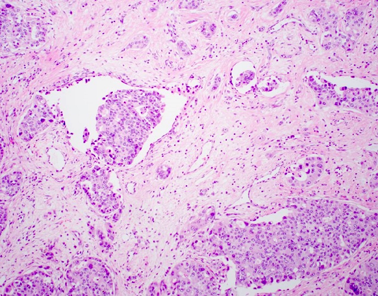
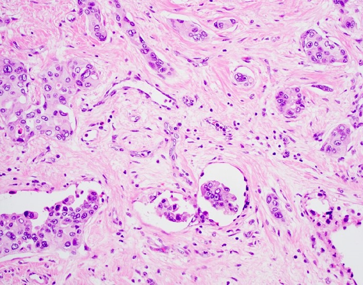
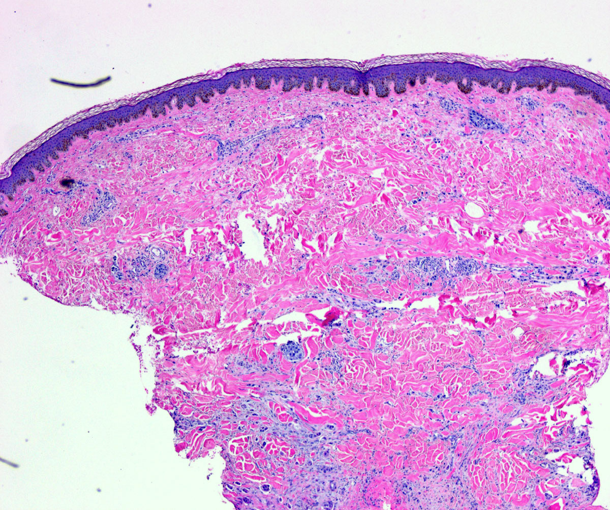
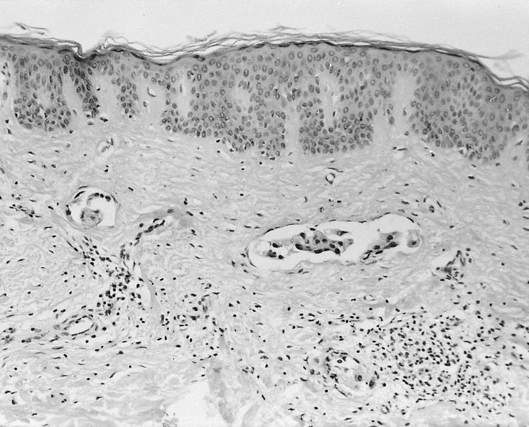
Contributed by Kiran Manjee, M.D., Julie M. Jorns, M.D., Weijie Li, M.D. and AFIP
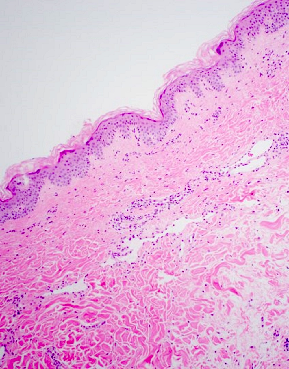
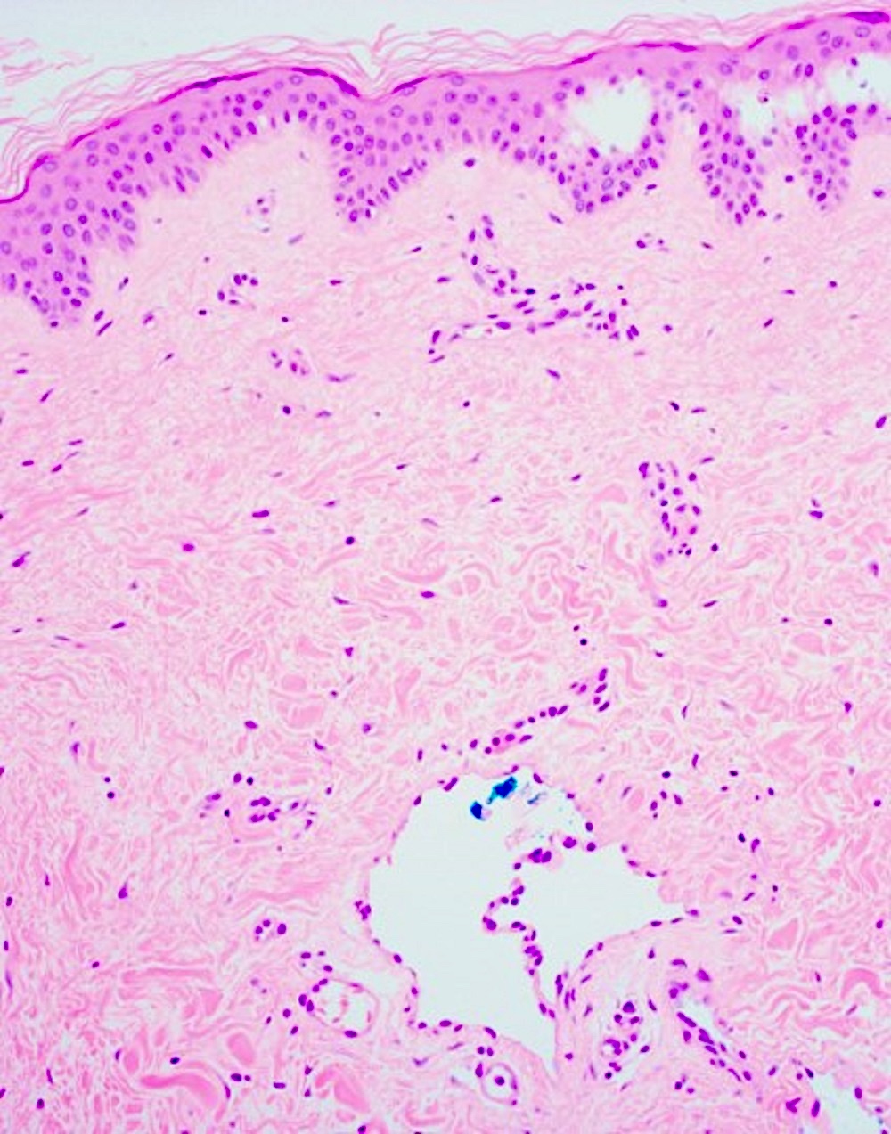
Dilated lymphatics
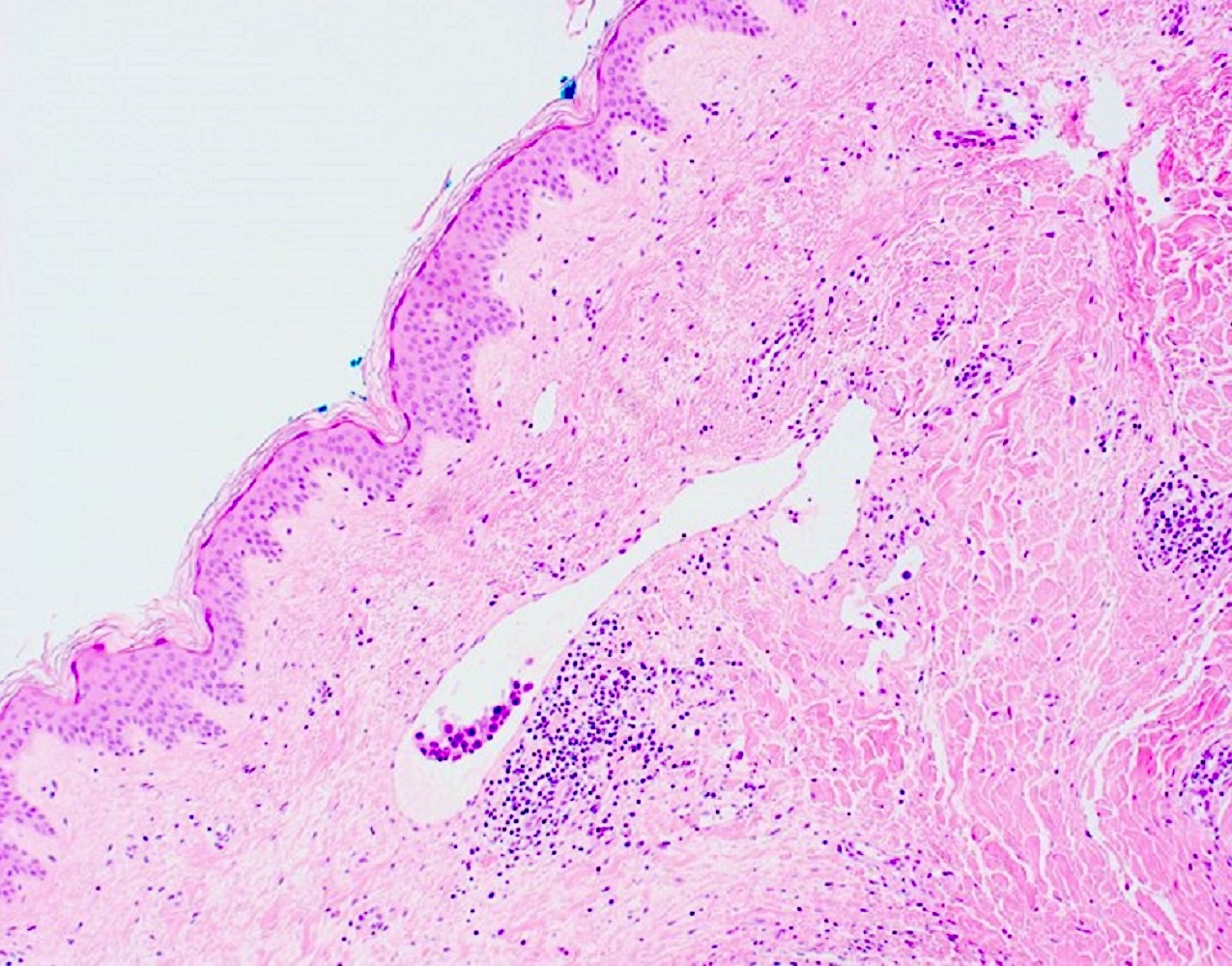
Dermal lymphovascular invasion
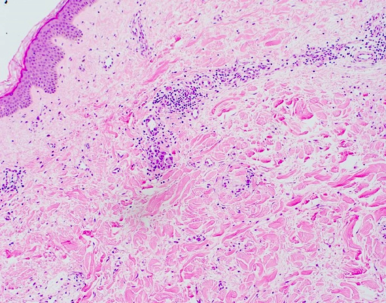
Dermal lymphovascular invasion
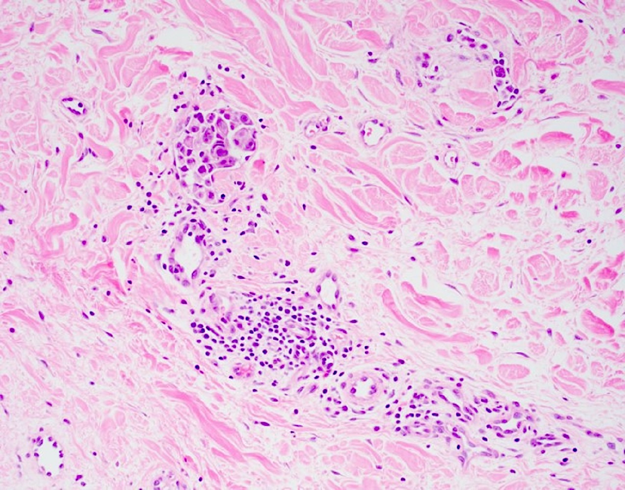
Dermal lymphovascular invasion
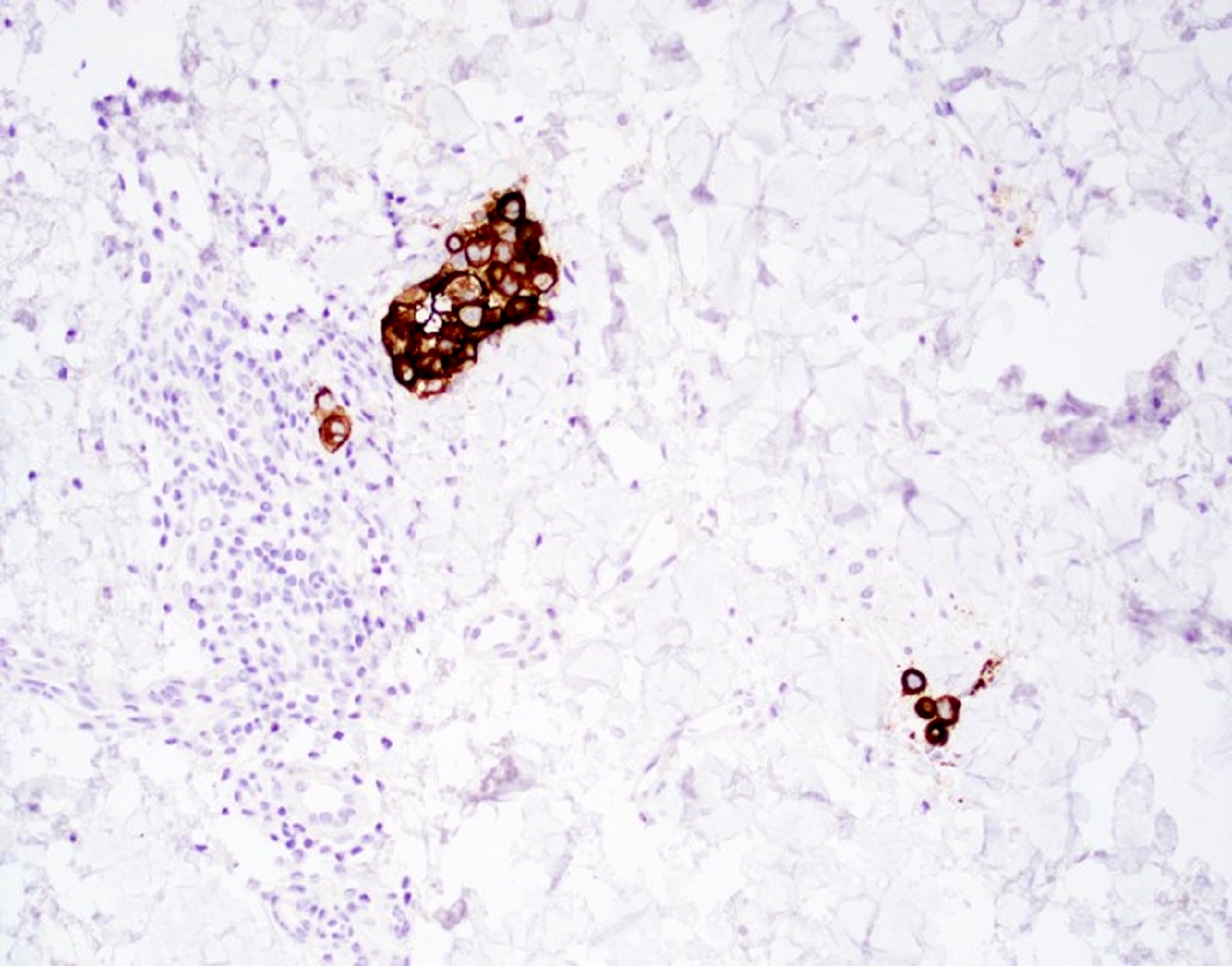
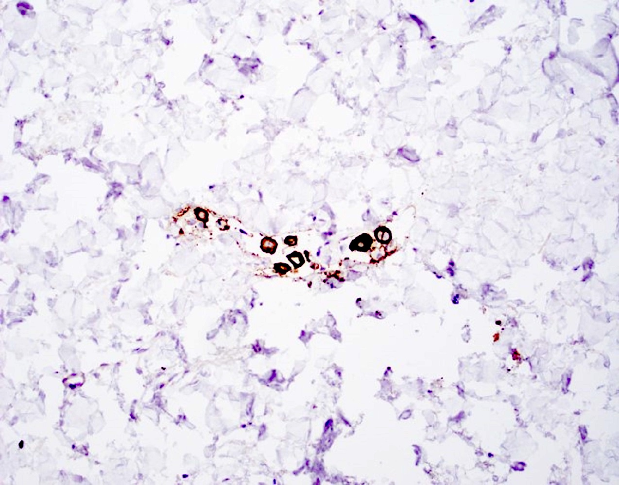
HER2 / neu
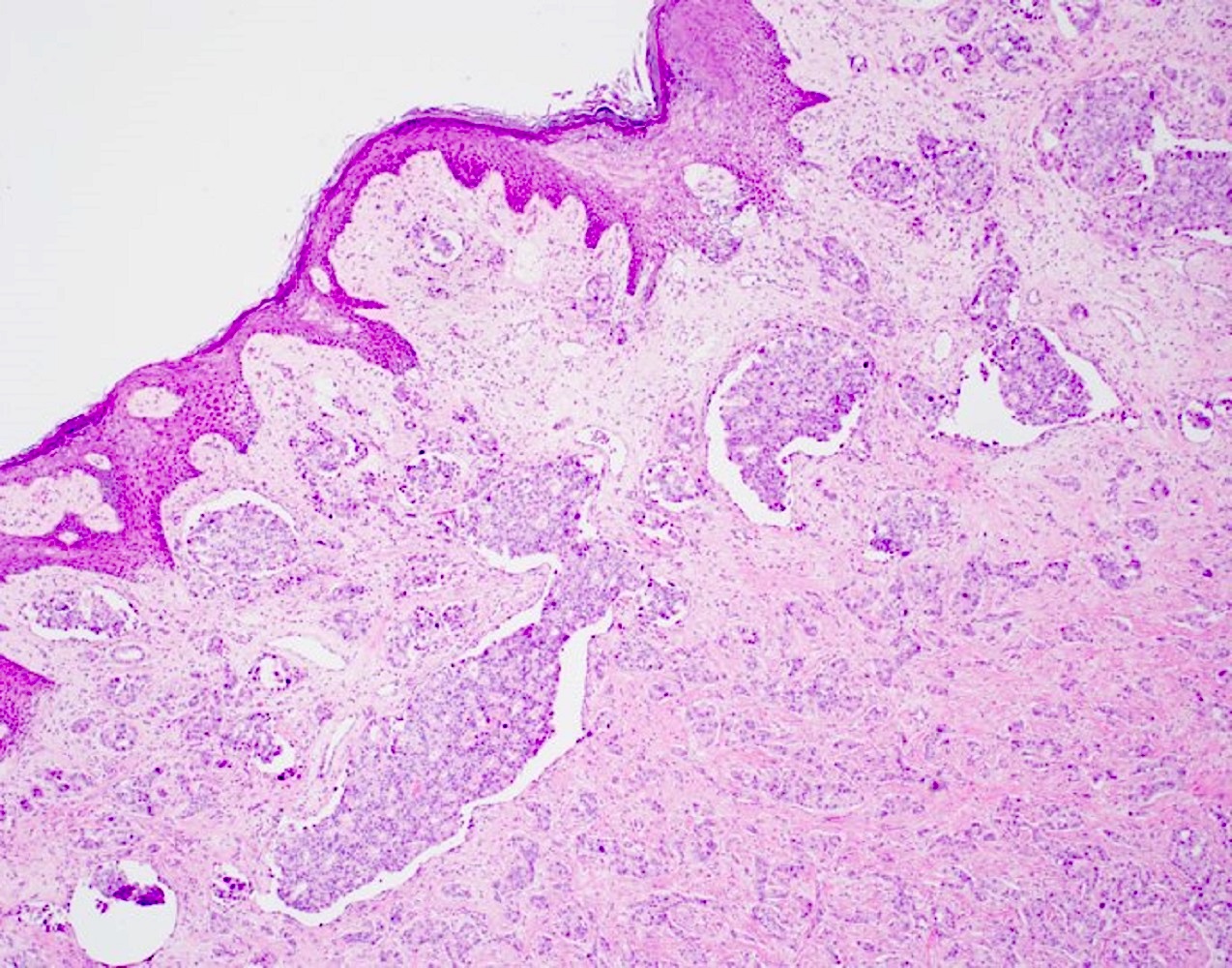
Locally advanced breast cancer
Skin punch biopsy
Infiltrative nests
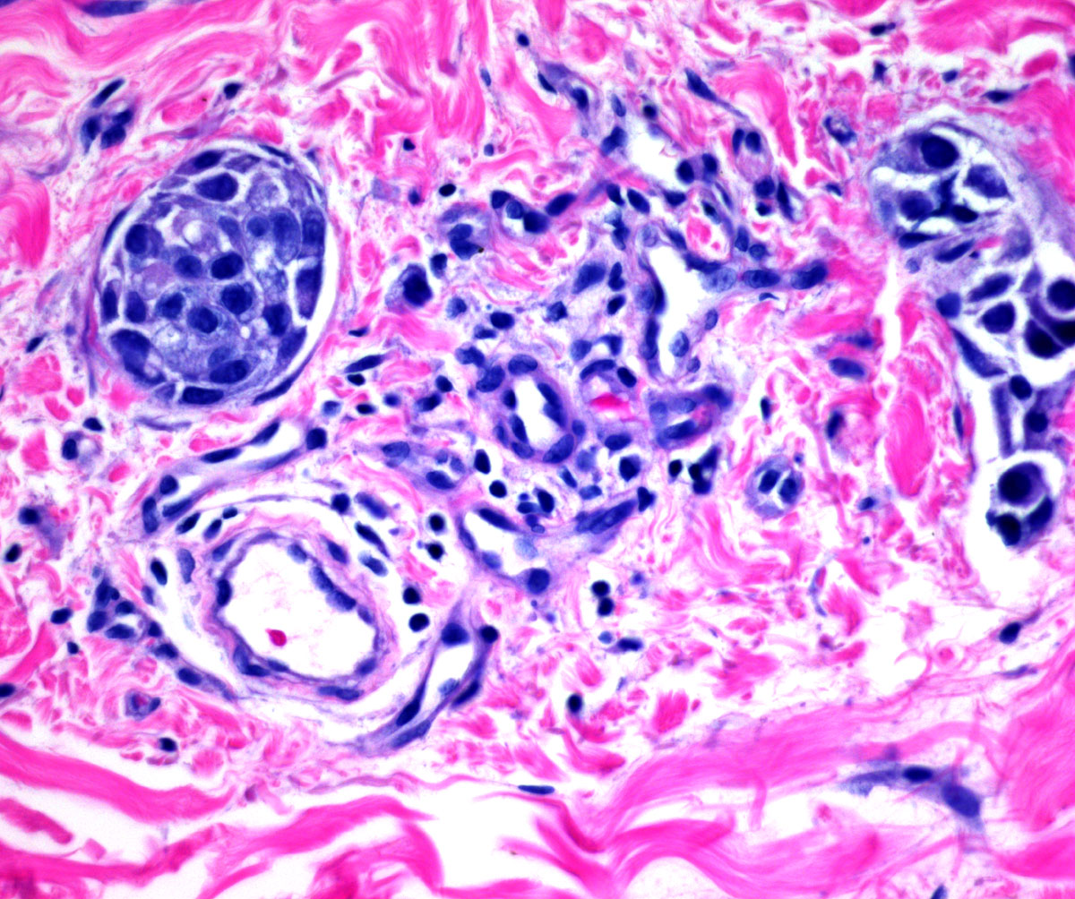
Tumor emboli
Tumor in dermal lymphatics
Virtual slides
Images hosted on other servers:

Inflammatory breast carcinoma
Positive stains
- E-cadherin positive tumor emboli (Mod Pathol 2001;14:458, Anticancer Res 2010;30:3903)
- HER2 (52%) (Breast 2004;13:97)
- ER (41 - 52%) (Breast 2004;13:97, Mod Pathol 2001;14:458)
- CD31, ERG highlight endothelial cells surrounding foci of lymphovascular invasion
Negative stains
- PR (35%) (Mod Pathol 2001;14:458)
Sample pathology report
- Skin, left breast, punch biopsy:
- Positive for high grade mammary (ductal) carcinoma within dermal lymphovascular spaces
- Comment: Immunohistochemistry shows carcinoma to be:
- GATA3 (subset, moderate) positive, supporting breast origin
- Estrogen receptor (< 1%) negative
- Progesterone receptor (< 1%) negative
- HER2 / neu (0) negative for overexpression
Differential diagnosis
- Invasive carcinoma involving dermis:
- Ductal carcinoma in situ (DCIS):
- Tumor clusters within myoepithelial lined spaces
- Infection (mastitis, cellulitis, abscess):
- More common in lactating women or in those with history of recent trauma or surgery
- Responds to antibiotic treatment
- Radiation dermatitis:
- Clinical history of radiation
- Benign biopsies
- Other causes of edema (lymphatic obstruction or surgical removal, congestive heart failure):
- Clinical history of medical condition, surgery, travel (parasitic infections)
- Benign biopsies
- Locally advanced carcinoma with skin involvement:
- Skin changes may be present due to dermal lymphatic involvement but clinical history is of longer duration (> 6 months), often with palpable mass
- Histologically bulky invasive carcinoma is present alongside lymphovascular space involvement
- Metastatic neoplasms from other sites:
- Unusual histology for breast
- Immunohistochemistry can help differentiate
Additional references
Board review style question #1
A middle aged woman presents with rapid onset (< 6 months) of breast skin thickening and discoloration. A punch biopsy is done and the patient is diagnosed with inflammatory breast cancer. She undergoes neoadjuvant chemotherapy and subsequent mastectomy reveals focal ductal carcinoma in situ (DCIS) but no residual invasive or metastatic carcinoma. How would she be staged?
- ypTX
- ypT0
- ypTis
- ypT4d
Board review style answer #1
D. ypT4d. In inflammatory breast cancer, the T stage is classified as T4d and does not change after neoadjuvant chemotherapy.
Comment Here
Reference: Inflammatory
Comment Here
Reference: Inflammatory
Board review style question #2
What feature best distinguishes inflammatory breast carcinoma from locally advanced breast cancer?
- Mammographic mass
- Peau d’orange
- Skin thickening and discoloration < 6 months
- Tumor emboli within lymphatic spaces
Board review style answer #2
C. Skin thickening and discoloration < 6 months. Inflammatory breast cancer is defined as breast cancer with rapid onset of clinical symptoms of skin thickening and discoloration / erythema involving at least 33% of the breast. There is frequent peau d’orange and tumor emboli within lymphatic spaces on skin punch biopsy; however, these findings are not necessary to make the diagnosis of inflammatory breast cancer nor are they unique to inflammatory breast cancer as they can also be seen in locally advanced breast cancer. A mass may or may not be seen on mammogram as in inflammatory breast cancer tumor quickly spreads to lymphatics (and beyond) and mammography has lower sensitivity in this setting.
Comment Here
Reference: Inflammatory
Comment Here
Reference: Inflammatory

