
Breast
Other nonneoplastic
Galactocele
Editorial Board Member: Kristen E. Muller, D.O.
Deputy Editor-in-Chief: Gary Tozbikian, M.D.


Last author update: 10 November 2022
Last staff update: 10 November 2022
Copyright: 2002-2024, PathologyOutlines.com, Inc.
PubMed Search: Galactocele breast
Table of Contents
Definition / general | Essential features | Terminology | ICD coding | Epidemiology | Sites | Pathophysiology | Etiology | Clinical features | Diagnosis | Laboratory | Radiology description | Radiology images | Prognostic factors | Case reports | Treatment | Gross description | Gross images | Microscopic (histologic) description | Microscopic (histologic) images | Cytology description | Cytology images | Sample pathology report | Differential diagnosis | Additional references | Board review style question #1 | Board review style answer #1 | Board review style question #2 | Board review style answer #2Cite this page: Khalid F, Anjum S, Kayani N. Galactocele. PathologyOutlines.com website. https://www.pathologyoutlines.com/topic/breastgalactocele.html. Accessed April 19th, 2024.
Definition / general
- Galactoceles are benign, milk filled cysts that occur almost exclusively in lactating and pregnant women (StatPearls: Galactocele [Accessed 21 September 2022], J Cytol 2020;37:149, Case Rep Obstet Gynecol 2017;2017:4807013, Ultrasonography 2020;39:298, Plast Reconstr Surg Glob Open 2021;9:e3943, J Comput Assist Tomogr 2022;46:282)
- ~4% of cases are among benign breast conditions (StatPearls: Galactocele [Accessed 21 September 2022])
Essential features
- Benign, milk filled cysts that occur almost exclusively in lactating and pregnant women, occurring anywhere along the milk line (StatPearls: Galactocele [Accessed 21 September 2022], J Cytol 2020;37:149, Case Rep Obstet Gynecol 2017;2017:4807013, J Comput Assist Tomogr 2022;46:282, J Pediatr Adolesc Gynecol 2017;30:499)
- Triad of secretory breast epithelium, prolactin stimulus and ductal obstruction results in cyst formation (StatPearls: Galactocele [Accessed 21 September 2022])
- Triple assessment (clinical examination, radiological and histologic correlation) is essential (StatPearls: Galactocele [Accessed 21 September 2022], J Cytol 2020;37:149, Ultrasonography 2020;39:298)
Terminology
- Lactocele / lacteal cyst (StatPearls: Galactocele [Accessed 21 September 2022])
ICD coding
- ICD-10: N64.89 - other specified disorders of breast
Epidemiology
- Age: occurs in second to fifth decade of life with a peak incidence in fourth to fifth decade; rare cases reported in male and female infants (StatPearls: Galactocele [Accessed 21 September 2022], Eur J Pediatr Surg 2012;22:246)
- Common in third trimester of pregnancy (StatPearls: Galactocele [Accessed 21 September 2022], Case Rep Obstet Gynecol 2017;2017:4807013, J Comput Assist Tomogr 2022;46:282)
- Gender: women are almost exclusively affected, with few reported cases in men (StatPearls: Galactocele [Accessed 21 September 2022], J Cytol 2020;37:149, Case Rep Obstet Gynecol 2017;2017:4807013, Eur J Breast Health 2021;18:102, Plast Reconstr Surg Glob Open 2021;9:e3943)
- No genetic risk factors have been identified to date (StatPearls: Galactocele [Accessed 21 September 2022])
Sites
- Most common in breast, unilateral / bilateral
- May occur anywhere along the milk line, with a predilection for retroareolar region (StatPearls: Galactocele [Accessed 21 September 2022])
Pathophysiology
- In women, mammary duct obstruction following trauma, nipple abnormalities or tumor (StatPearls: Galactocele [Accessed 21 September 2022])
- In men, small silent retention cysts in neonatal period (StatPearls: Galactocele [Accessed 21 September 2022])
- Drugs such as metoclopramide / domperidone can increase the risk of galactocele formation (StatPearls: Galactocele [Accessed 21 September 2022])
Etiology
- Triad of secretory breast epithelium, prolactin stimulus and ductal obstruction results in cyst formation
- Secretory breast epithelium: estrogen and progesterone contribute to mammogenesis
- Prolactin stimulus: transplacental passage of prolactin, pituitary adenoma, hyperprolactinemia due to prolactinomas
- Ductal obstruction: postaugmentation following periareolar incisions (Ann Plast Surg 2021;86:115, StatPearls: Galactocele [Accessed 21 September 2022], J Cytol 2020;37:149, Case Rep Obstet Gynecol 2017;2017:4807013)
- Difficulty in breastfeeding: infants with cleft palate
- Breastfeeding contraindications, such as in congenital disease-like phenylketonuria, congenital diaphragmatic hernia, mothers with HIV, human T lymphotropic virus (HTLV) and taking radioactive agents
- Oral contraceptive pills (StatPearls: Galactocele [Accessed 21 September 2022], Ann Plast Surg 2021;86:115)
Clinical features
- Solitary / multiple, palpable, nontender, firm, discrete, movable, often associated with a milky discharge (StatPearls: Galactocele [Accessed 21 September 2022], Case Rep Obstet Gynecol 2017;2017:4807013, Ultrasonography 2020;39:298, J Comput Assist Tomogr 2022;46:282)
- Does not demonstrate inflammation unless secondarily infected (StatPearls: Galactocele [Accessed 21 September 2022])
Diagnosis
- Triple assessment (clinical examination, radiological and histologic correlation) (StatPearls: Galactocele [Accessed 21 September 2022], J Cytol 2020;37:149, Ultrasonography 2020;39:298)
Laboratory
- Presence of milk is confirmed chemically by a positive mucic acid test (StatPearls: Galactocele [Accessed 21 September 2022])
Radiology description
- Ultrasound:
- Solitary, well defined, anechoic lesion with thin, echogenic walls and some distal acoustic enhancement
- Simple cyst if centrally located or thinly septated, multilocular cyst in peripheral location (StatPearls: Galactocele [Accessed 21 September 2022], Radiographics 2013;33:2003)
- Mammogram:
- Indeterminate mass or a circumscribed mass with high radiolucency due to high fat content and water fat level (StatPearls: Galactocele [Accessed 21 September 2022])
Radiology images
Images hosted on other servers:

Cyst with circumscribed rounded margins

Oval shaped mass with hypoechoic rim
Prognostic factors
- Benign condition; carries excellent prognosis (StatPearls: Galactocele [Accessed 21 September 2022])
Case reports
- 3 year old prepubertal girl presented with galactocele in right breast (J Pediatr Adolesc Gynecol 2017;30:499)
- 22 year old woman presented with fine needle aspiration cytology (FNAC) diagnosis of crystallizing galactocele (J Cytol 2020;37:149)
- 32 year old woman presented with axillary accessory breast mimicking suspicious solid mass on ultrasound (Case Rep Obstet Gynecol 2017;2017:4807013)
- 48 year old woman presented with nipple eczema causing galactorrhea by reactive hyperprolactinemia complicated by a galactocele (Eur J Breast Health 2021;18:102)
Treatment
- Conservative; resolves spontaneously on cessation of lactation (StatPearls: Galactocele [Accessed 21 September 2022])
- Aspiration and surgical excision can be performed in complicated cases (StatPearls: Galactocele [Accessed 21 September 2022])
- Dopamine agonists help in postaugmentation galactocele (StatPearls: Galactocele [Accessed 21 September 2022])
- Treat underlying condition such as prolactinoma (StatPearls: Galactocele [Accessed 21 September 2022])
Gross description
- Circumscribed to oval lesions usually ranging in size from 1 - 6 cm; cut section showing cyst filled with thin yellow fluid
Gross images
Contributed by Nasir Ud Din, M.B.B.S.
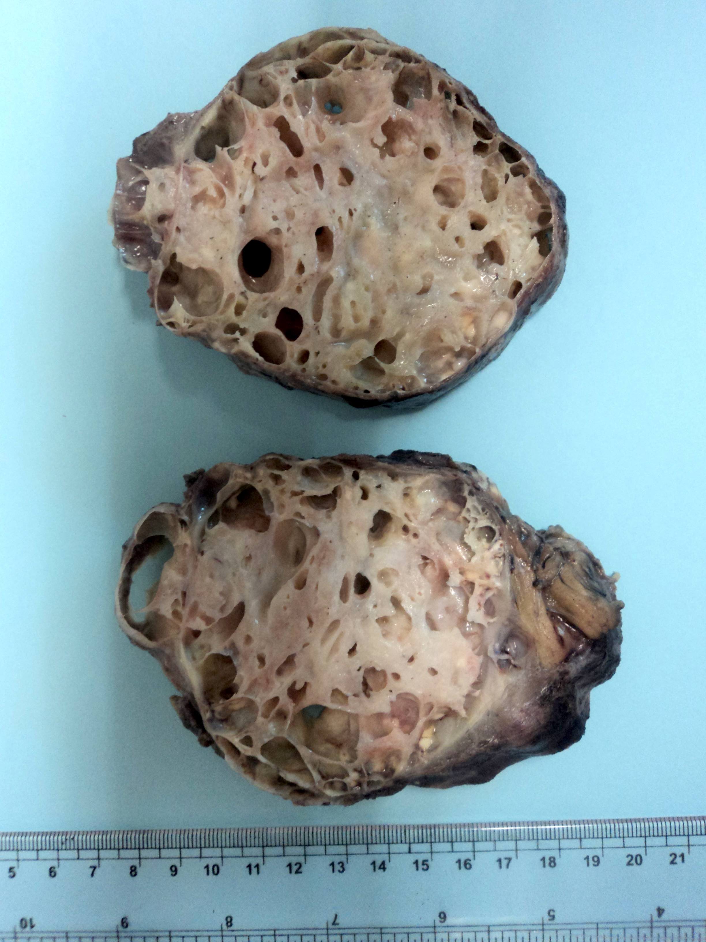
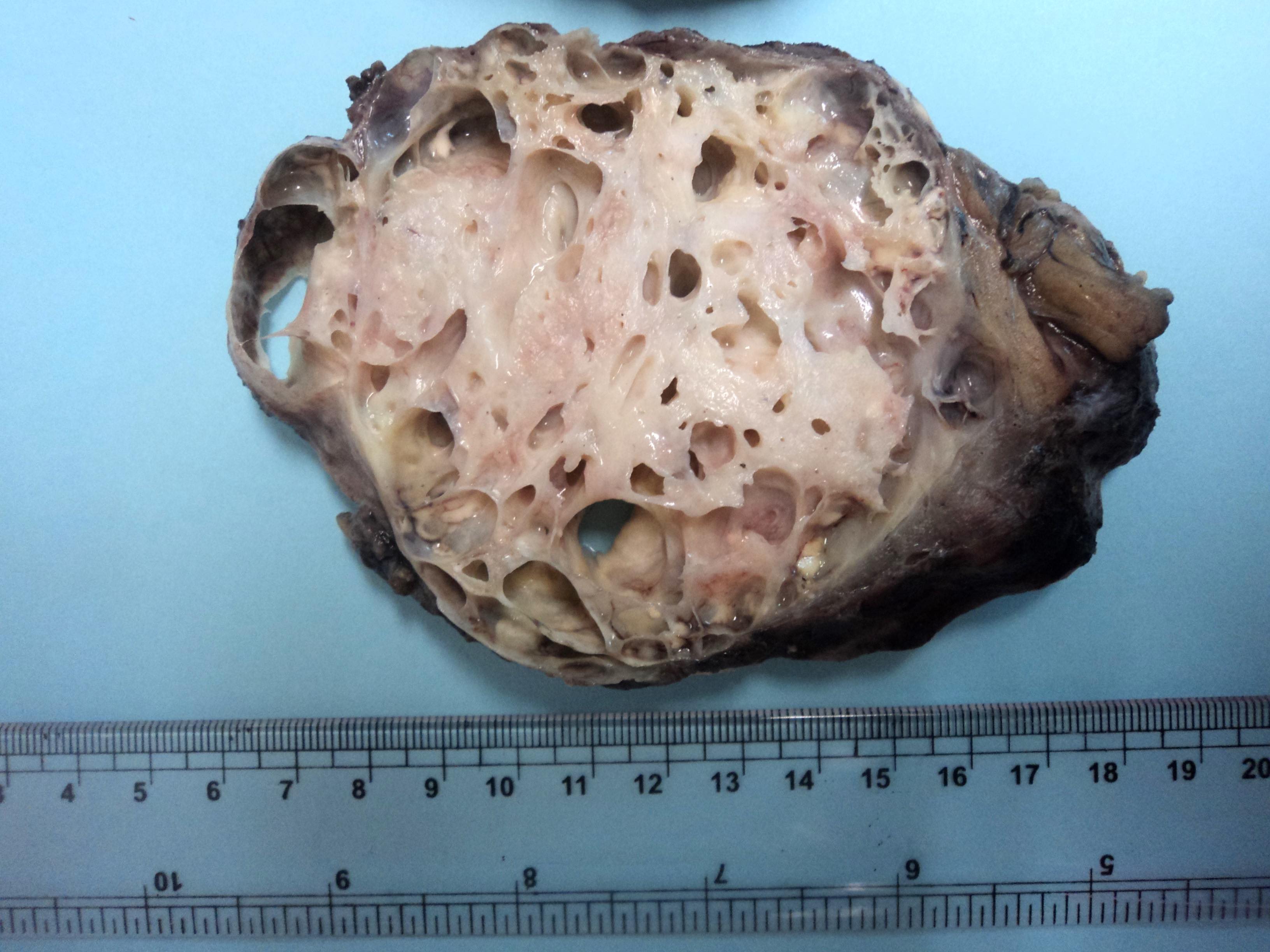
Oval multicystic lesion with well circumscribed borders
Microscopic (histologic) description
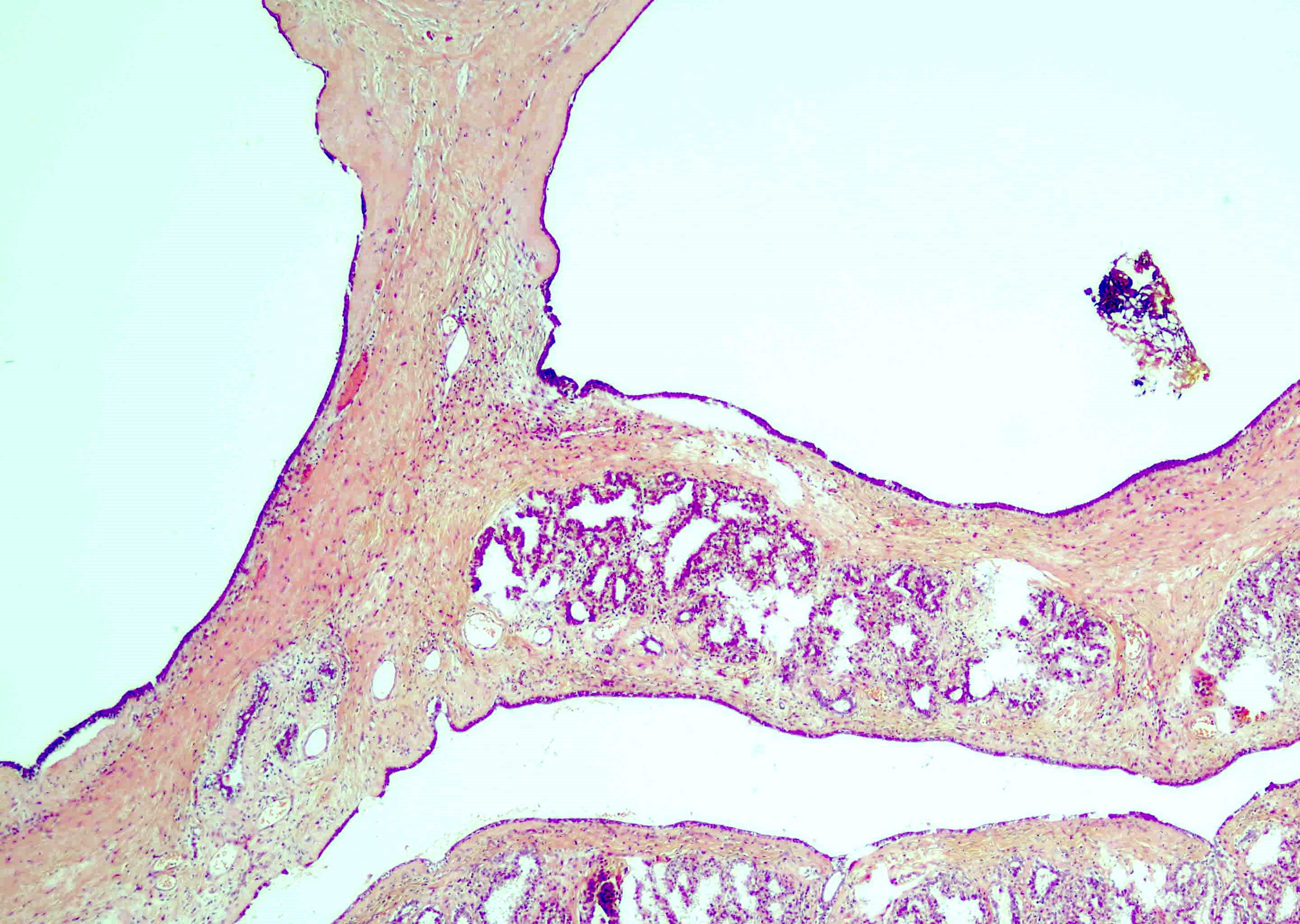
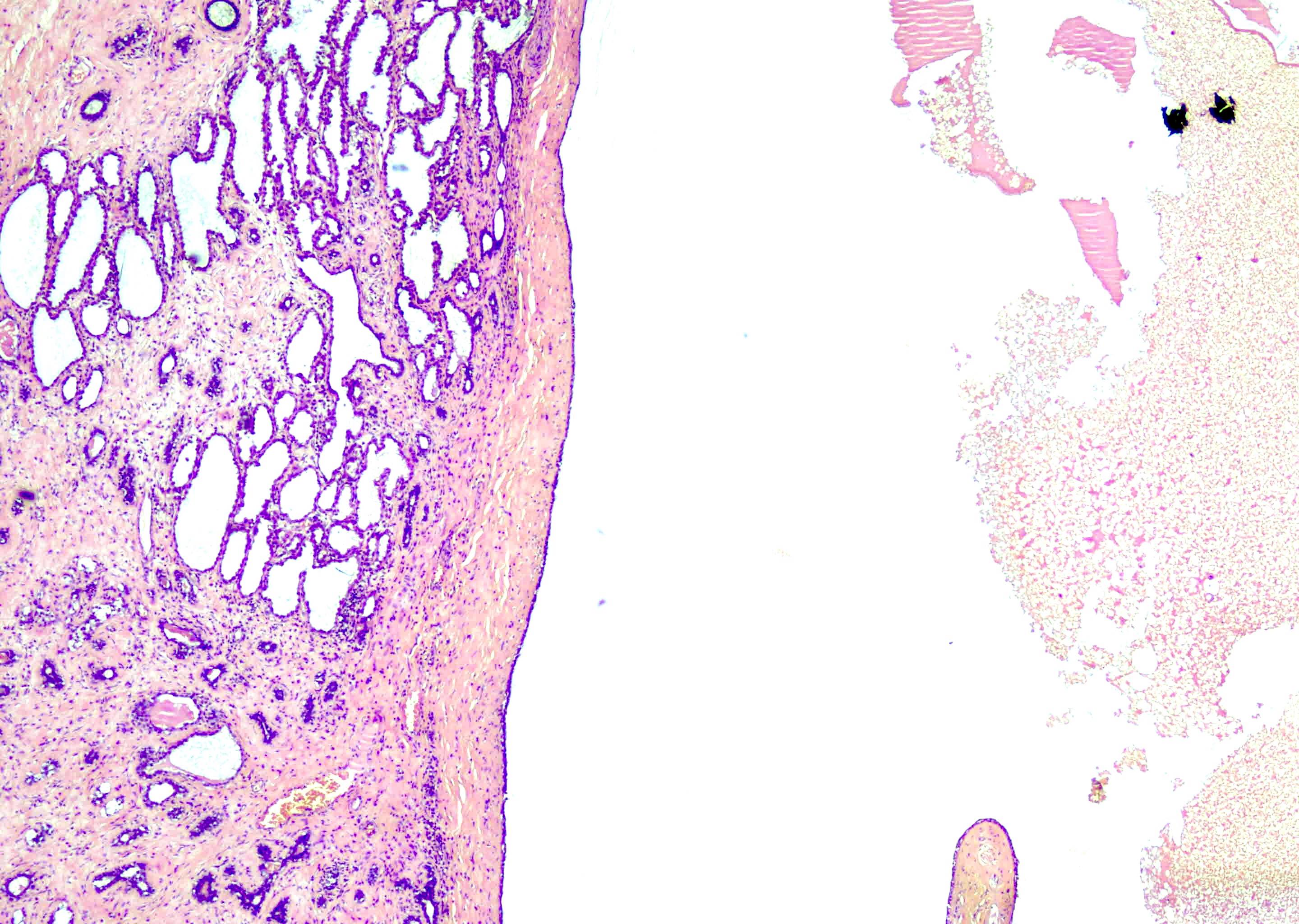
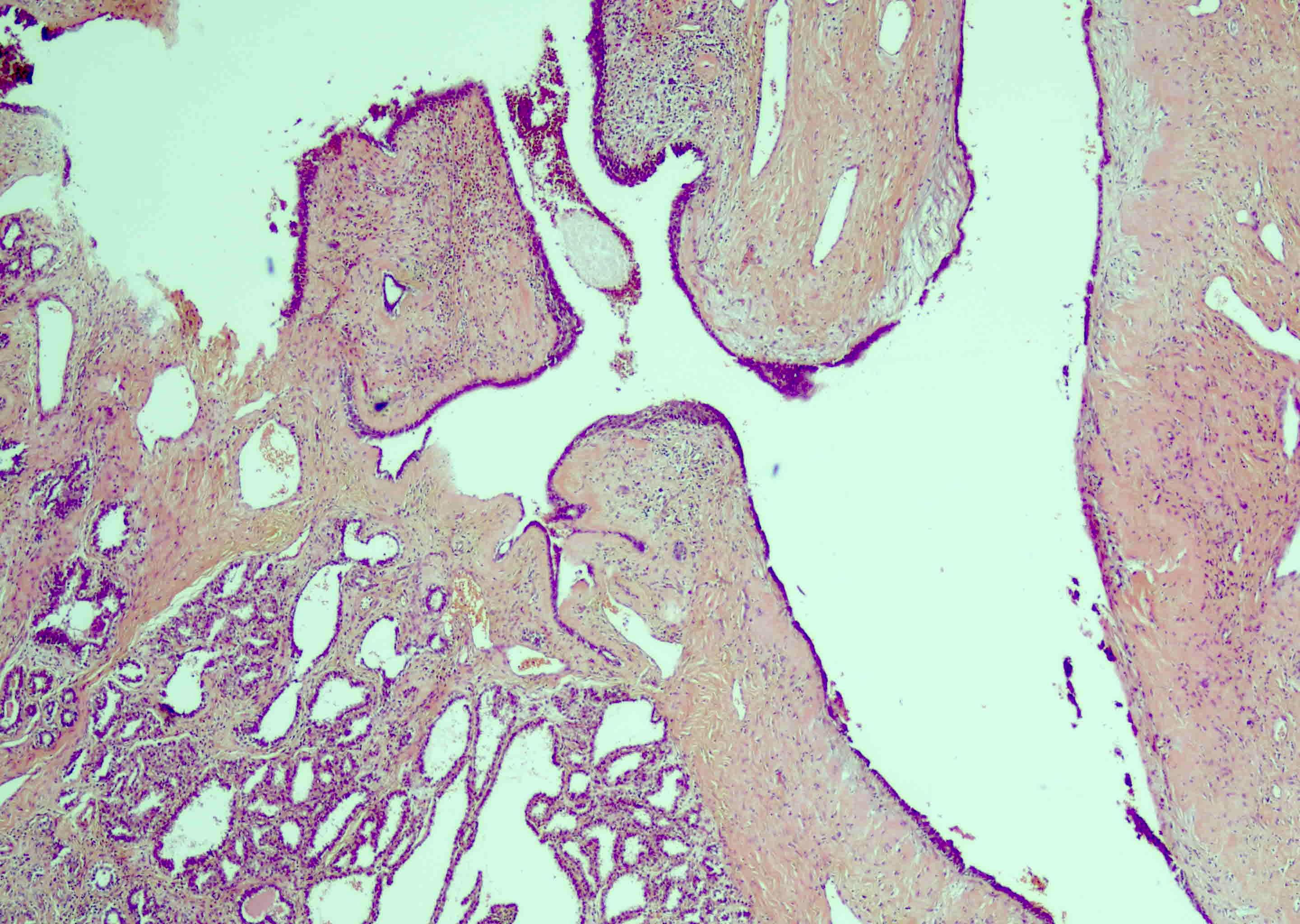
- Multiple variable sized anastomosing cysts that are lined by cuboidal or flat epithelial cells
- Lining epithelium exhibits regular nuclei with cytoplasmic vacuolations due to lipid accumulation
- Apocrine metaplasia may be seen
- Intact cysts are surrounded by fibrous wall of varying thickness
- Little to no inflammatory reaction, unless leakage of cyst contents occurs
- Fat necrosis can be present (Hoda: Rosen's Breast Pathology, 5th Edition, 2020)
Microscopic (histologic) images
Contributed by Saba Anjum, M.B.B.S.
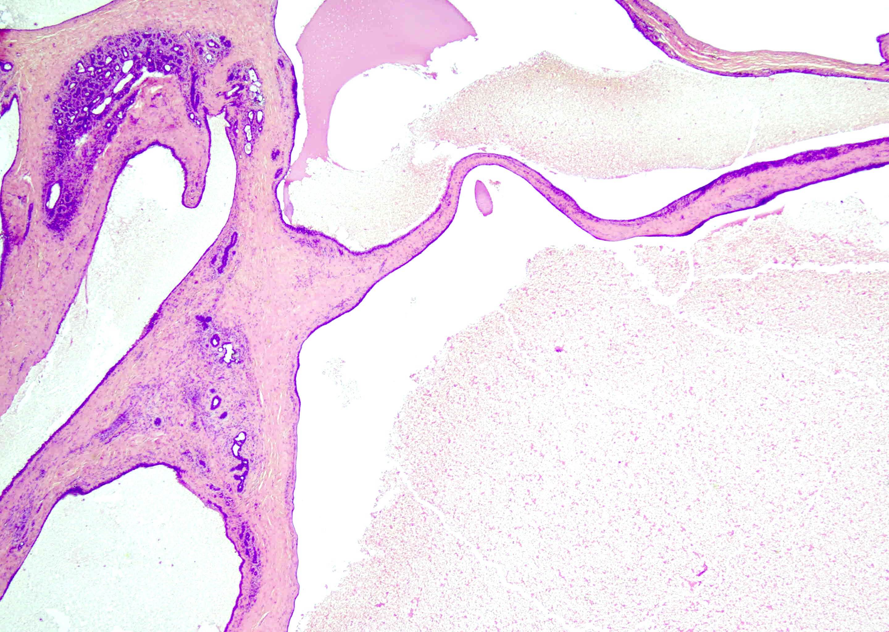
Multiple variable sized anastomosing cysts
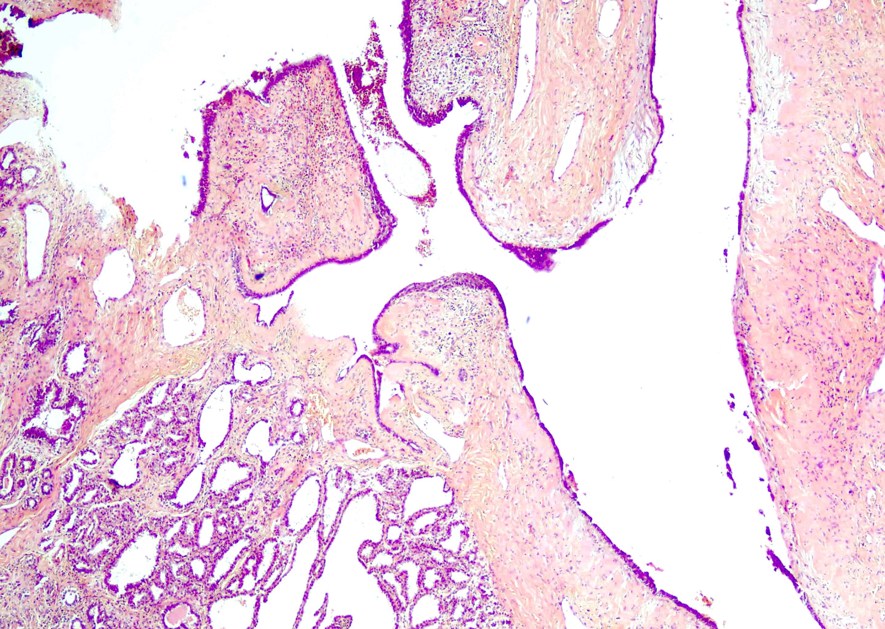
Cystic lesion lined by cuboidal epithelium
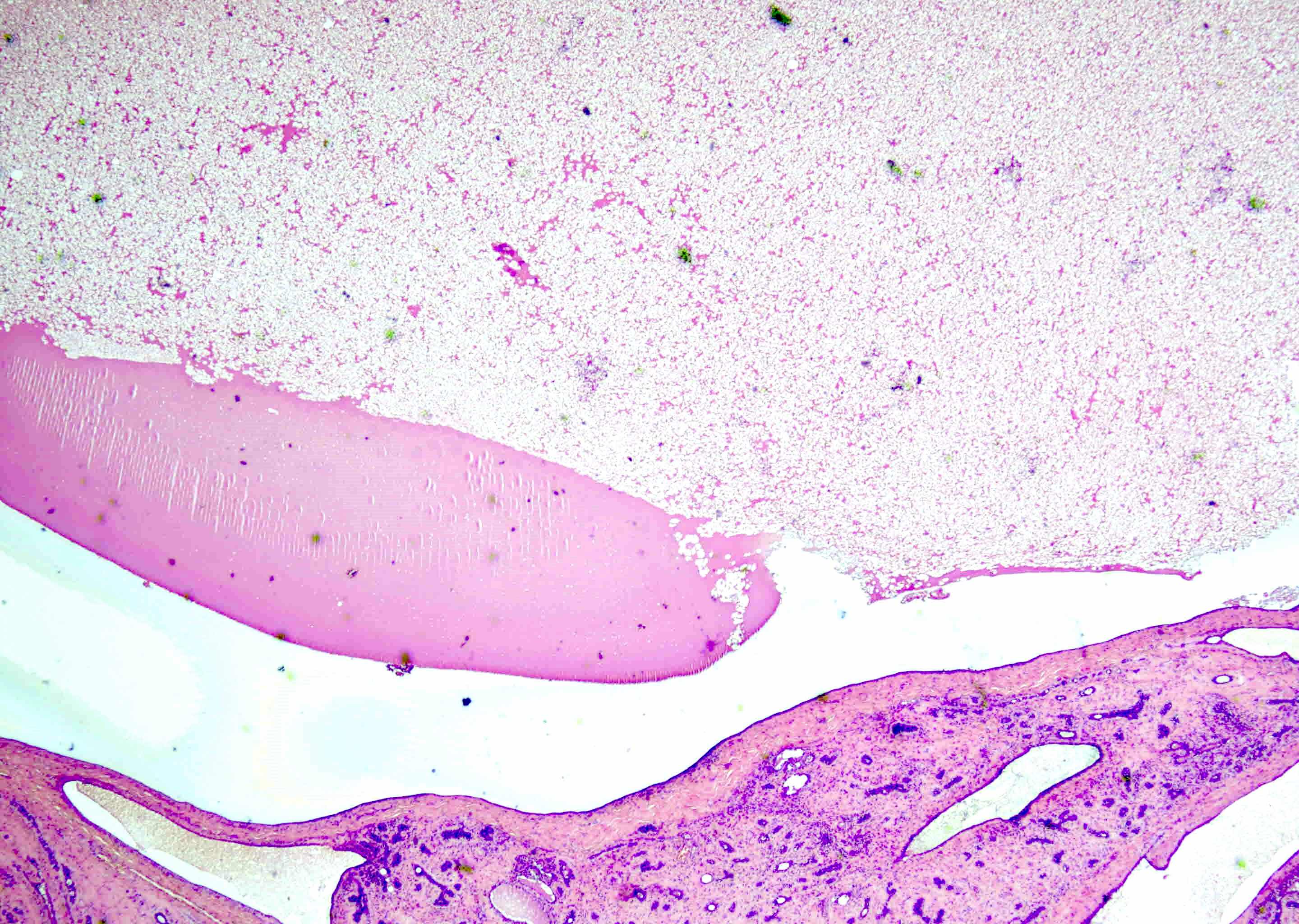
Proteinaceous material
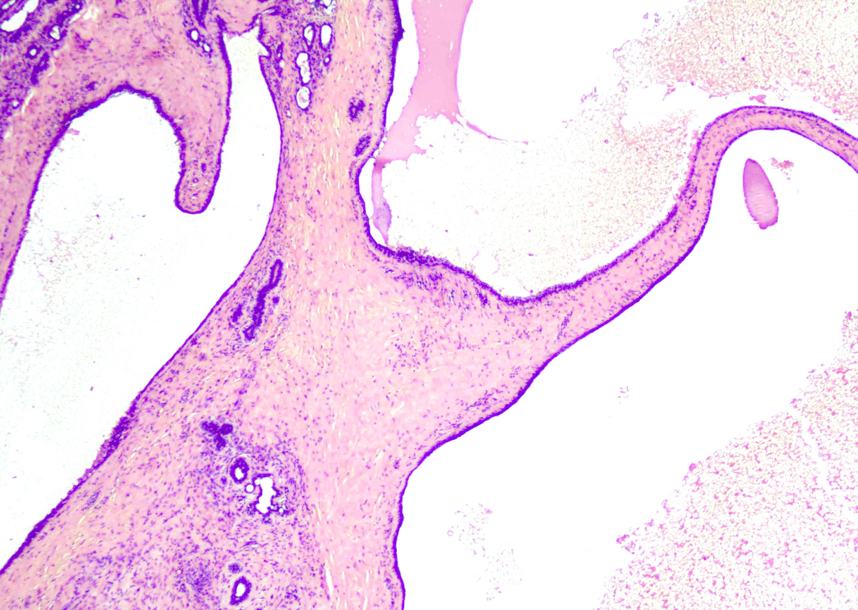
Cyst lined by cuboidal epithelium
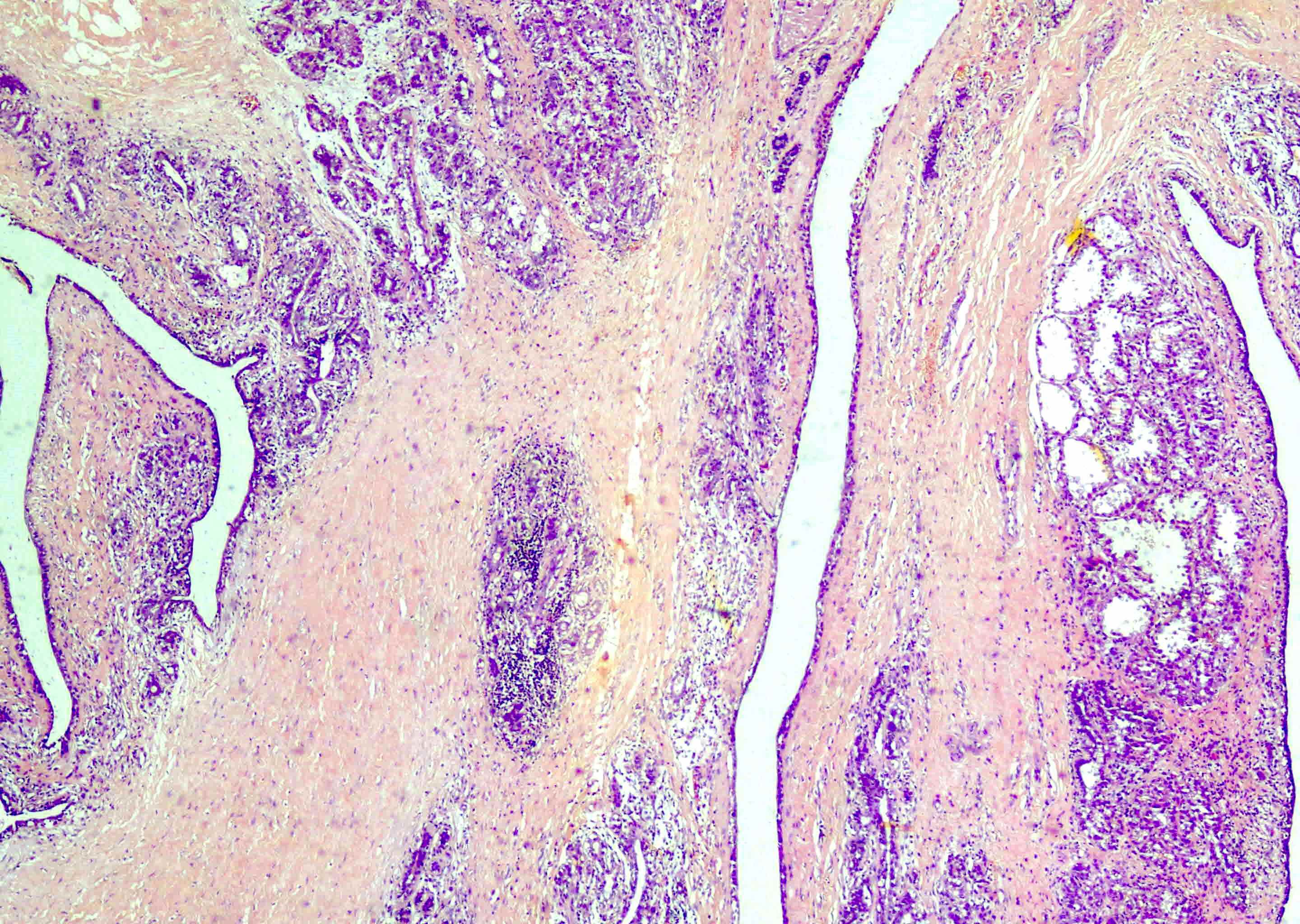
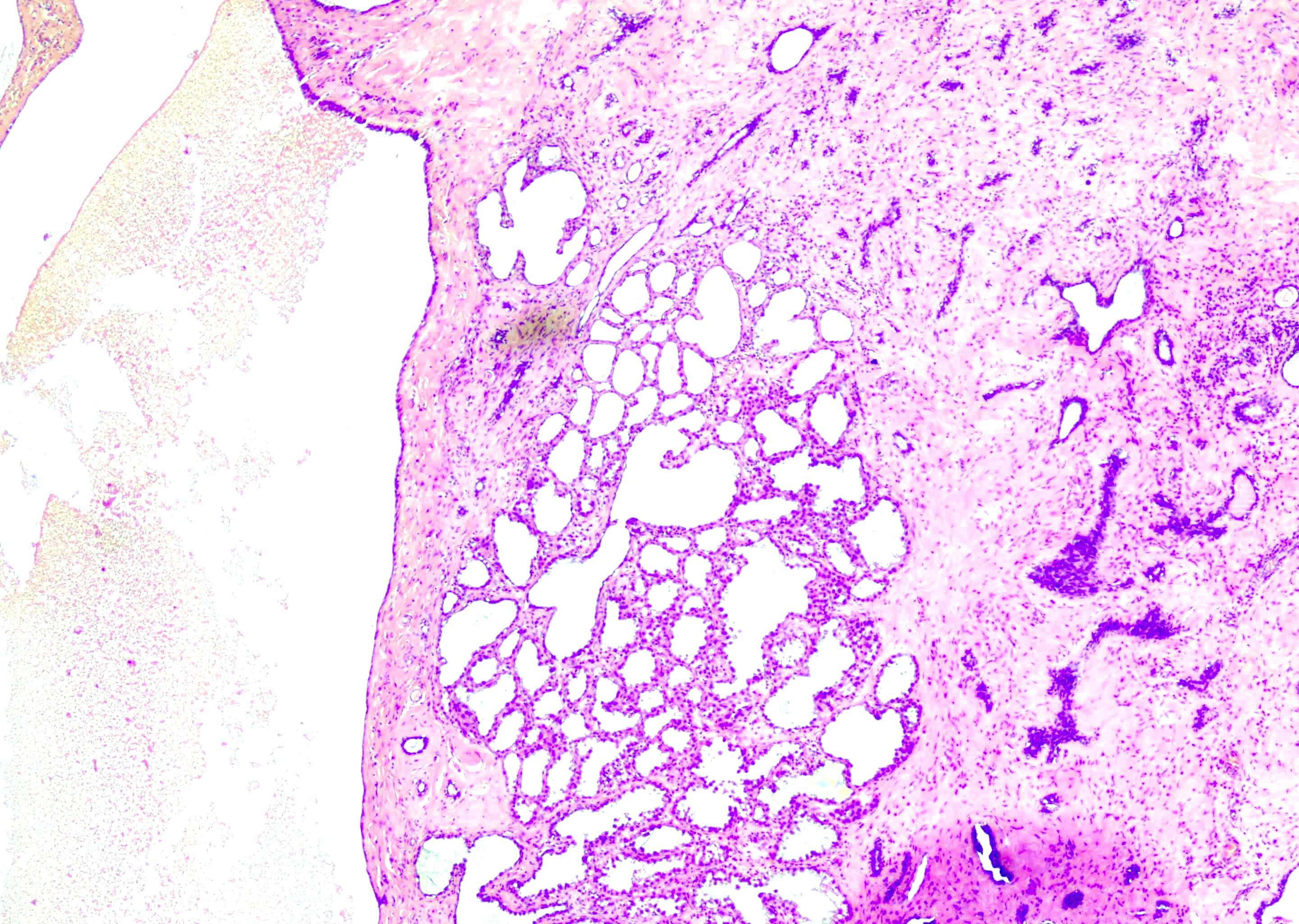
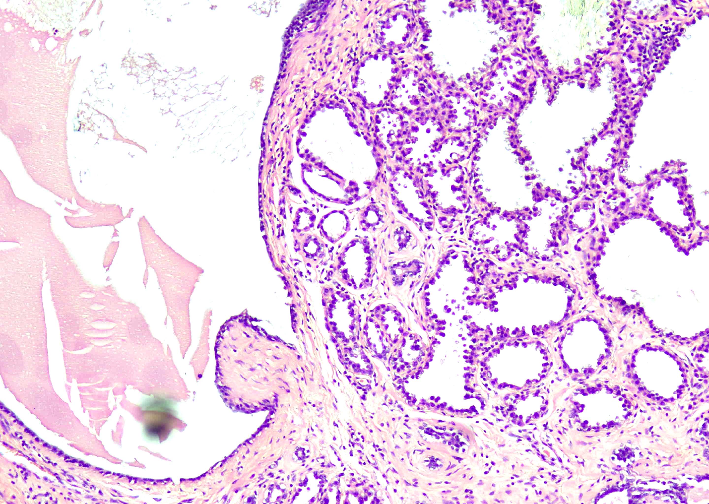
Galactocele with lactational change and mild fibrosis
Cytology description
- Thick, chalky white material with a gritty sensation during aspiration; well defined purple crystals, which show positive birefringence
- Granular, amorphous, proteinaceous material in the background admixed with frothy appearing lipid micelles
- Absence of distinct epithelial fragments, bipolar nuclei and fibrous stroma (J Cytol 2020;37:149, StatPearls: Galactocele [Accessed 21 September 2022])
Cytology images
Contributed by Dhiraj B. Nikumbh, M.D.
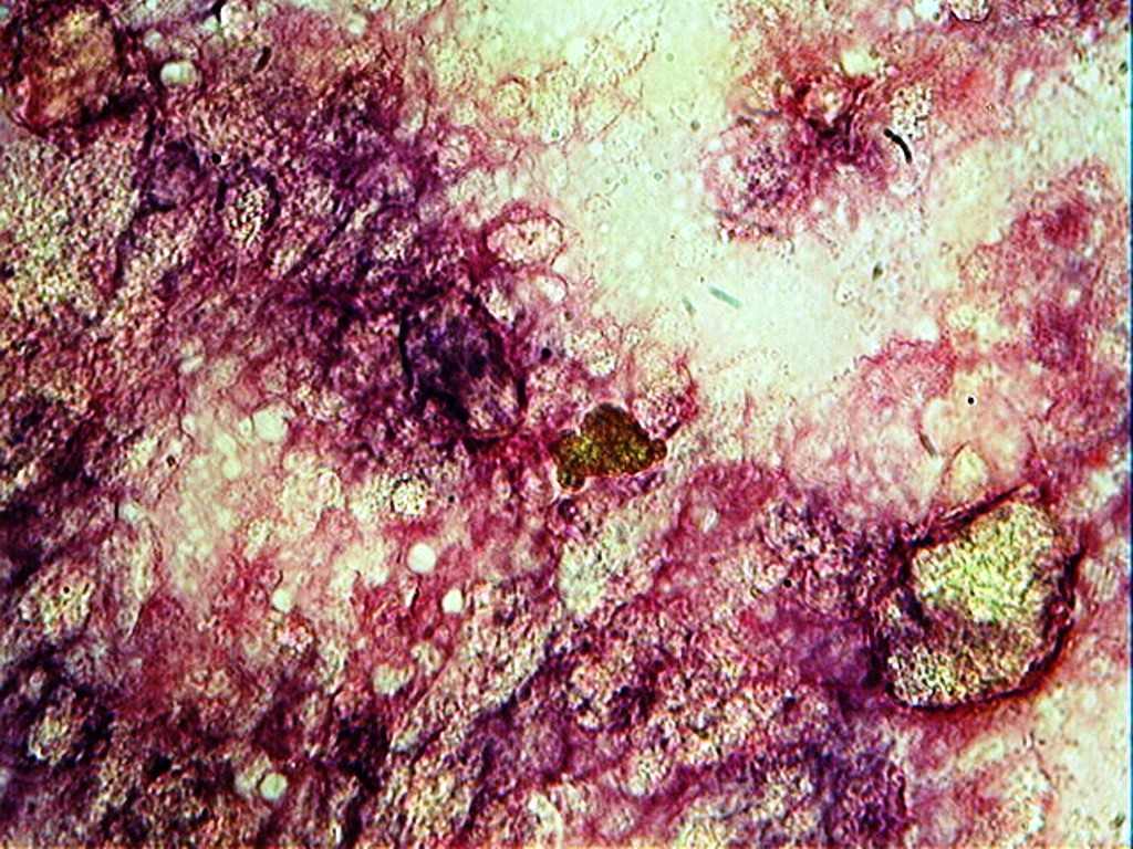

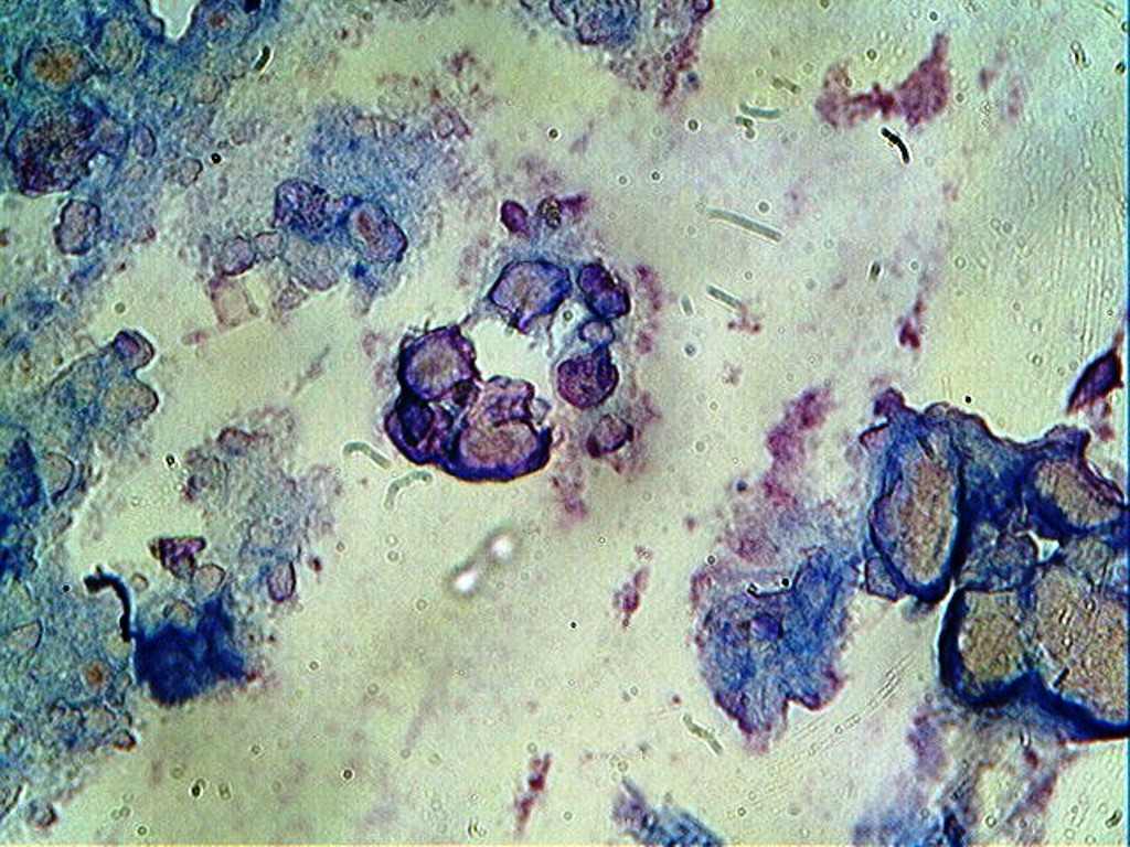
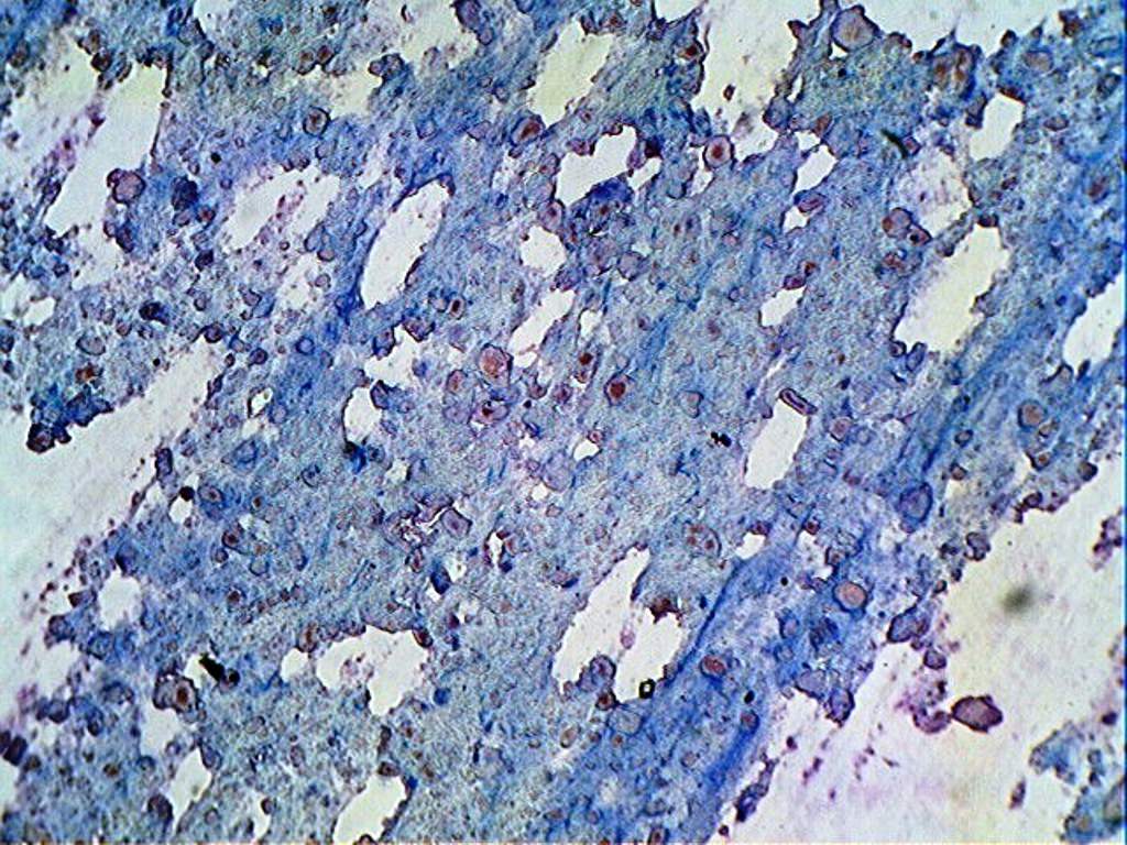
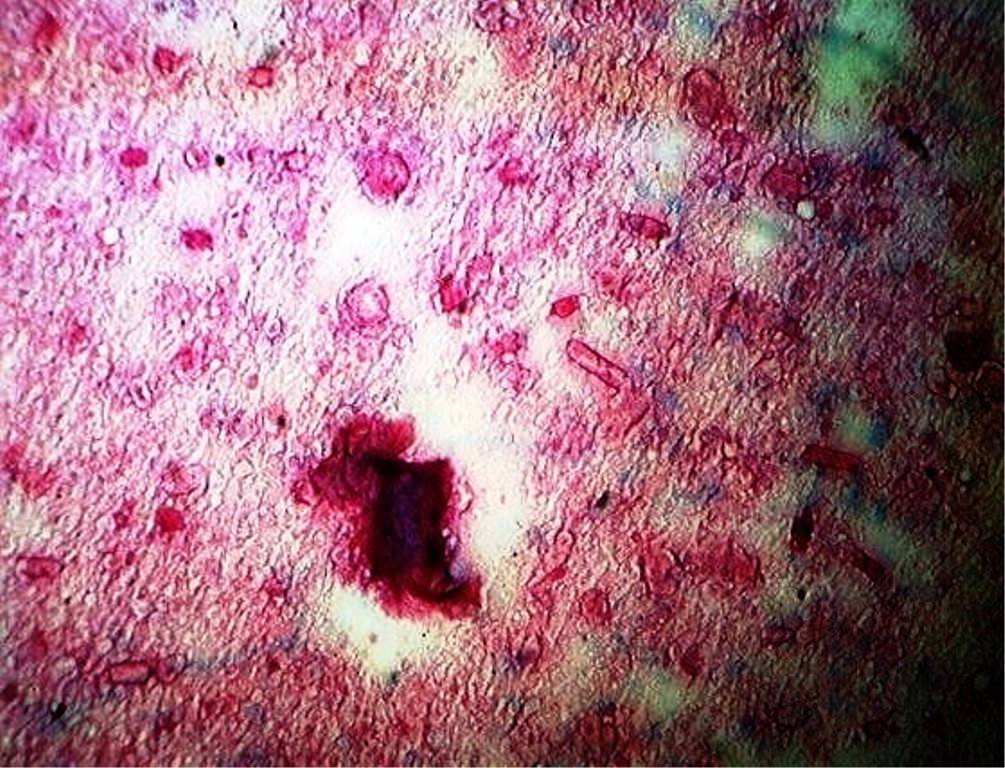
27 year old woman with 1.5 cm nontender breast nodule and fine needle aspiration cytology
Images hosted on other servers:

Dark purple crystals in a proteinaceous background
Sample pathology report
- Breast, excision:
- Galactocele (see comment)
- Comment: It is a benign condition with no recurrence.
- Microscopic description: Breast tissue exhibiting dilated anastomosing channels lined by cuboidal epithelium with secretory activity. Adjacent tissue shows secretory change and fibroinflammatory reaction with foamy macrophages. No evidence of granulomatous or neoplastic processes are seen.
Differential diagnosis
- Lactating adenoma:
- Composed of collections of lobules exhibiting lactational changes
- Fibroadenoma:
- Biphasic lesion exhibiting epithelial and stromal proliferations with intracanacular or pericanacular pattern
- Hamartoma:
- Circumscribed lesion showing lobulated architecture composed of ducts, lobules, fibrous tissue and adipose tissue in varying proportions
- Duct ectasia:
- Presents in premenopausal women and women 45 - 55 years of age (StatPearls: Mammary Duct Ectasia [Accessed 9 November 2022])
- May present with nipple discharge, soft to firm swelling with mild erythema or asymptomatic (StatPearls: Mammary Duct Ectasia [Accessed 9 November 2022])
- Radiology may show microcalcifications, lobulated partially smooth masses, nipple retraction, retroareolar duct dilatation and rarely speculated looking mass (StatPearls: Mammary Duct Ectasia [Accessed 9 November 2022])
- Histologically shows dilation of large ducts, lipid rich detritus accumulation in lumen, fibrous thickening of walls, florid inflammatory reaction on leakage of luminal contents (StatPearls: Mammary Duct Ectasia [Accessed 9 November 2022])
- Mucocele-like lesion:
- Rare, usual age of presentation is between 25 - 61 years (Am J Surg Pathol 1986;10:464)
- Radiology may show multiple aggregated cystic lesions, calcifications can be present (Am J Case Rep 2019;20:926)
- Histologically shows multiple cysts lined by cytologically uniform flat or cuboidal to columnar epithelium with extravasated mucin (Ultrasonography 2015;34:133)
Additional references
Board review style question #1
34 year old lactating mother presented with the history of a palpable lump in the left breast. Breast ultrasound examination showed a circumscribed cystic lesion. Histology is shown in the picture above. What is the diagnosis?
- Fibroadenoma
- Galactocele
- Hamartoma
- Invasive breast carcinoma of no special type
- Lactating adenoma
Board review style answer #1
Board review style question #2
Galactocele formation is related to which of the following conditions?
- Age of the mother
- Duct obstruction
- Estrogen effect
- Family history
- Number of pregnancies
Board review style answer #2
