
Bone marrow nonneoplastic
Normal
Osteoclasts
Author: Dragos C. Luca, M.D.

Last author update: 1 September 2012
Last staff update: 10 December 2021
Copyright: 2002-2024, PathologyOutlines.com, Inc.
PubMed Search: Bone marrow [title] osteoclasts [title]
Table of Contents
Definition / general | Diagrams / tables | Microscopic (histologic) description | Microscopic (histologic) images | Positive stains | Electron microscopy description | Electron microscopy images | Additional referencesCite this page: Luca DC. Osteoclasts. PathologyOutlines.com website. https://www.pathologyoutlines.com/topic/bonemarrowosteoclasts.html. Accessed April 19th, 2024.
Definition / general
- Cells derived from hematopoietic progenitor cells involved in bone resorption, primarily due to remodeling and not calcium homeostasis
- Along endosteal surface of bony trabeculae (reside in Howship lacunae) or along margins in marrow smears
- Common in children (active bone remodelling); in adults associated with metabolic or neoplastic diseases
- Some overlapping immunophenotypic features with monocytes / macrophages
- Relatively recently identified osteoclast colony stimulating factor and transcription factors involved in osteoclastogenesis
- Activated by parathyroid hormone and by cytokines RANKL and macrophage colony stimulating factor (Arthritis Res Ther 2006;8:201)
- Osteoclasts use their ruffled borders (with villous extensions) to bind to matrix adhesion proteins, produce resorption pits / bays (shallow concavities) called Howship lacunae
- Plasma membrane forms a seal with bone; osteoclast acidifies extracellular area, which solubilizes the mineral and releases enzymes which dissolve the matrix
Diagrams / tables
Images hosted on other servers:

Cross section

Bone resorptive cycle
Microscopic (histologic) description
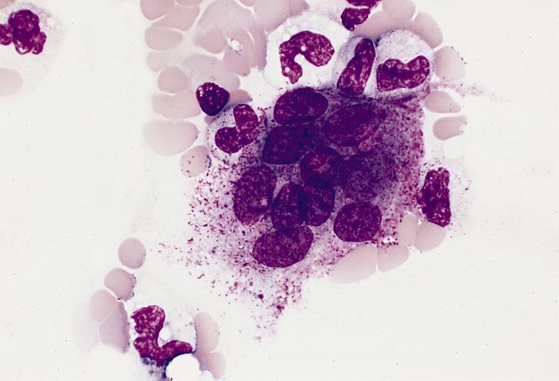
- Very large (up to 100 microns), multinucleated (2 - 12 nuclei) giant cells associated with bone surface
- Abundant, blue purple pale pink cytoplasm containing many fine, red purple granules
- Also contain bone sand in the cytoplasm
- Multiple, relatively uniform but widely separated nuclei, each with one nucleolus and dense chromatin
- Generally, less mature osteoclasts have basophilic cytoplasm and mature forms have brightly eosinophilic cytoplasm
Microscopic (histologic) images
AFIP images
Very large cell
Images hosted on other servers:


Osteoclasts
Positive stains
Electron microscopy description
- Numerous mitochondria, rare lysosomes
- Ruffled edge in area of cell membrane is associated with bone resorption
Electron microscopy images
Images hosted on other servers:

Osteoclast
Additional references
