
Bone marrow nonneoplastic
Normal
Osteoblasts
Author: Dragos C. Luca, M.D.

Last author update: 1 September 2012
Last staff update: 13 December 2021
Copyright: 2002-2025, PathologyOutlines.com, Inc.
PubMed Search: Bone marrow [title] osteoblasts [title]

Table of Contents
Definition / general | Diagrams / tables | Microscopic (histologic) description | Microscopic (histologic) images | Positive stains | Negative stains | Electron microscopy description | Electron microscopy images | Additional referencesCite this page: Luca DC. Osteoblasts. PathologyOutlines.com website. https://www.pathologyoutlines.com/topic/bonemarrowosteoblasts.html. Accessed April 2nd, 2025.
Definition / general
- Bone forming cell that arises from marrow mesenchymal (stromal) cells, unlike osteoclasts which are of hematopoietic origin
- Along endosteal surface of bony trabeculae or along margins in marrow smears
- Common in children; in adults associated with various diseases and healing biopsy sites
- Synthesize angiopoietin and osteopontin, which inhibit hematopoietic stem cell proliferation (Br J Haematol 2006;134:467, J Clin Invest 2006;116:1195)
- Synthesize and transport collagenous matrix, initiate and regulate mineralization, control removal of bone via osteoclasts
- When active are plump and present on bone surface; eventually are encased within the collagen they produce and get flattened (and become osteocytes)
- Activity is promoted by physical activity (Wolff's law of bone adaptation)
- Express parathormone receptors (mediates the activation of osteoclasts) and vitamin D receptor
- Control osteoclast activity via parathyroid hormone (parathormone), PHRP (parathyroid hormone related protein), IL1 and TNF alpha
- Digestion of bone by osteoclasts releases cytokines and growth factors for osteoblasts
Diagrams / tables
Images hosted on other servers:

Interaction of osteoblasts and hematopoietic stem cells
Microscopic (histologic) description
- Large (25 - 50 microns), often oval with abundant blue gray cytoplasm and perinuclear hof separated from the nucleus (unlike plasma cells)
- Nucleus is markedly eccentric and round / ovoid with one or more nucleoli
Microscopic (histologic) images
AFIP images
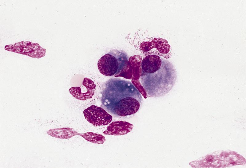
Osteoblasts in marrow smear
Images hosted on other servers:


Osteoblasts
Positive stains
Electron microscopy description
- Resemble fibroblasts due to well developed rough endoplasmic reticulum and Golgi zone
Electron microscopy images
Images hosted on other servers:

Colorized SEM
Additional references
