
Bone & joints
Other tumors
Lymphoma
Author: Sheren Younes, M.D., Ph.D.

Last author update: 1 October 2013
Last staff update: 18 January 2022
Copyright: 2003-2025, PathologyOutlines.com, Inc.
PubMed search: lymphoma [title] bone, general [all fields]

Table of Contents
Definition / general | Epidemiology | Sites | Pathophysiology | Clinical features | Diagnosis | Laboratory | Radiology description | Prognostic factors | Case reports | Treatment | Clinical images | Gross description | Microscopic (histologic) description | Microscopic (histologic) images | Cytology description | Peripheral smear description | Positive stains | Negative stains | Flow cytometry description | Electron microscopy description | Molecular / cytogenetics description | Molecular / cytogenetics images | Differential diagnosis | Staging / staging classificationsCite this page: Younes S. Lymphoma. PathologyOutlines.com website. https://www.pathologyoutlines.com/topic/bonelymphomageneral.html. Accessed March 27th, 2025.
Definition / general
- 40% of bone tumors are hematologic neoplasms, usually myeloma or lymphoma
- Primary bone lymphoma (PBL): defined as lymphoma presenting in an osseous site with no evidence of disease elsewhere for at least 6 months
- Presence of regional lymph nodes does not exclude the diagnosis of PBL (Joint Bone Spine 2000;67:446)
Epidemiology
- PBL accounts for < 5% of extranodal lymphoma and < 1% of all non-Hodgkin lymphoma (Am J Surg Pathol 1990;14:329)
- Slight male preponderance
- Can occur at any age, common in adults, mean age 48 years (range 11 - 83 years)
- 80% are diffuse large B cell lymphoma
- PBL is classified into four groups:
- Group 1: solitary bone lymphoma
- Group 2: multifocal bony lesions
- Group 3: cases with distant nodal disease
- Group 4: cases with visceral disease
- Secondary involvement of bone by lymphoma is more common than primary, considered to be stage IV
Sites
- Any bone can be involved
- Usually sites of bone marrow, axial skeleton (spine), femur
- Common in metaphysis; presence in diaphysis or epiphysis probably means progressive disease
Pathophysiology
- Unknown why bone marrow develops into PBL; may be due to osteoclast activating factor has been
- Strong tendency to spread and relapse suggests homing properties of lymphoma
Clinical features
- Most commonly bone pain
- Also palpable mass, pathologic fracture, neurologic symptoms with spine involvement
- LDH may be elevated
- B symptoms are uncommon
- May relapse and involve other bones, lymph nodes, adjacent soft tissue, lung, bone marrow, CNS
Diagnosis
- Clinical, radiologic, and biopsy
Laboratory
- Elevated LDH and B2 microglobulin
- CBC, ESR and CRP
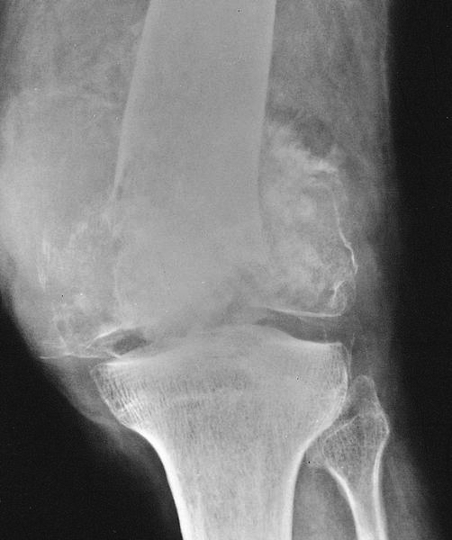
Radiology description
- Most are permeative, mixed lytic sclerotic bone lesions
- Usually large portion of bone is affected
- May be unifocal, monostotic, or polystotic
- Cortex is destroyed without reactive periosteal new bone formation
- Variable sclerosis
- Cystic, commonly mixed
- Lymph node and soft tissue involvement is common
- MRI shows signal abnormalities in bone marrow
Prognostic factors
- Relatively good prognosis: 5 years survival > 80%
- Factors associated with poor prognosis include: polystotic type, advanced disease, age > 60 years, site of mandible / maxilla
- Also high IPI score, performance status < 2, normal LDH
- Also combined modality therapy, higher radiation dose, > 3 cycles of chemotherapy, non germinal center phenotype of diffuse large B cell lymphoma
Case reports
- 23 year old woman with primary bone diffuse large B cell lymphoma of calcaneus (J Foot Ankle Surg 2013;52:666)
- 47 year old man with primary bone anaplastic large cell lymphoma (Acta Ortop Mex 2009;23:142)
- 85 year old man with primary sacral non germinal center type diffuse large B cell lymphoma with MYC translocation (Int J Clin Exp Pathol 2013;6:1919)
- Iliac bone (JBR-BTR 2012;95:375)
- Six cases of anaplastic large cell lymphoma (Mod Pathol 2000;13:1143)
Treatment
- Combination of radiotherapy and chemotherapy (Clin Orthop Relat Res 2013;471:2684)
- Surgery has limited role - may delay start of chemotherapy
- In children, chemotherapy alone is treatment of choice - gives better response
- Radiotherapy may cause bone sarcoma
Clinical images
AFIP images
Femur
Images hosted on other servers:

Imaging of sacral lymphoma
Gross description
- Usually small biopsy to avoid pathologic fracture disabilities
- Fish flesh appearance of lymphoma
- Extraosseous extension and indistinct margins
Microscopic (histologic) description
- Most common type is diffuse large B cell lymphoma (80%)
- Similar to morphology at other sites: diffuse growth pattern, infiltrating between bone trabeculae
- Large atypical cells, abundant cytoplasm
- Centroblasts, immunoblasts, or large bizarre cells
- Nuclei show clumped chromatin, prominent nucleoli
- Component of small lymphocytes is admixed with large cells
- May have marked spindling and fibrosis
- Also Burkitt lymphoma, lymphoblastic lymphoma, follicular lymphoma, other low grade B cell lymphoma, Hodgkin lymphoma, T cell lymphomas
Microscopic (histologic) images
AFIP images

Dense nuclei and histiocyte-like cells
Images hosted on other servers:

Sacral diffuse large B cell lymphoma
Cytology description
- Variable combination of centroblasts, immunoblasts
- May show large bizarre cells
Peripheral smear description
- Lymphoma cells are rarely present in peripheral blood; when centroblasts are present, cells are very large and pleomorphic with abundant cytoplasm, often lobulated nucleus containing one or more fairly prominent nucleoli
Positive stains
Flow cytometry description
- Shows clonal rearrangement, helps determine phenotype
Electron microscopy description
- Centroblasts have cleaved nuclei, immunoblasts have large, round nuclei with coarse chromatin and one to three prominent nucleoli
- Rough endoplasmic reticulum with dilated cisternae and prominent Golgi apparatus are also apparent
- The degree to which these features are present reflects the cellular degree of plasmacytoid differentiation
Molecular / cytogenetics description
- Clonally rearranged immunoglobulin genes (BCL2, BCL6, MYC)
- 80% of Non Hodgkin lymphoma have chromosomal abnormalities
- Usually Epstein-Barr virus negative
Molecular / cytogenetics images
Images hosted on other servers:

FISH: diffuse
large B cell
lymphoma with
MYC translocation
Differential diagnosis
- Sample size, fibrosis, crush artifacts, and admixed small cells can cause diagnostic problems
- Metastatic carcinoma:
- Cells may have cytoplasmic clearing or be arranged in Indian file pattern; keratin+ (other epithelial markers), negative for lymphoid markers
- Osteosarcoma:
- Due to reactive bone formation associated with lymphoma
- Reactive inflammatory conditions:
- Polymorphous infiltrate, no large neoplastic cells, mixed B and T cells, not clonal
- Spindle cell sarcoma:
- When neoplastic cells are spindled, or caused by fibrosis; are negative for CD45, lymphoid markers
- Other hematopoietic neoplasms, including poorly differentiated plasmacytoma, myeloid sarcoma
- Other round blue cell tumors of bone, all lack immunoreactivity to lymphoid markers
- Neuroectodermal tumor: rosettes, positive for chromogranin and synaptophysin
- Metastatic neuroblastoma: young age, Homer-Wright rosettes, fibrillary background, neuroendocrine marker immunoreactivity
- Osteosarcoma, small cell variant: osteoid is present
- Mesenchymal chondrosarcoma foci of chondroid differentiation, S100+
- Metastatic small cell carcinoma: positive for keratin, neuroendocrine markers
Staging / staging classifications
- Ann Arbor staging system is preferred until a better system is available
