
Bone & joints
Fibrogenic / fibrohistiocytic tumors
Fibrosarcoma of bone
Author: Nat Pernick, M.D.

Last author update: 1 June 2005
Last staff update: 10 January 2022
Copyright: 2003-2025, PathologyOutlines.com, Inc.
PubMed search: fibrosarcoma [title] bone

Table of Contents
Definition / general | Radiology description | Radiology images | Prognostic factors | Treatment | Gross description | Gross images | Microscopic (histologic) description | Microscopic (histologic) images | Differential diagnosisCite this page: Pernick N Fibrosarcoma of bone. PathologyOutlines.com website. https://www.pathologyoutlines.com/topic/bonefibrosarcoma.html. Accessed March 29th, 2025.
Definition / general
- Age 40 years or older, no gender preference
- Sites: medulla of metaphysis of long bones, usually distal femur or proximal tibia, jaw
- Often secondary to infarct, Paget disease, radiation
- Occasionally is multicentric, but metastatic sarcomatoid carcinoma (kidney or other sites) is more likely
Radiology description
- Osteolytic, soap bubble appearance
- Invasive or well defined margins depending on differentiation of tumor
Radiology images
Contributed by Mark R. Wick, M.D. and AFIP images
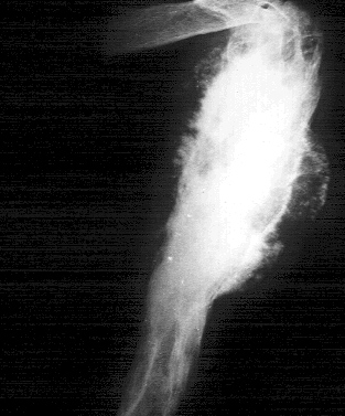
Humerus Xray
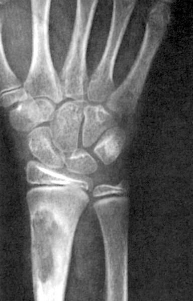
Radius
Prognostic factors
- High grade cytology (10 year survival 34% versus 83% for low grade)
Treatment
- Amputation, wide local excision
Gross description
- Fish flesh appearance of sarcomas
- May destroy cortex and extend into soft tissue
Gross images
AFIP images

Well circumscribed tumor
Microscopic (histologic) description
- Resembles soft tissue fibrosarcoma with herringbone pattern of spindle cells with variable anaplasia
- No malignant osteoid
- Classify as malignant fibrous histiocytoma if prominent pleomorphism
- Well differentiated tumors are hypo- or hypercellular with mitotic figures and atypia
- High grade tumors have more hyperchromasia and mitotic figures
- May have small cells simulating Ewing / PNET
- Other variants are sclerosing epithelioid and myofibroblastic
Microscopic (histologic) images
Contributed by Mark R. Wick, M.D. and AFIP images

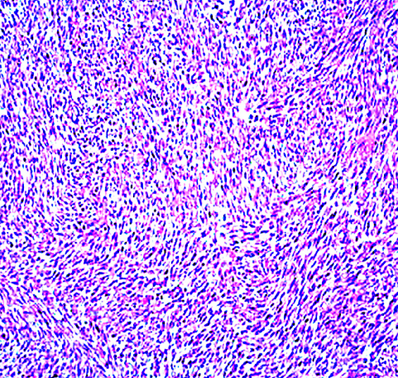
Various images

Well differentiated
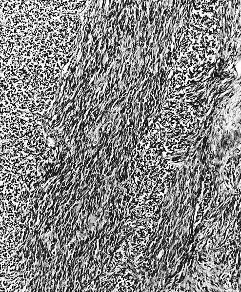
Grade 2

Grade 3
Differential diagnosis
