

Bone & joints
Chondrosarcoma
Dedifferentiated chondrosarcoma
Resident / Fellow Advisory Board: Erna Forgó, M.D.
Deputy Editor-in-Chief: Borislav A. Alexiev, M.D.


Last author update: 1 February 2022
Last staff update: 1 February 2022
Copyright: 2003-2024, PathologyOutlines.com, Inc.
PubMed Search: Bone chondrosarcoma dedifferentiated[TI] free full text[sb]
Table of Contents
Definition / general | Essential features | Terminology | ICD coding | Epidemiology | Sites | Pathophysiology | Etiology | Clinical features | Diagnosis | Radiology description | Radiology images | Prognostic factors | Case reports | Treatment | Gross description | Gross images | Microscopic (histologic) description | Microscopic (histologic) images | Positive stains | Molecular / cytogenetics description | Sample pathology report | Differential diagnosis | Additional references | Board review style question #1 | Board review style answer #1 | Board review style question #2 | Board review style answer #2Cite this page: Kao E, Belzarena C. Dedifferentiated chondrosarcoma. PathologyOutlines.com website. https://www.pathologyoutlines.com/topic/bonedediffchondrosarcoma.html. Accessed April 26th, 2024.
Definition / general
- High grade malignant neoplasm defined by the presence of a conventional chondrosarcoma component juxtaposed to a high grade noncartilaginous sarcoma component
- Poor prognosis with 5 year overall survival of 7 - 24% (Eur J Cancer 2007;43:2060)
Essential features
- Development of a high grade sarcoma in association with a preexisting conventional chondrosarcoma; theoretically, may occur in approximately 1 of 10 - 20 untreated chondrosarcomas (Hum Pathol 1982;13:36, Rev Chir Orthop Reparatrice Appar Mot 1994;80:669)
- Defined histologically by areas of lower grade chondrosarcoma adjacent to a high grade sarcoma
- IDH mutations present
Terminology
- Dedifferentiated chondrosarcoma
ICD coding
- ICD-O: 9243/3 - dedifferentiated chondrosarcoma
- ICD-11: 2B50.Z & XH6E77 - chondrosarcoma of bone or articular cartilage of unspecified sites & dedifferentiated chondrosarcoma
Epidemiology
- Mean age 60 years, slight male predominance (Sarcoma 2019;2019:9069272)
- Development of a high grade sarcoma in association with a preexisting conventional chondrosarcoma; theoretically, may occur in approximately 1 of 10 - 20 untreated chondrosarcomas (Hum Pathol 1982;13:36, Rev Chir Orthop Reparatrice Appar Mot 1994;80:669)
- Dedifferentiation occurs in 9% of peripheral chondrosarcomas and 14% of central chondrosarcomas (Front Med (Lausanne) 2021;8:746909)
- As with conventional chondrosarcoma, it is associated with Ollier disease, multiple hereditary exostoses and enchondromas (Sarcoma 2019;2019:9069272)
Sites
- Most common sites of involvement: femur, pelvis, humerus and scapula (Eur J Cancer 2007;43:2060)
- Preferred site of involvement follows that of conventional chondrosarcoma
Pathophysiology
- Common mutations with conventional chondrosarcoma (IDH and TP53) suggest similar pathogenesis with conventional type (J Pathol 2011;224:334)
Etiology
- Unknown
Clinical features
- Localized pain and palpable mass
- Occasionally a clinical history of prolonged local discomfort with recent change of a rapidly enlarging mass
- Pathological fractures can occur in 22% of the patients (Eur J Cancer 2007;43:2060)
Diagnosis
- Histologic evaluation necessary, although it can be difficult to make the diagnosis on a limited amount of biopsy tissue if only the high grade sarcoma component is sampled and requires correlation with imaging findings for a cartilaginous component (Skeletal Radiol 1995;24:409)
- Sample must show both components, conventional chondrosarcoma and high grade noncartilaginous sarcoma; otherwise, misdiagnosis can occur (Skeletal Radiol 1995;24:409)
- IDH mutation helpful when present (J Pathol 2011;224:334)
Radiology description
- Low grade component manifests as mineralized area with rings and arcs
- High grade component is lytic and aggressive, with permeation and destruction of underlying bone
Radiology images
Contributed by Mark R. Wick, M.D. and AFIP images
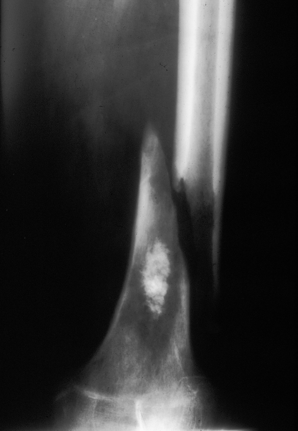
Pathological fracture
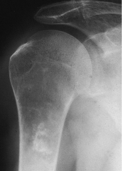
Cartilaginous matrix
Prognostic factors
- Poor prognostic factors include tumor size (> 8 cm), pelvic location, metastatic disease and surgical resection with inadequate margins (Clin Sarcoma Res 2018;8:23, Eur J Cancer 2007;43:2060)
- Pathological fracture at diagnosis has been considered a negative prognostic factor but recent literature suggests it does not alter prognosis (J Orthop Sci 2021;26:473)
- Metastases are present at diagnosis in 20% of patients with median survival of 5 months (J Orthop Res 2020;38:311)
- Overall survival at 5 years is dismal, ranging from 7 - 24% (Eur J Cancer 2007;43:2060)
Case reports
- 29 year old man with nonmetastatic right proximal humerus dedifferentiated chondrosarcoma (Int J Surg Case Rep 2020;72:590)
- 54 year old man with a femur pathological fracture due to dedifferentiated chondrosarcoma and metastasis to the thyroid gland (Oman Med J 2021;36:e283)
- 55 year man with dedifferentiated chondrosarcoma of the temporomandibular joint with extra-articular involvement of the infratemporal space (J Clin Diagn Res 2014;8:ZD09)
- 81 year old man with a cervical spine dedifferentiated chondrosarcoma (World J Surg Oncol 2013;11:32)
Treatment
- Surgical resection with adequate margins
- Chemotherapy and radiotherapy can be used as adjuvants but have no effect on prognosis
Gross description
- Cartilaginous and noncartilaginous components grossly evident in varying proportions (Skeletal Radiol 1995;24:409)
- Blue-gray lobulated cartilage component usually located centrally and a fleshy, yellow or tan high grade sarcoma component is located near the site of pathologic fracture or is located extraosseously in the surrounding soft tissues (Hum Pathol 1982;13:36)
- High grade sarcomatous component may show hemorrhage and necrosis (Skeletal Radiol 1995;24:409)
Gross images
Contributed by Mark R. Wick, M.D.
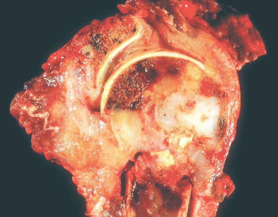
Gray-white cartilage next to tan sarcomatous component
Microscopic (histologic) description
- Conventional chondrosarcoma juxtaposed with an abrupt transition to high grade pleomorphic or spindle cell sarcoma
- Cartilaginous component ranges from an enchondroma-like appearance to grade 1 or grade 2 chondrosarcoma
- High grade dedifferentiated component usually has the appearance of high grade undifferentiated pleomorphic sarcoma or osteosarcoma
- Less frequently, it may have features of angiosarcoma, leiomyosarcoma, rhabdomyosarcoma
- Can even have epithelial differentiation to include squamous and adamantinoma-like morphologies (Oncol Res Treat 2018;41:456, Am J Surg Pathol 1996;20:293, Pathol Res Pract 2017;213:698)
- Amount of dedifferentiation is highly variable and can range from 2 - 98% (Cancer 2006;106:2682)
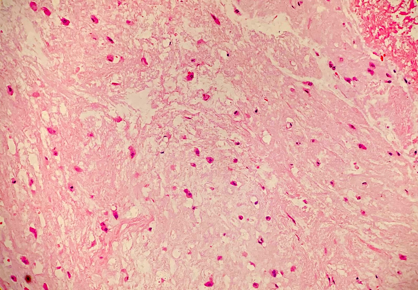
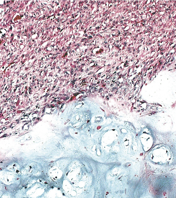
Microscopic (histologic) images
Contributed by Erica Kao, M.D. and Mark R. Wick, M.D.
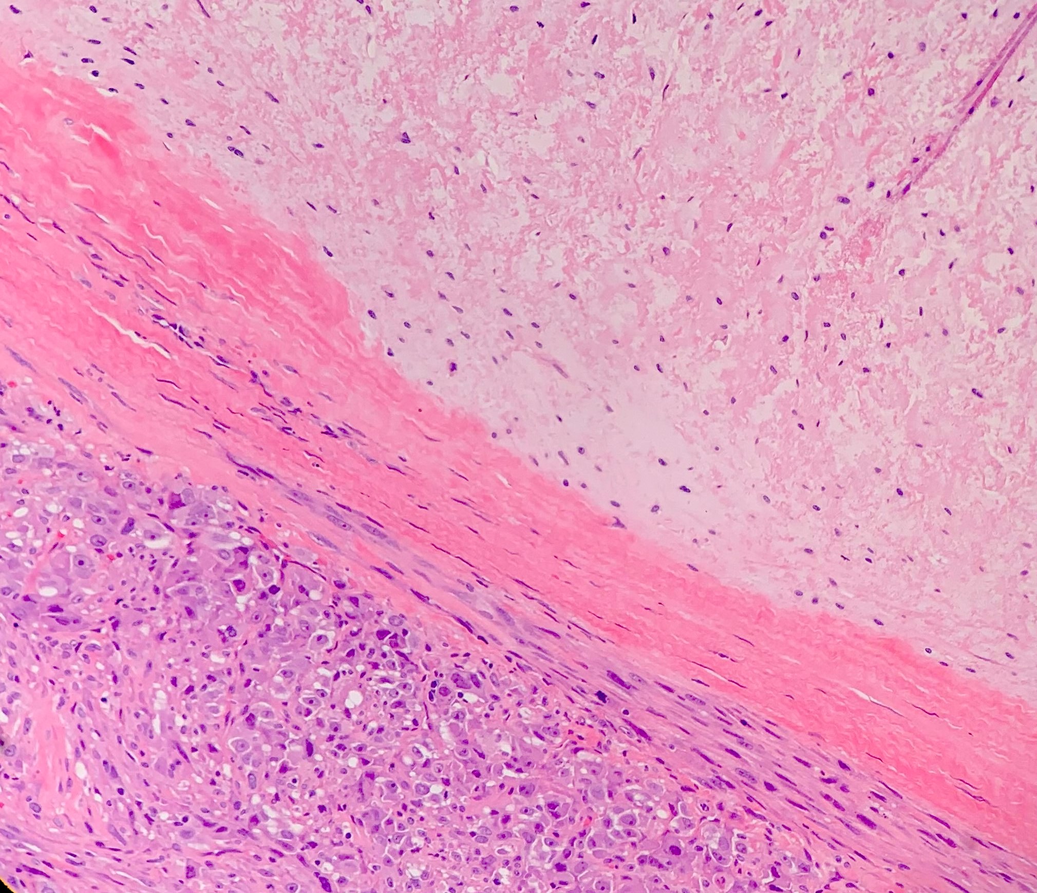
Abrupt demarcation
Low grade cartilage
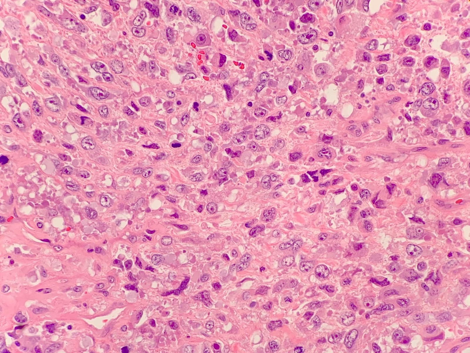
High grade component
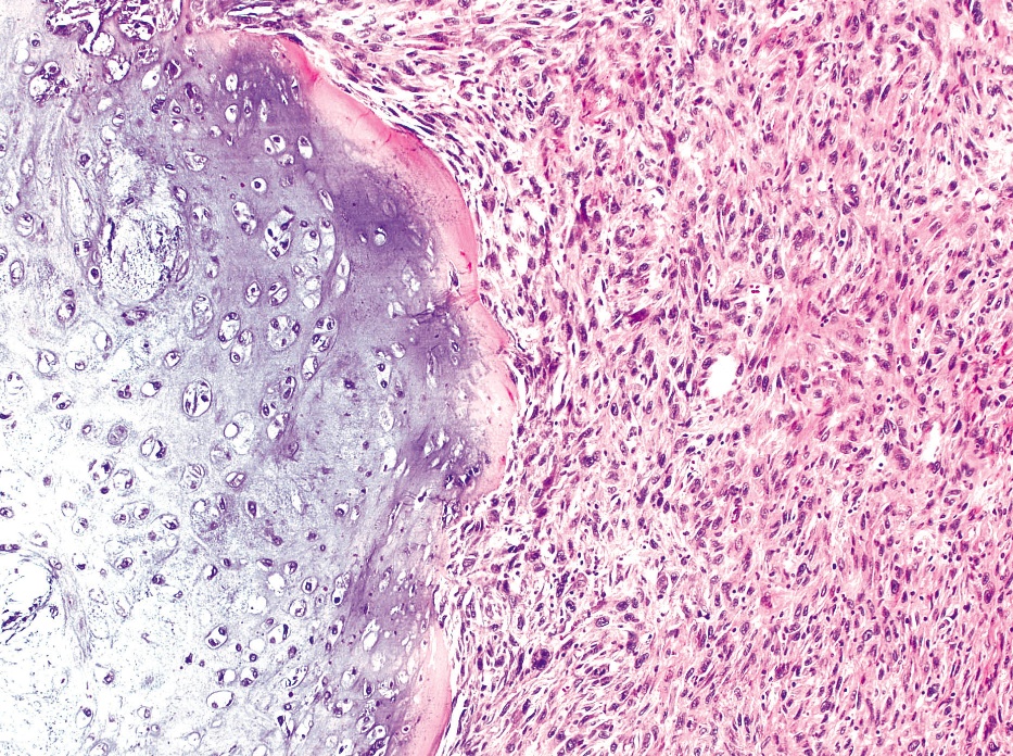
Low grade cartilage juxtaposed with high grade sarcoma
Positive stains
- IDH1 and IDH2 overexpression but of limited use
- Only a small percentage of mutations can be captured using the mutation specific antibody (J Pathol 2011;224:334)
- SOX9 and S100 may be expressed as markers of cartilage lineage differentiation in the low grade component
- p53 overexpression, although not useful in practice
Molecular / cytogenetics description
- Dedifferentiated chondrosarcomas have complex karyotypes (Genes Chromosomes Cancer 2012;51:899)
- 50 - 87% carry mutations in IDH1 or IDH2, which can be found in both low grade and high grade components, supporting a common precursor lesion (J Pathol 1999;189:454, Genes Chromosomes Cancer 2012;51:899)
- Mutations in TP53 are frequently found (Genes Chromosomes Cancer 2012;51:899)
Sample pathology report
- Bone, left femur, core biopsy:
- High grade malignant spindle cell neoplasm (see comment)
- Comment: The patient's reported history of atypical cartilaginous tumor of the distal femur is noted. In that context, the morphologic findings in this biopsy could be consistent with dedifferentiated chondrosarcoma, although there is no cartilaginous component present in this biopsy.
Differential diagnosis
- Chondroblastic osteosarcoma:
- Younger patient population
- Neoplastic bone formation
- Gradual transition from high grade cartilaginous tumor to spindle cell sarcoma
- High grade (grade 3) conventional chondrosarcoma:
- May show spindling of tumor cells at the periphery of lobules
- Gradual transition from chondrocytes to spindled cells without the sharp demarcation characteristic of dedifferentiated chondrosarcoma
- Undifferentiated pleomorphic sarcoma of bone:
- Especially problematic on small biopsies when only the high grade component is sampled
- Requires clinical and radiographic correlation for a cartilage component
- Metastatic sarcomatoid carcinoma:
- Especially problematic on small biopsies when only the high grade component is sampled
- Should express p63, at least focally
- Clinical presentation (mass in lung or kidney) and imaging studies are important for the final diagnosis
Additional references
Board review style question #1
What mutation can be frequently found in the tumor pictured above?
- EXT1
- IDH
- MDM2
- NF1
- RB
Board review style answer #1
B. IDH. 50 - 87% of dedifferentiated chondrosarcomas carry mutations in IDH1 or IDH2, which can be found in both the low grade and high grade components.
Comment Here
Reference: Dedifferentiated chondrosarcoma
Comment Here
Reference: Dedifferentiated chondrosarcoma
Board review style question #2
What benign lesion can be associated with the tumor pictured above?
- Bone island
- Enchondroma
- Nonossifying fibroma
- Osteoblastoma
- Osteoid osteoma
Board review style answer #2
B. Enchondroma. As with conventional chondrosarcoma, dedifferentiated chondrosarcoma is associated with Ollier disease, multiple hereditary exostoses and enchondromas.
Comment Here
Reference: Dedifferentiated chondrosarcoma
Comment Here
Reference: Dedifferentiated chondrosarcoma
