
Anus & perianal area
Other nonneoplastic
Inflammatory cloacogenic polyp
Editorial Board Member: Naziheh Assarzadegan, M.D.
Deputy Editor-in-Chief: Catherine E. Hagen, M.D.


Last author update: 8 June 2023
Last staff update: 8 June 2023
Copyright: 2002-2024, PathologyOutlines.com, Inc.
PubMed Search: Inflammatory cloacogenic polyp
Table of Contents
Definition / general | Essential features | ICD coding | Epidemiology | Sites | Pathophysiology | Clinical features | Diagnosis | Laboratory | Prognostic factors | Case reports | Treatment | Clinical images | Microscopic (histologic) description | Microscopic (histologic) images | Sample pathology report | Differential diagnosis | Additional references | Board review style question #1 | Board review style answer #1 | Board review style question #2 | Board review style answer #2Cite this page: Khorsandi N, Akarca FG, Wen KW. Inflammatory cloacogenic polyp. PathologyOutlines.com website. https://www.pathologyoutlines.com/topic/anusinflamcloaco.html. Accessed April 20th, 2024.
Definition / general
- Benign polyp with colorectal, squamous and transitional epithelium with thickened and prolapsed muscularis mucosa associated with rectal prolapse
Essential features
- Benign polyp with colorectal, squamous and transitional epithelium and prolapsed muscularis mucosae
- Thought to be associated with mucosal prolapse or chronic inflammatory diseases
- Located at anorectal transition zone
Epidemiology
- Wide age range from pediatric to adult population
Sites
- Anorectal transition zone (Eur J Gastroenterol Hepatol 2000;12:247)
Pathophysiology
- Rectal mucosal prolapse due to repeated mucosal ischemia and regeneration are thought to contribute to the development (Histopathology 2008;53:91)
Clinical features
- Symptoms range from asymptomatic to rectal bleeding to constipation, tenesmus, anal swelling or anal itching (Cureus 2022;14:e22014)
- On endoscopy, these polyps can mimic hemorrhoids, solitary rectal ulcer, villous adenoma or anorectal carcinoma (Cureus 2022;14:e22014)
- Associated with solitary rectal ulcer syndrome, mucosal prolapse or chronic inflammatory conditions such as Crohn's disease and colorectal tumors (Cureus 2022;14:e22014, Eur J Gastroenterol Hepatol 2000;12:247)
Diagnosis
- Colonoscopy or sigmoidoscopy typically identifies a sessile or pedunculated polyp (Eur J Gastroenterol Hepatol 2000;12:247)
Laboratory
- Patients sometimes present with a positive fecal immunochemical test (e.g., Cologuard)
Prognostic factors
- Benign entity with no increased risk for malignancy (Eur J Gastroenterol Hepatol 2000;12:247)
Case reports
- 15 year old boy with Marshall-Smith syndrome and perianal subcutaneous mass (Pediatr Pathol 1993;13:409)
- 34 year old woman with anal intraepithelial neoplasia in an inflammatory cloacogenic polyp (J Clin Pathol 1999;52:393)
- 38 year old woman and 41 year old woman with squamous cell carcinoma in situ arising in inflammatory cloacogenic polyp (Histopathology 2008;53:91)
Treatment
- Endoscopic polypectomy or mucosal resection (Am J Gastroenterol 1994;89:438)
- High fiber diet (Cureus 2022;14:e22014)
Clinical images
Images hosted on other servers:

Cloacogenic polyps on rectal retroflexion
Microscopic (histologic) description
- Tubulovillous architecture with elongated crypts stretching into the submucosa
- Areas of stratified squamous, colorectal or transitional epithelium that are often eroded and contain mixed inflammation and granulation tissue (Hum Pathol 1987;18:1120, Am J Surg Pathol 1981;5:761)
- Abundant regenerative epithelial changes can be seen
- Thickened muscularis mucosa with extension into the lamina propria
- Fibromuscular obliteration of the lamina propria can be seen
Microscopic (histologic) images
Contributed by Nikka Khorsandi M.D., M.P.H., Kwun Wah Wen, M.D., Ph.D. and Yvonne Bury, M.D.
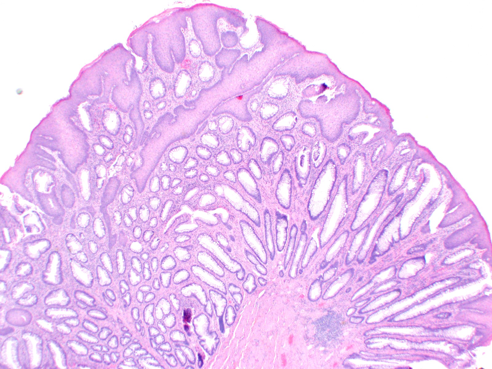
Squamous and glandular epithelium
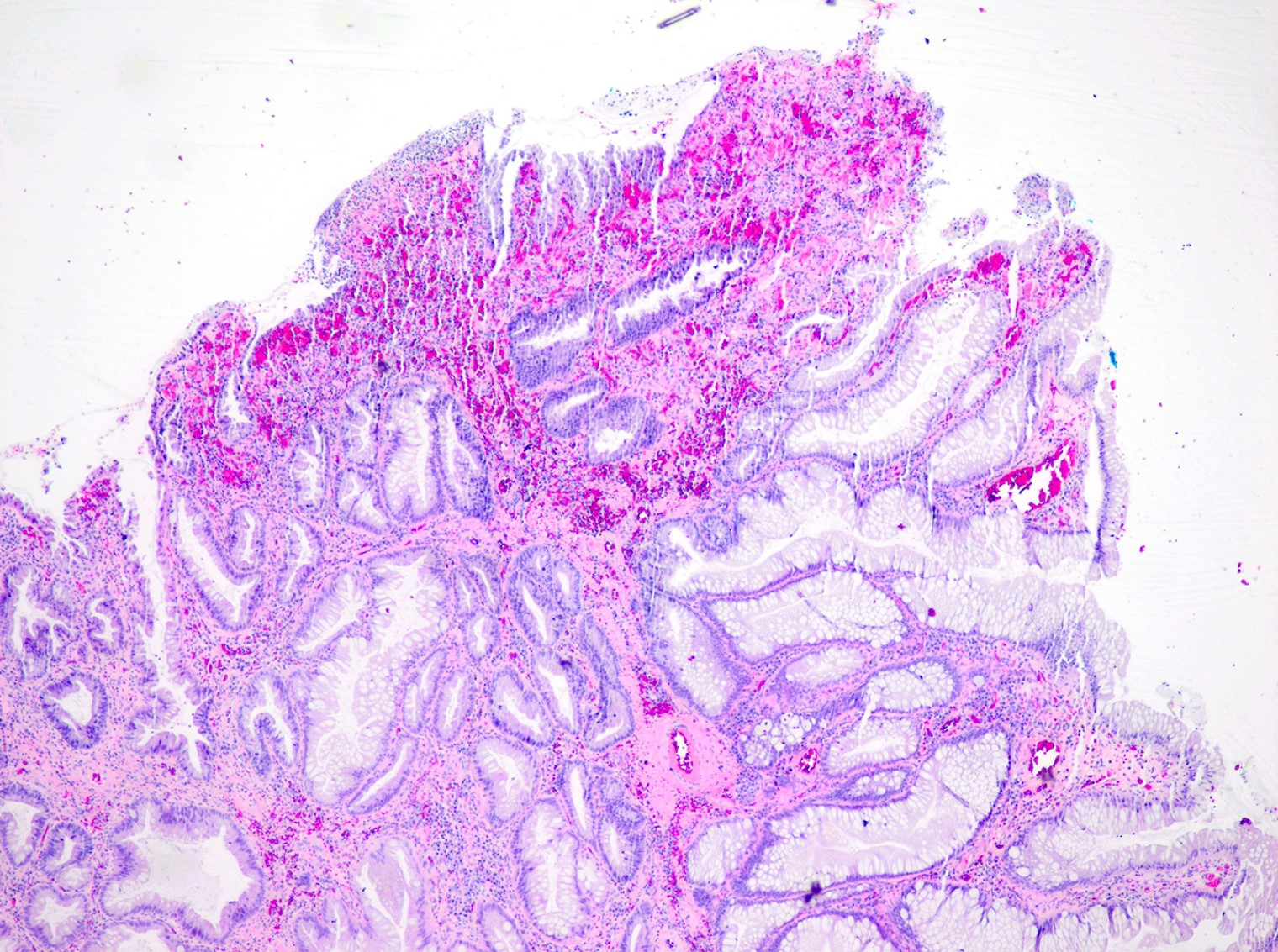
Surface erosion

Mucosal erosion
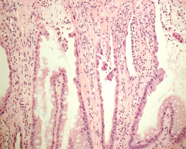
Regenerative epithelial changes
Sample pathology report
- Anus, polypectomy:
- Inflammatory cloacogenic polyp. Negative for dysplasia.
Differential diagnosis
- Solitary rectal ulcer syndrome:
- Should be at anterior rectum, proximal to anorectal junction and is usually flat and ulcerated
- Inflammatory cloacogenic polyp with adenomatous dysplasia:
- Often not necessary but p53 and Ki67 overexpression can assist in identifying areas of dysplasia (Histopathology 2008;53:91)
Additional references
Board review style question #1
Where is the lesion shown in the above image located?
- Anorectal transition zone
- Ascending colon
- Duodenum
- Rectum
- Stomach
Board review style answer #1
A. Inflammatory cloacogenic polyp is located in the anorectal transition zone.
Comment Here
Reference: Inflammatory cloacogenic polyp
Comment Here
Reference: Inflammatory cloacogenic polyp
Board review style question #2
What is the preferred treatment for an inflammatory cloacogenic polyp?
- Chemotherapy
- Colectomy
- Endoscopic / surgical resection
- Fecal immunohistochemical test
- Routine endoscopic surveillance
Board review style answer #2
C. Endoscopic / surgical resection is the preferred treatment for an inflammatory cloacogenic polyp. Routine endoscopic surveillance is necessary after resection because of the risk of recurrence but it is not the treatment modality.
Comment Here
Reference: Inflammatory cloacogenic polyp
Comment Here
Reference: Inflammatory cloacogenic polyp
