

Small intestine & ampulla
General
Histology-ampulla
Author: Raul S. Gonzalez, M.D.
Editor-in-Chief: Debra L. Zynger, M.D.

Last author update: 10 December 2020
Last staff update: 21 July 2023
Copyright: 2002-2025, PathologyOutlines.com, Inc.
PubMed Search: Ampulla of vater [title] normal histology


Table of Contents
Definition / general | Essential features | Diagrams / tables | Microscopic (histologic) description | Microscopic (histologic) images | Positive stains (IHC and special stains) | Differential diagnosis | Additional references | Board review style question #1 | Board review style answer #1 | Board review style question #2 | Board review style answer #2Cite this page: Gonzalez RS. Histology-ampulla. PathologyOutlines.com website. https://www.pathologyoutlines.com/topic/ampullahistology.html. Accessed January 19th, 2025.
Definition / general
- Formed by the union of the distal pancreatic duct and the intrapancreatic distal common bile duct
- Opens into the major duodenal papilla
Essential features
- Complex anatomical structure generally lined by pancreatobiliary-type cells
Diagrams / tables
Images hosted on other servers:

Ampulla of Vater and related structures

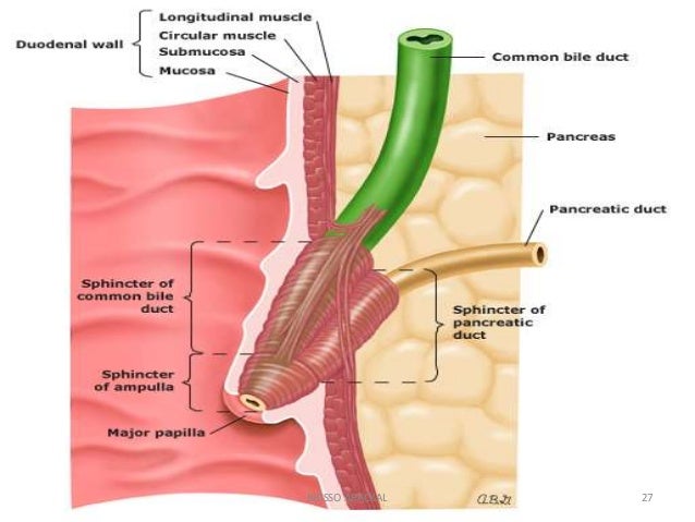
Ampulla, papilla and sphincters
Microscopic (histologic) description
- Ampullary mucosa forms prominent papillary folds
- Epithelium transitions from intestinal type (duodenal surface) to foveolar-like mucosa with occasional goblet cells (papilla) to pancreatobiliary epithelium (distal ducts) (Am J Surg Pathol 2012;36:1592)
- Lamina propria has only occasional lymphocytes, plasma cells and mast cells
- Small mucous glands / ductules underlie the mucosa
- Smooth muscle fibers (representing sphincter of Oddi) may extend into epithelial folds
- May have adjacent pancreatic acini, but usually no islets around major papillae
Microscopic (histologic) images
Contributed by Raul Gonzalez, M.D.
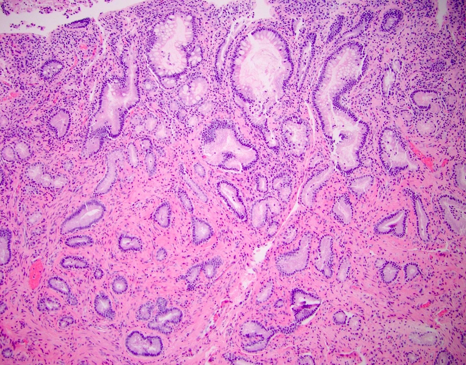
Normal ampullary mucosa and submucosa
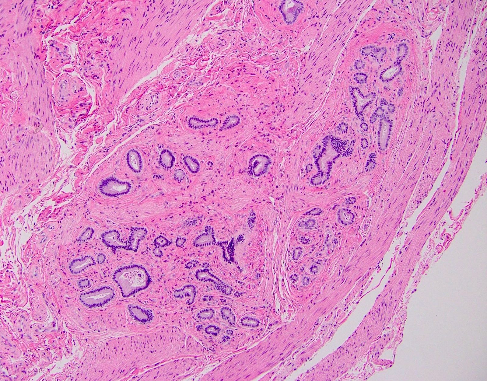
Ampullary submucosal glands / ductules
Positive stains (IHC and special stains)
- Ampullary biopsy performed in autoimmune pancreatitis can help to differentiate from other "mass forming" pancreatitis by IgG4+ to IgG+ ratio (Am J Surg Pathol 2008;32:1770)
Differential diagnosis
- Adenocarcinoma:
- Submucosal ducts / glands may appear malignant, especially if crushed or secondarily involved by an adenoma
Additional references
Board review style question #1
- Which of the following may be seen or around in normal ampullary mucosa?
- Ciliated epithelium
- Islets of Langerhans
- Pancreatic acini
- Squamous epithelium
Board review style answer #1
Board review style question #2
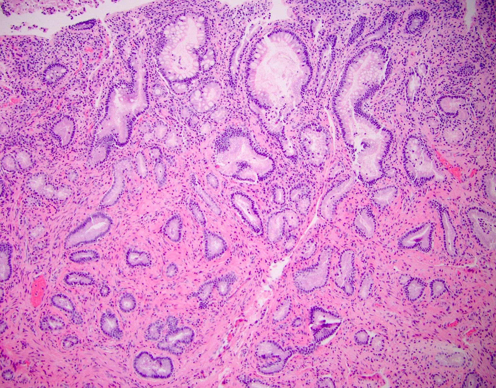
- A patient with a bile duct stricture undergoes esophagogastroduodenoscopy. A biopsy is taken of the ampulla and is shown above. What is the correct diagnosis for this sample?
- Adenocarcinoma
- Gastric heterotopic
- Intestinal metaplasia
- Normal ampulla
Board review style answer #2

