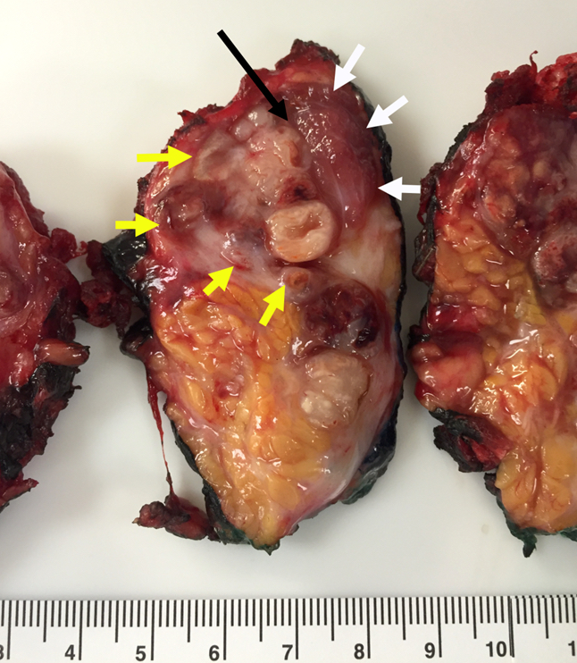
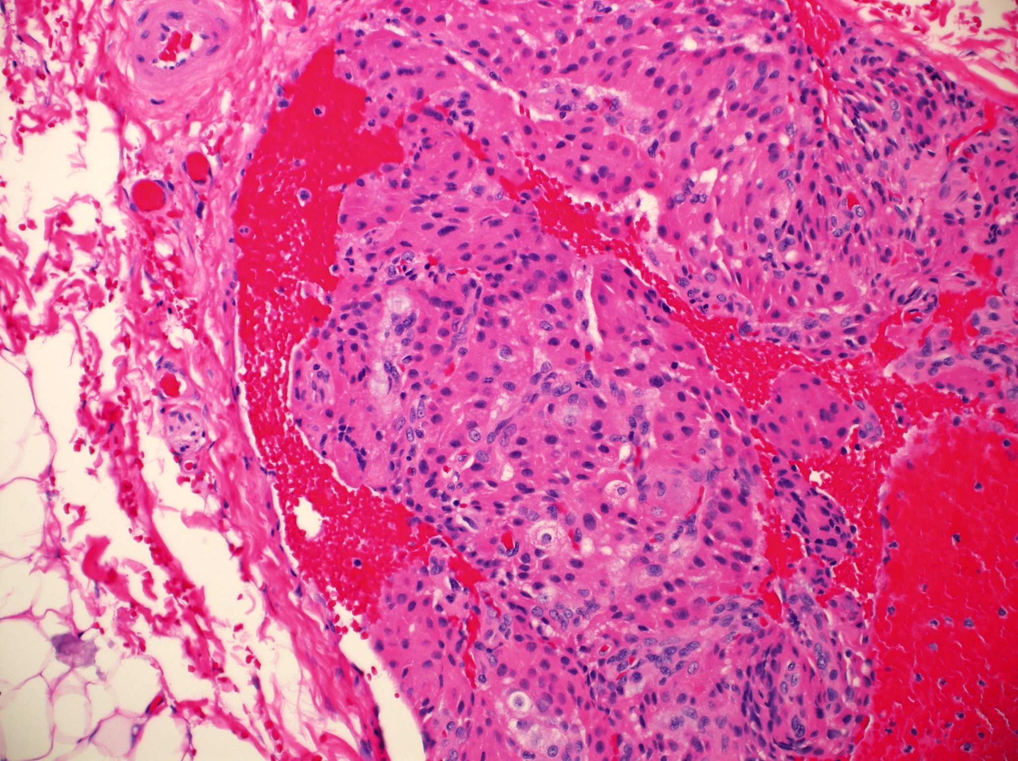
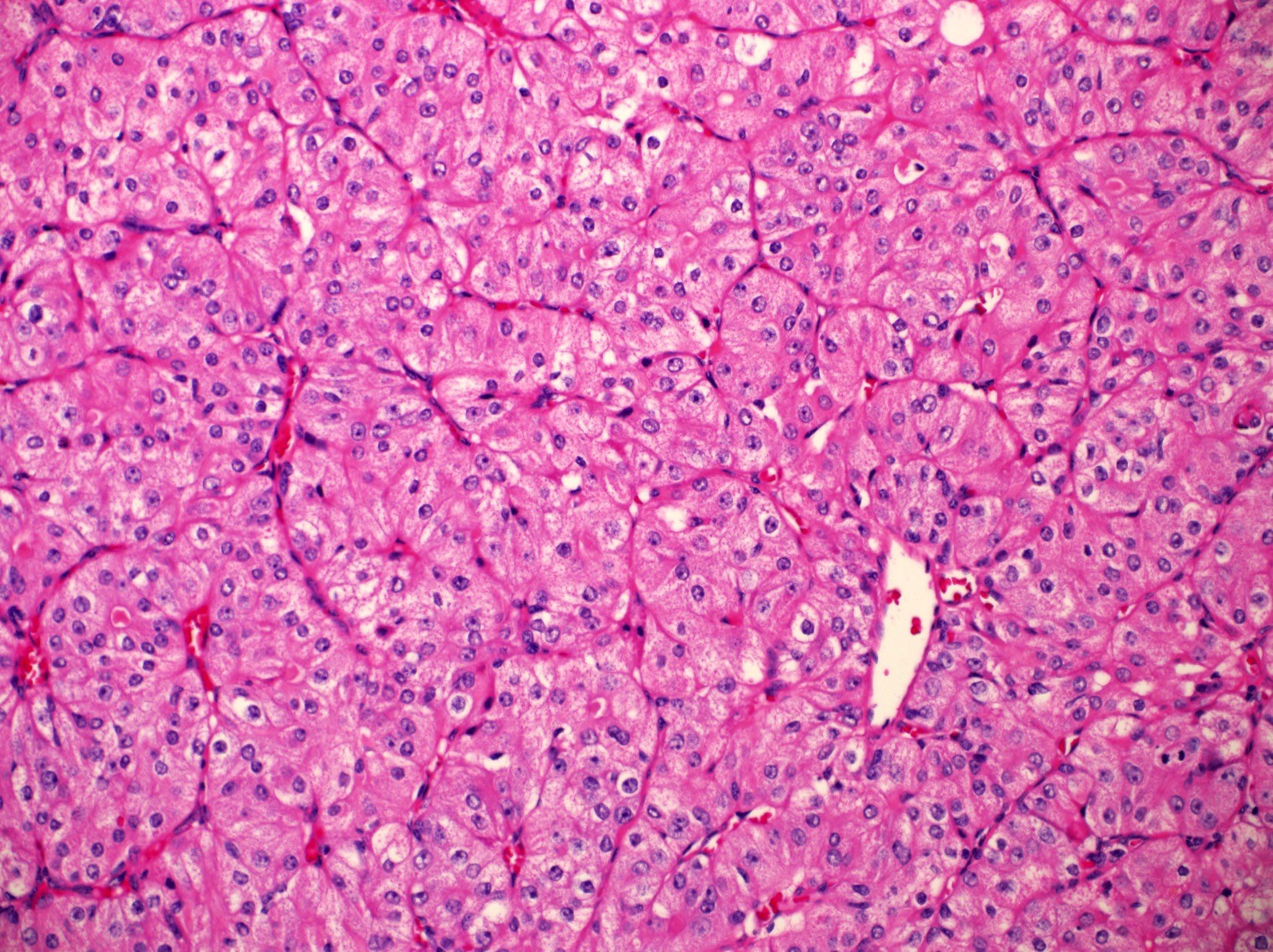
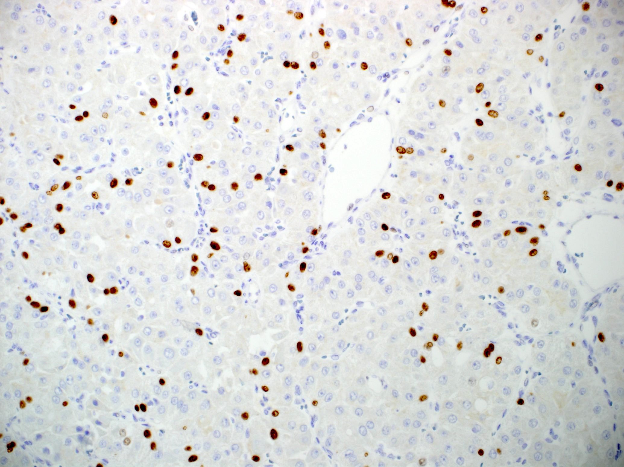
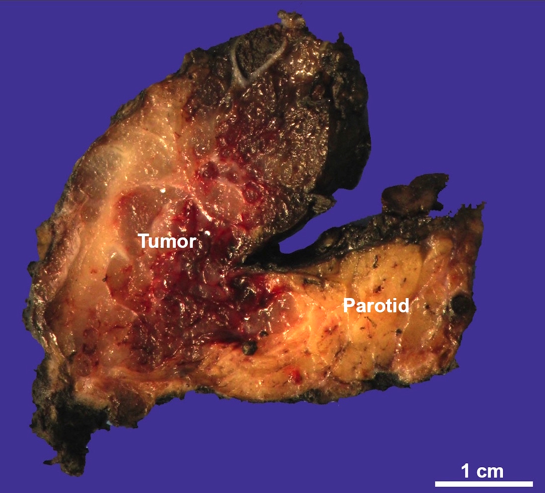
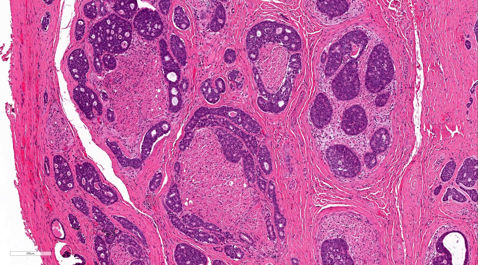
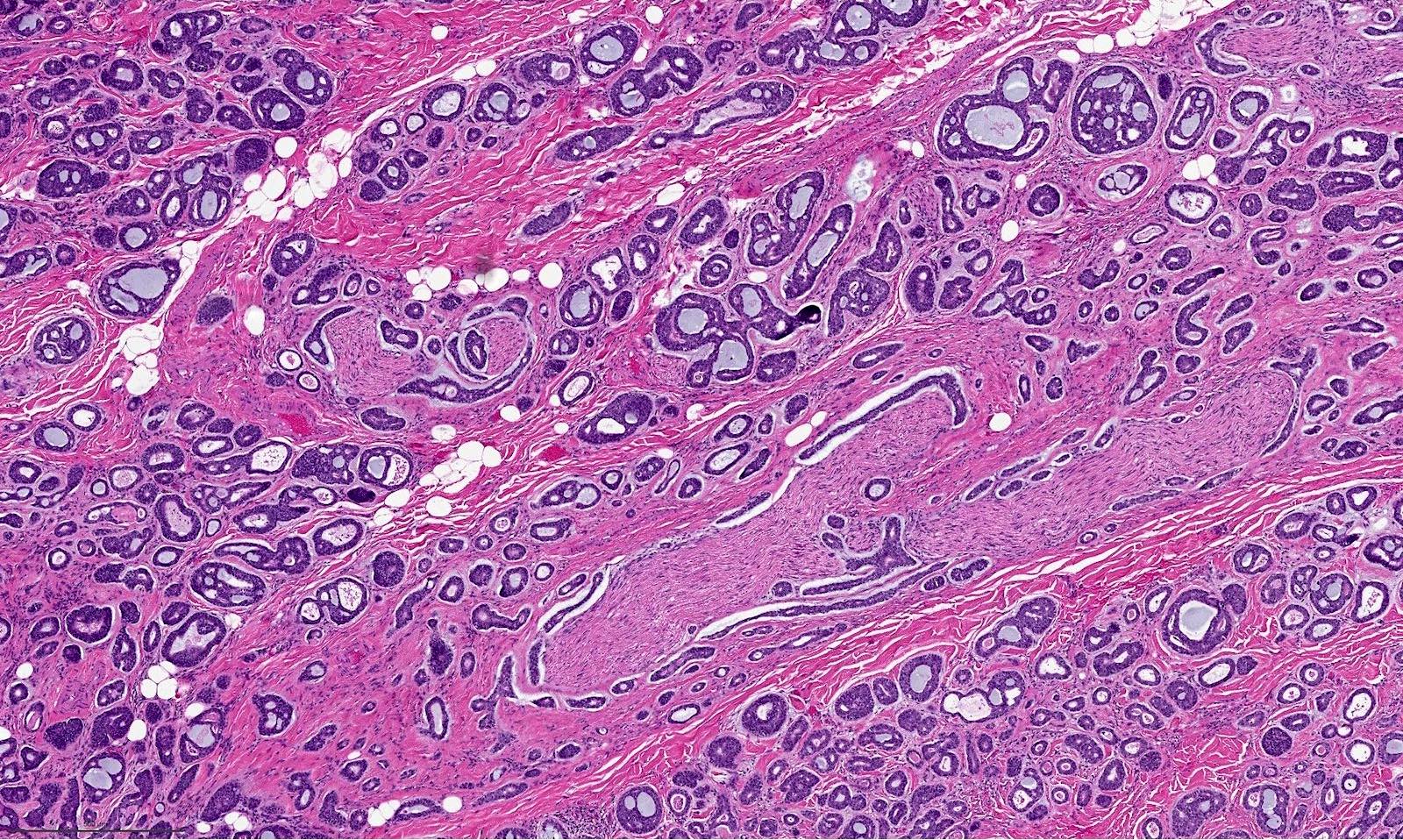
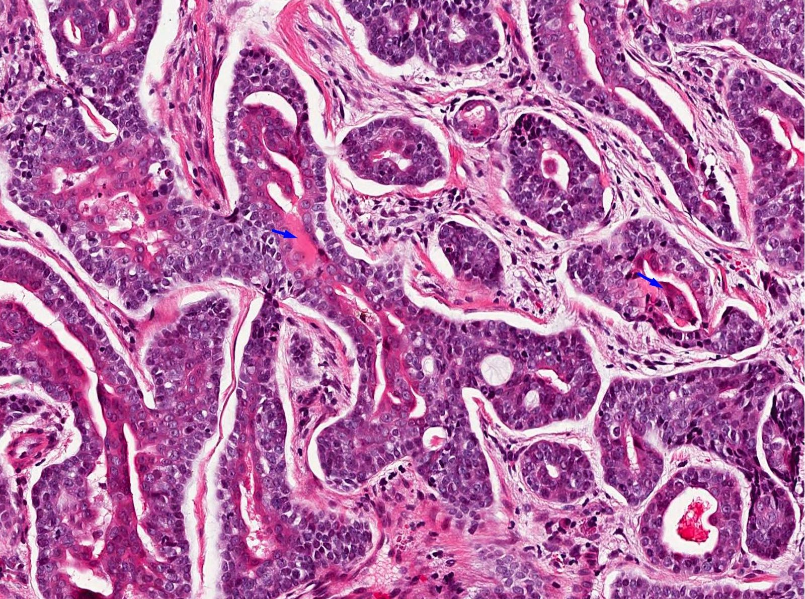
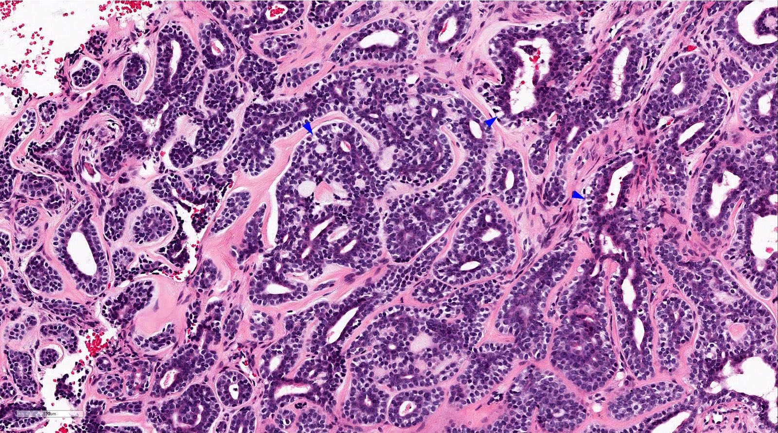
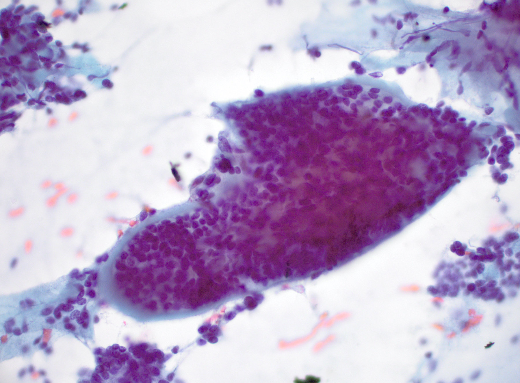
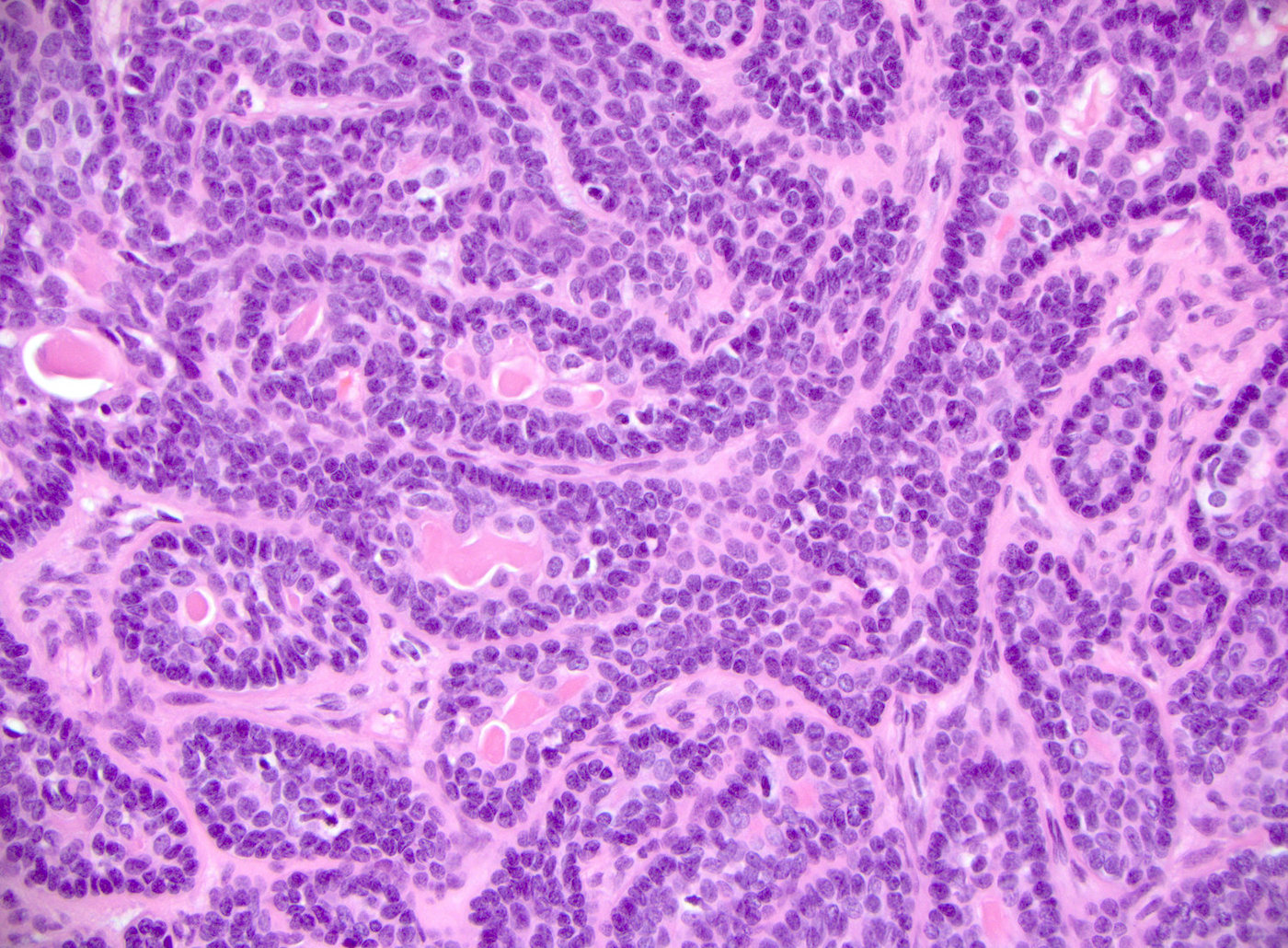
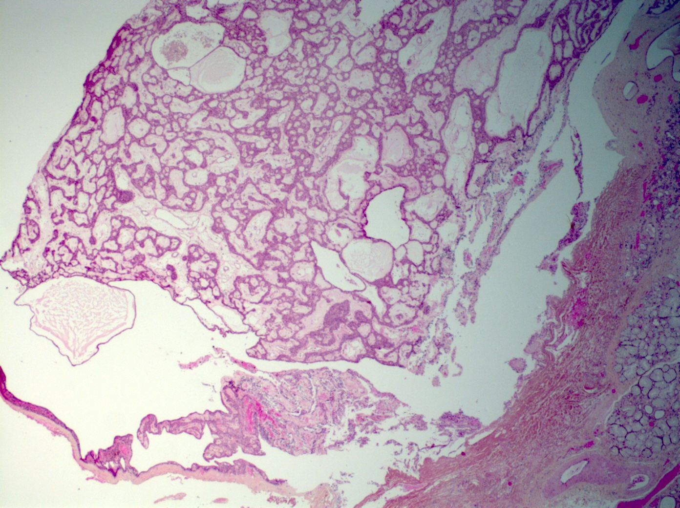
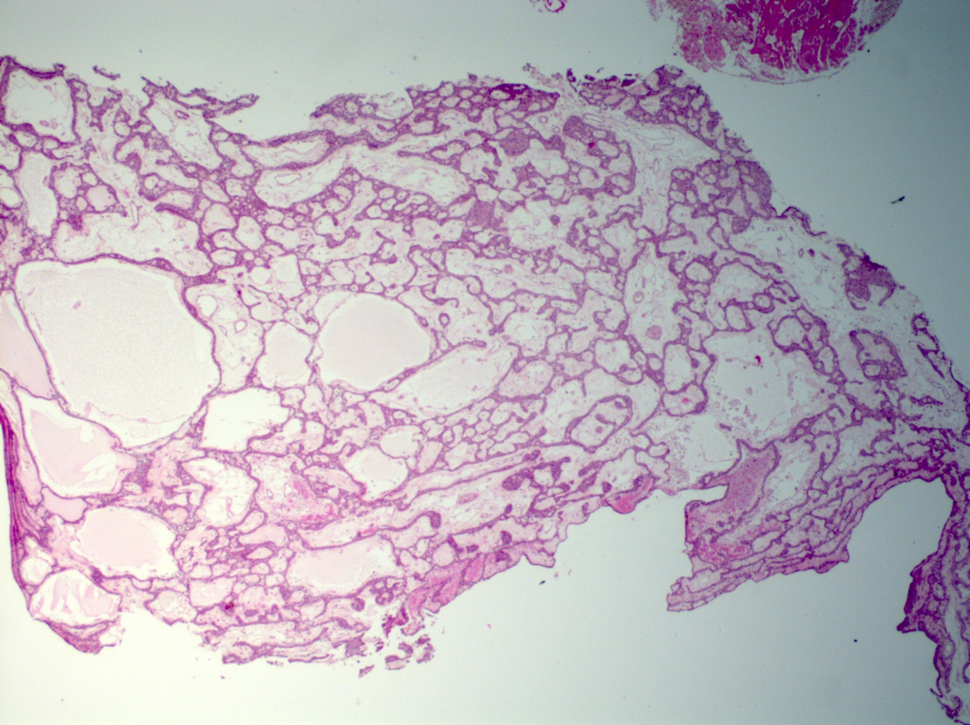
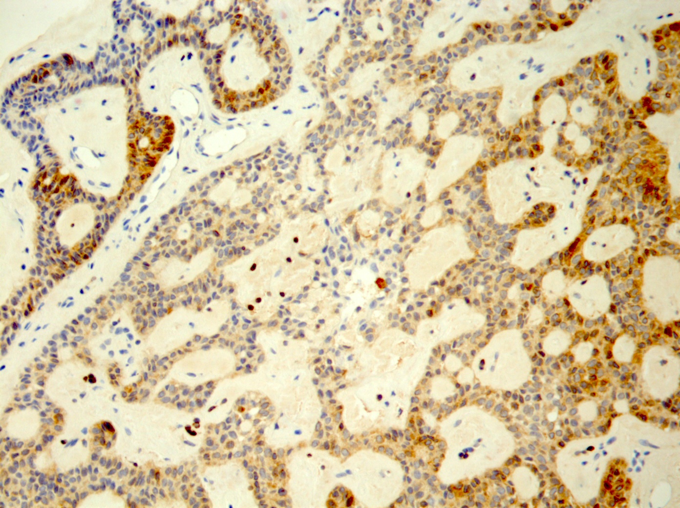
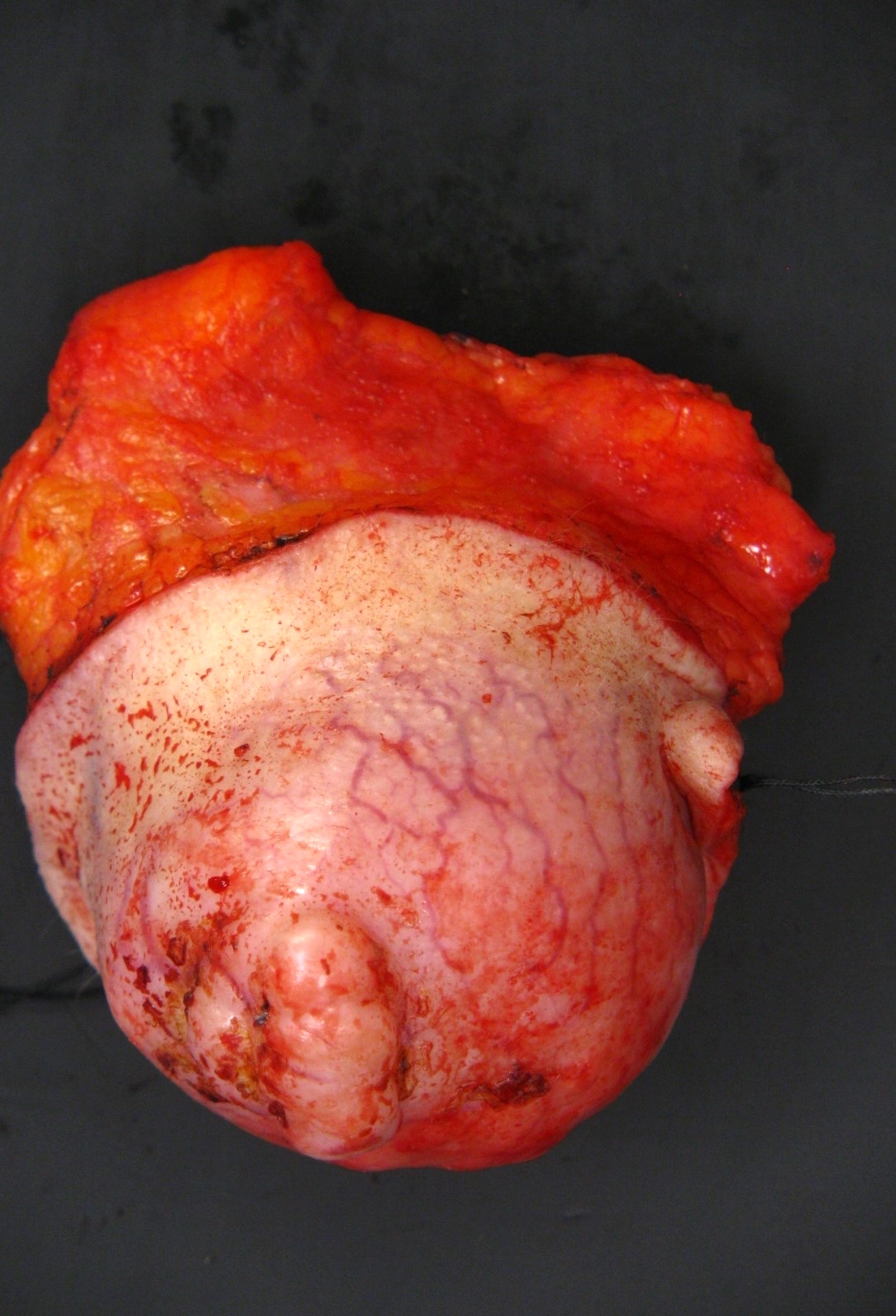
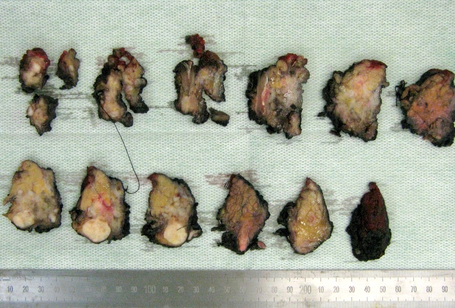
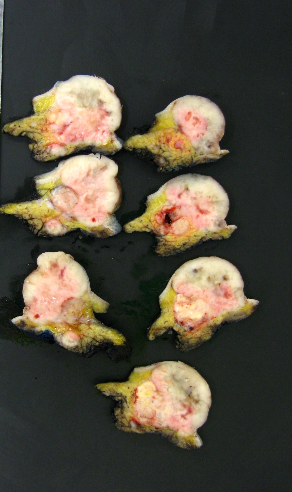
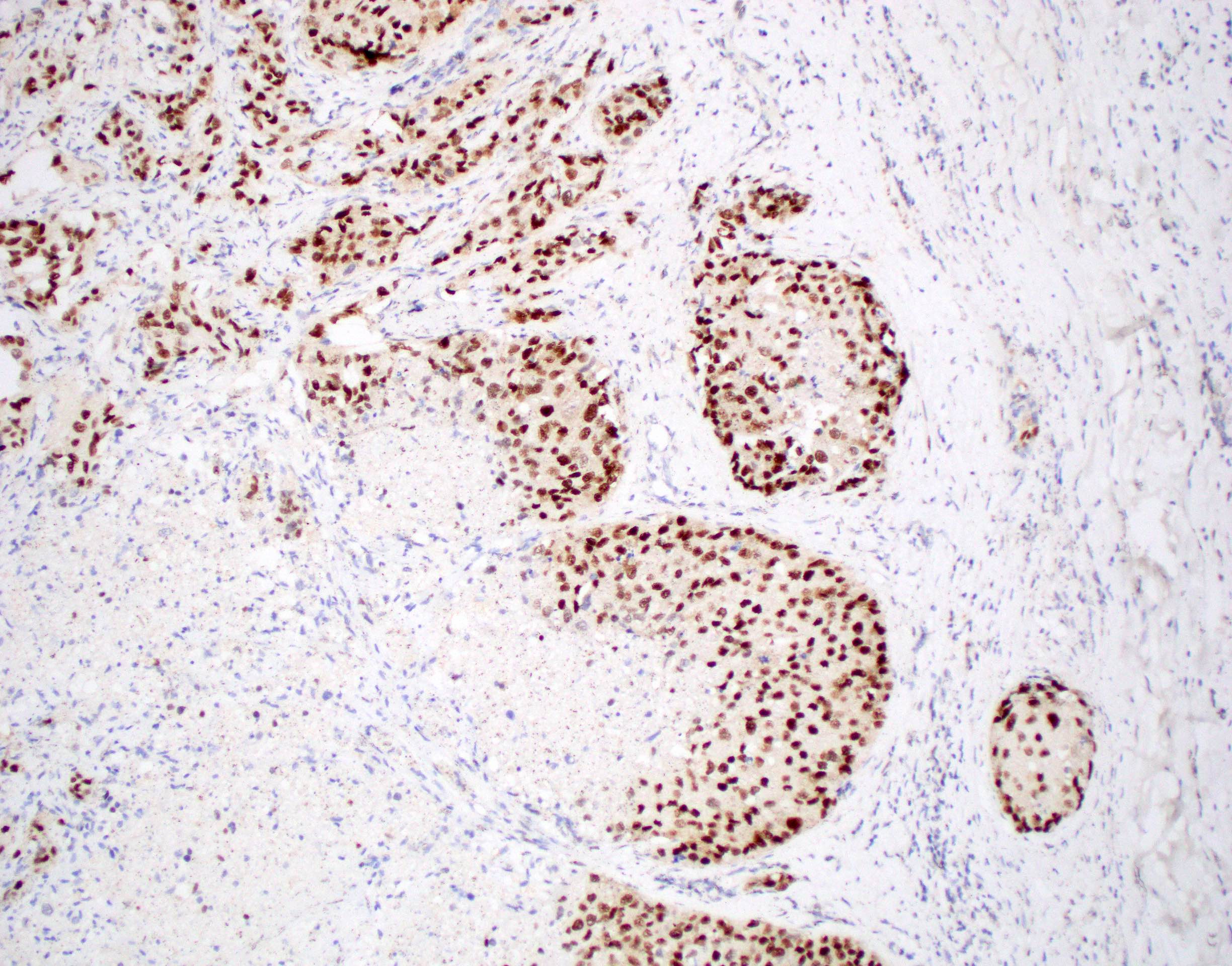
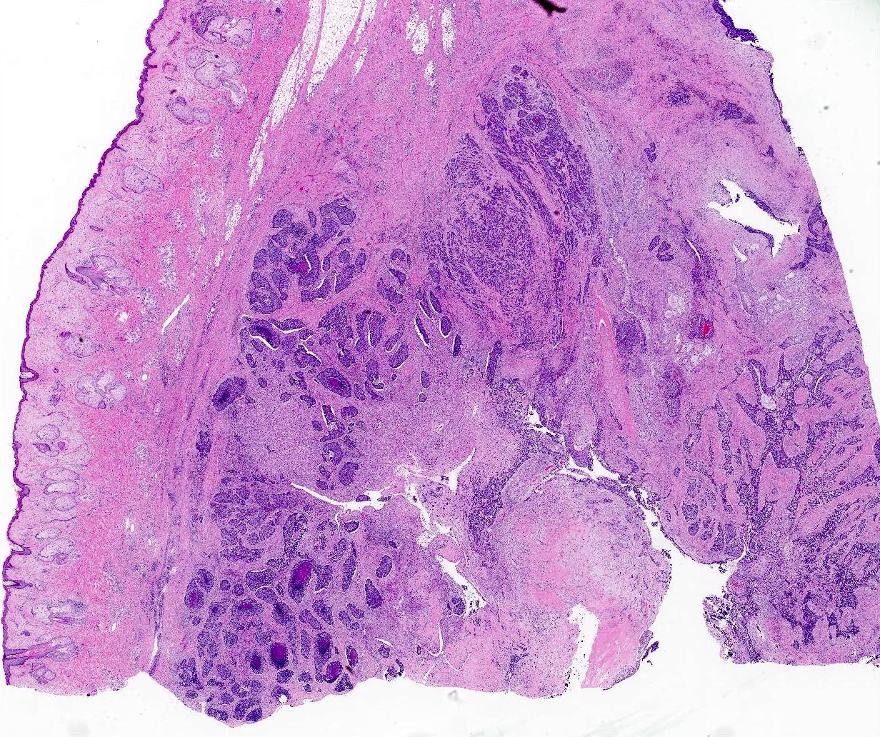
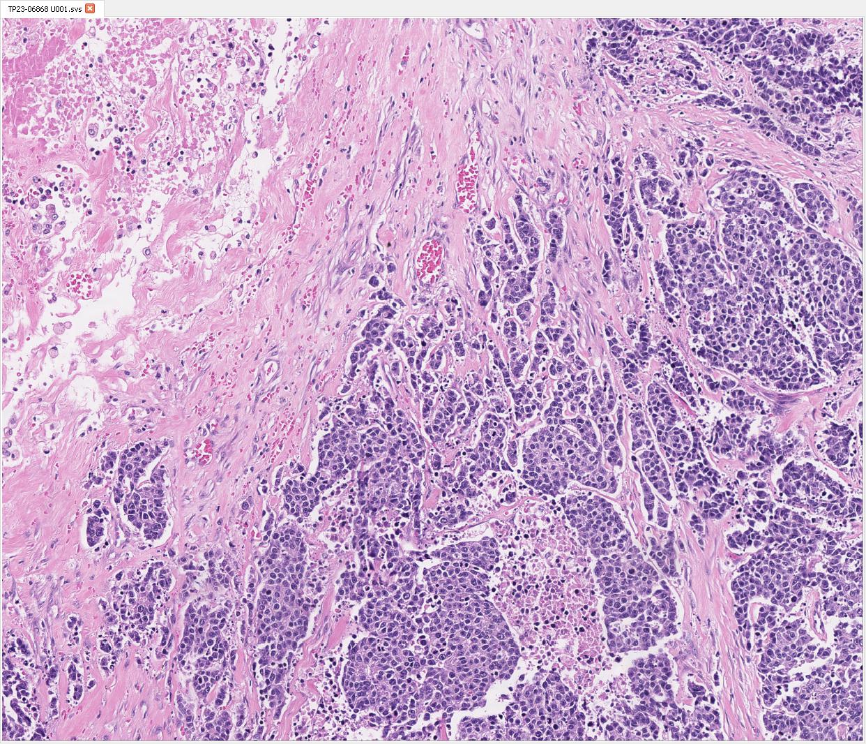
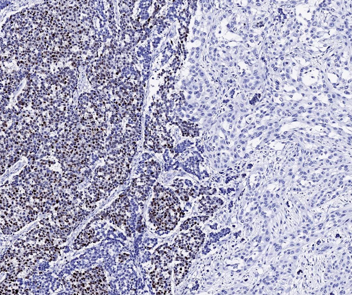
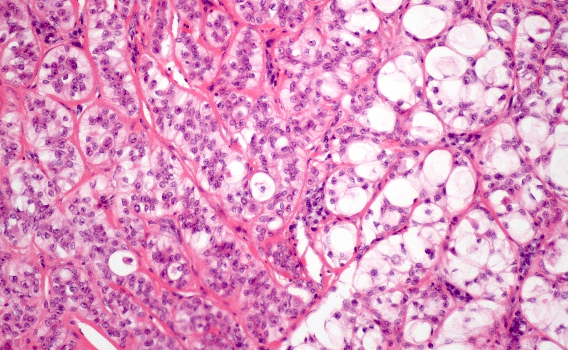
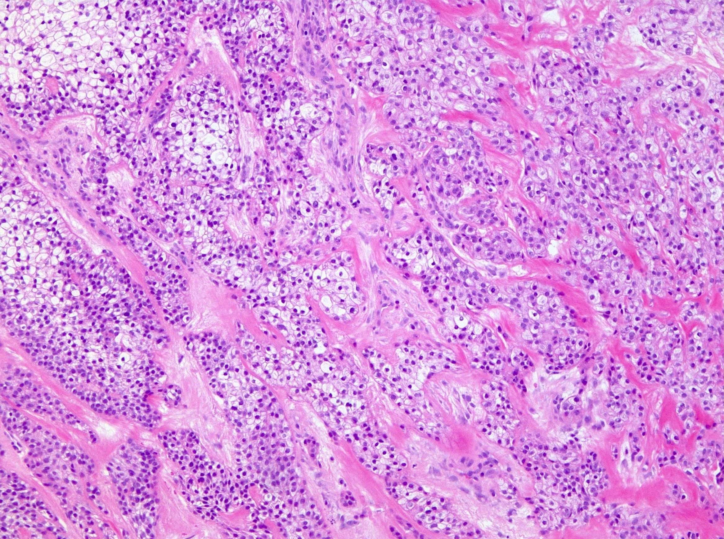
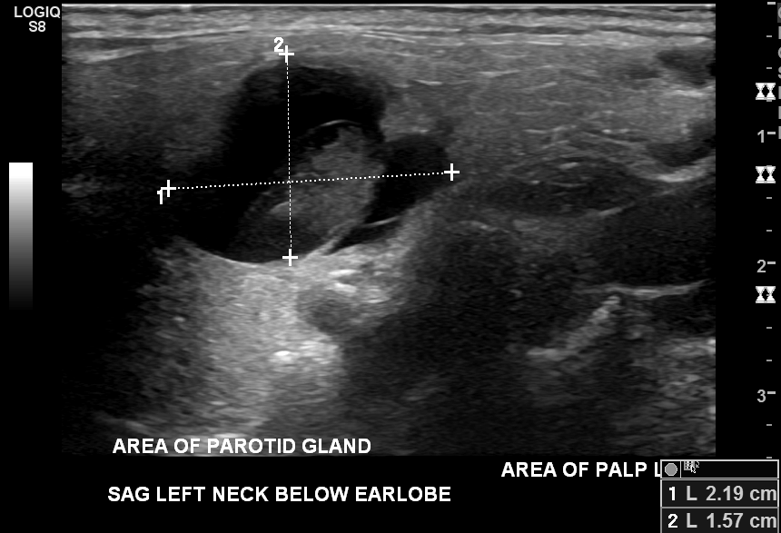
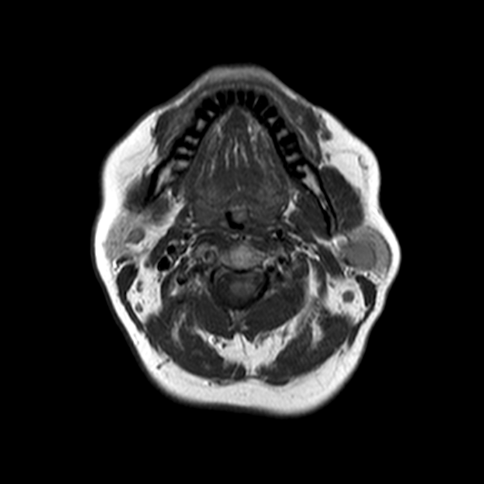
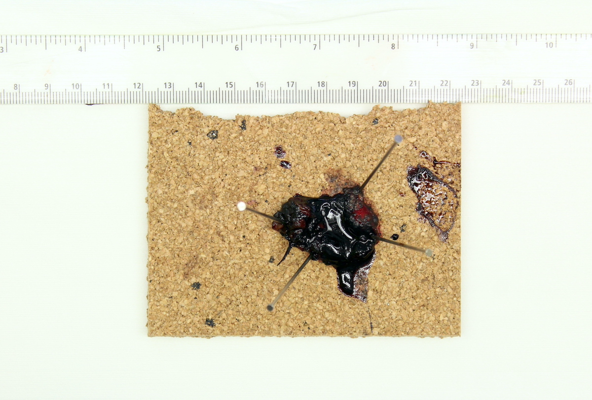
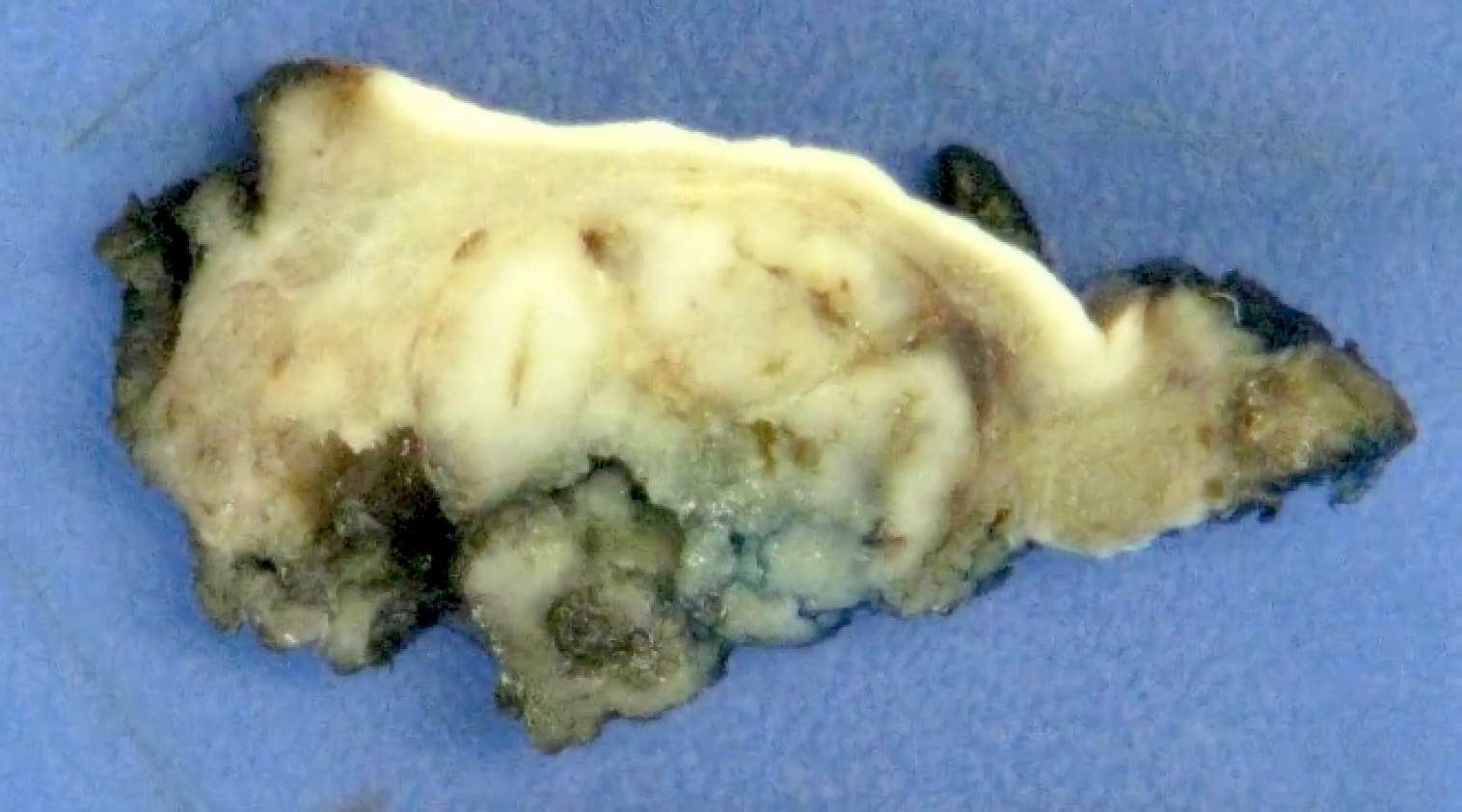
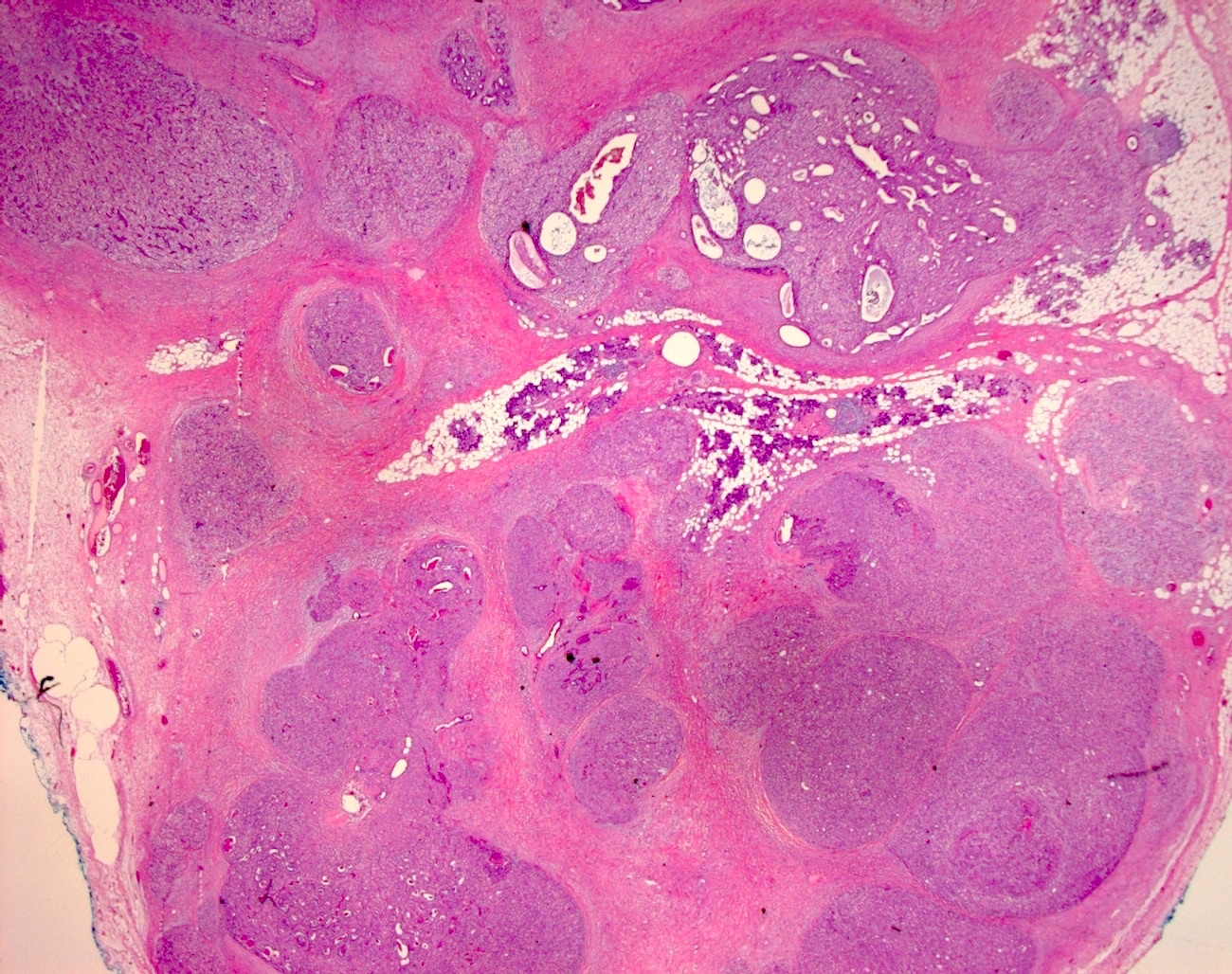
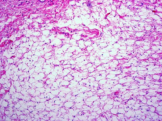
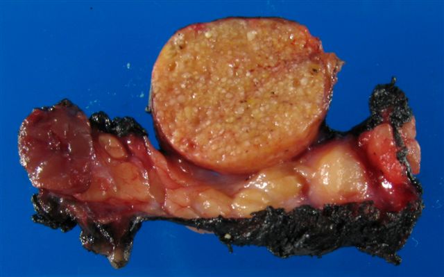
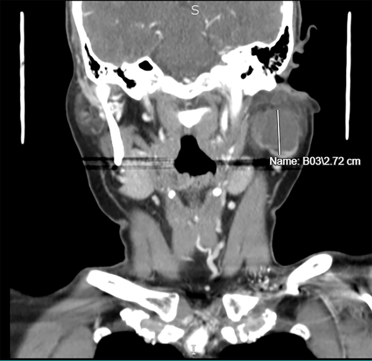
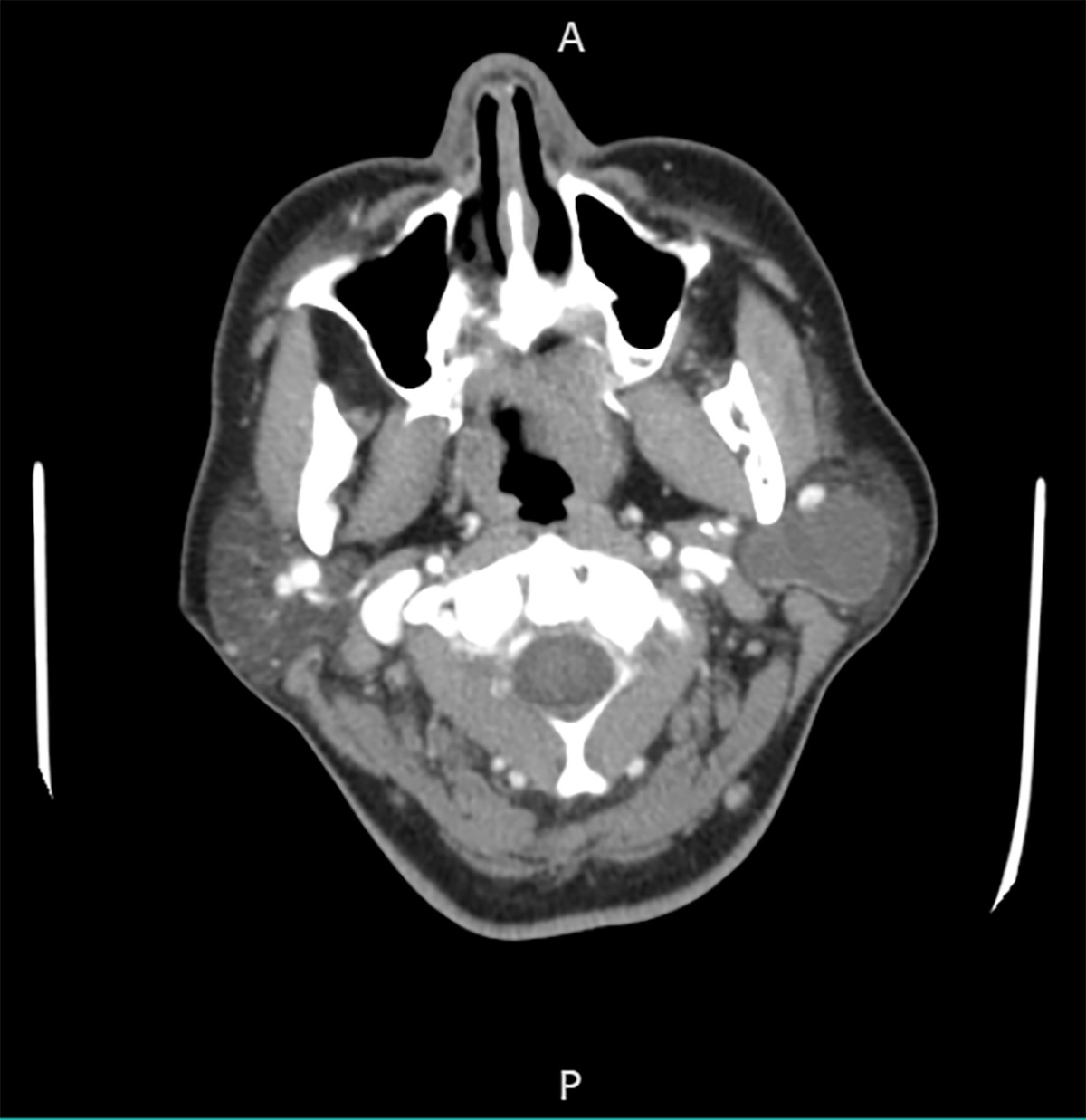
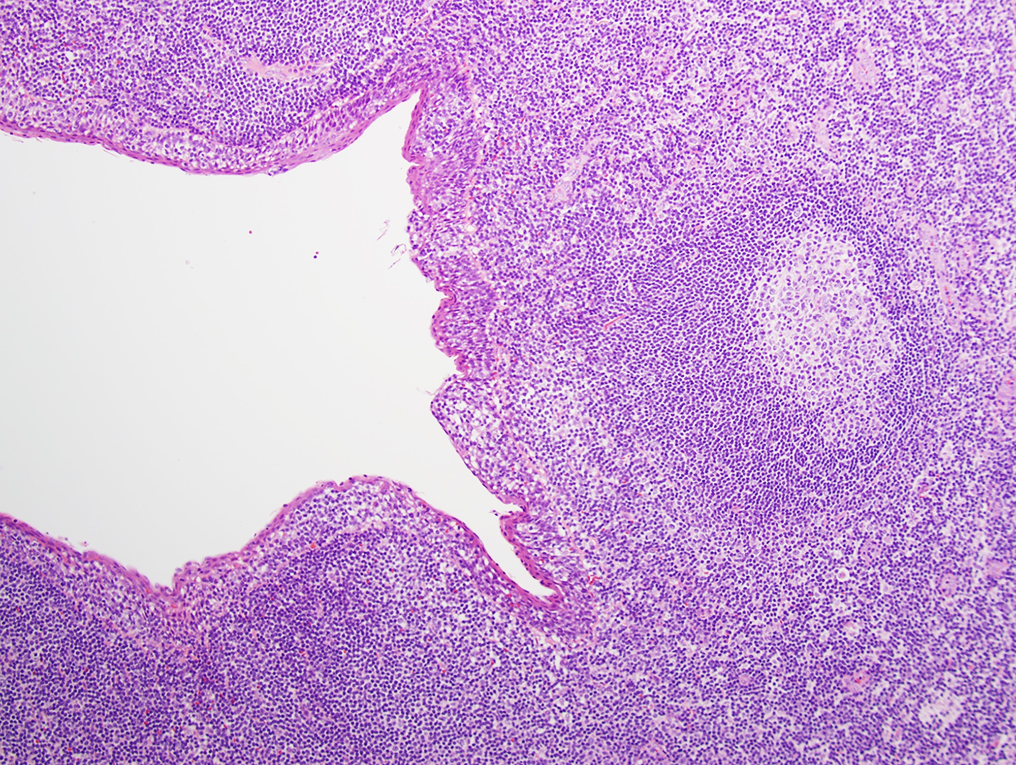
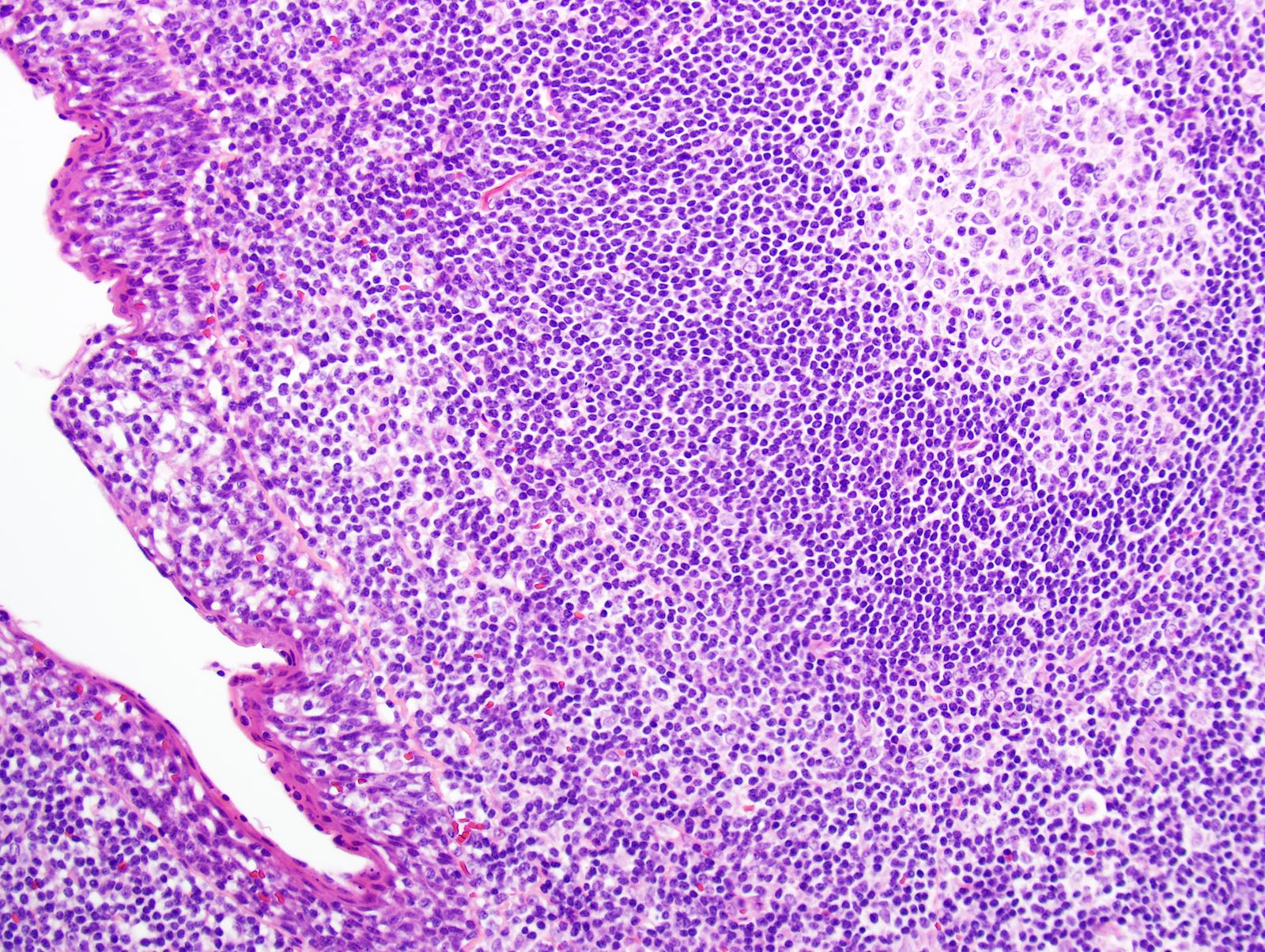
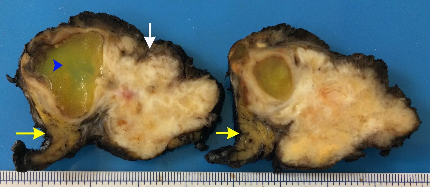
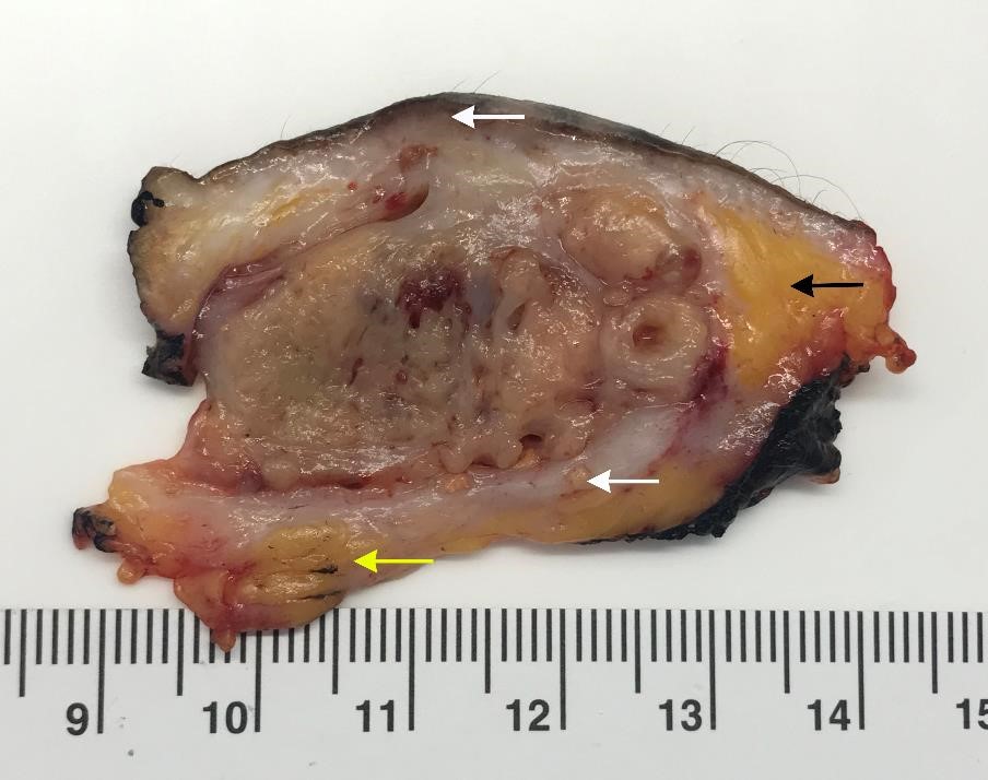
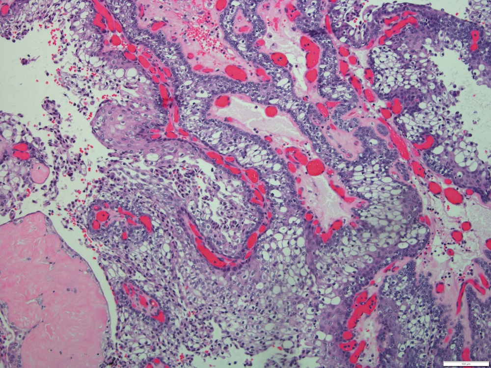
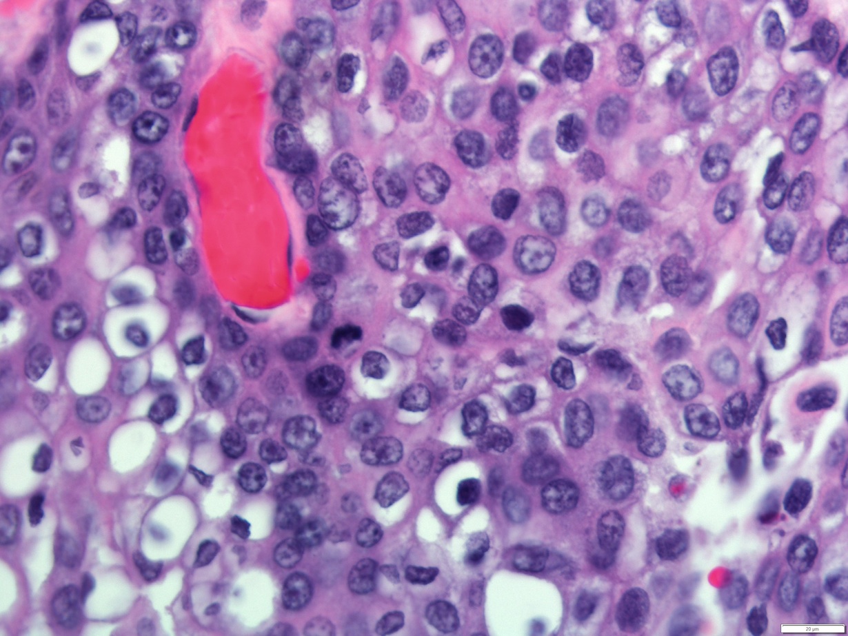
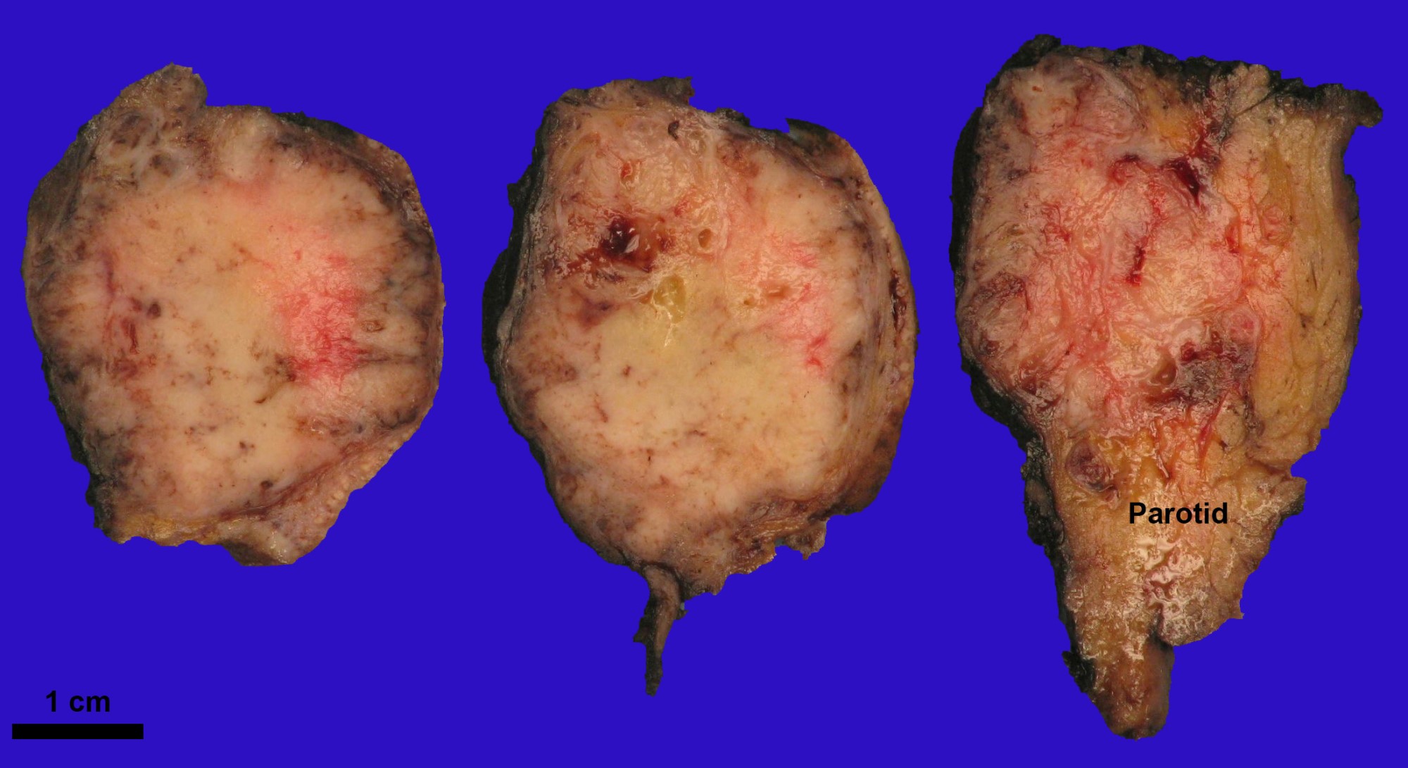
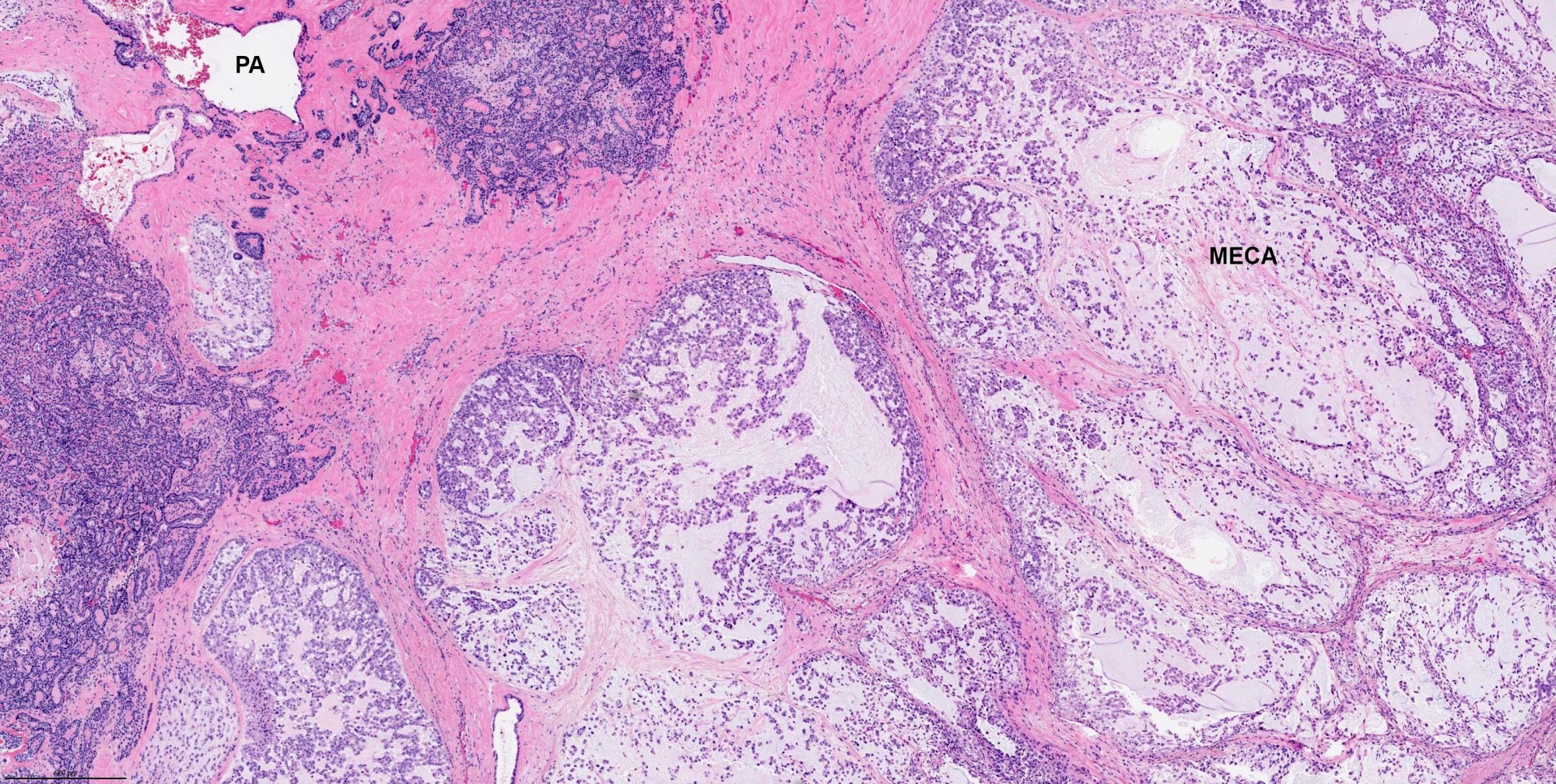
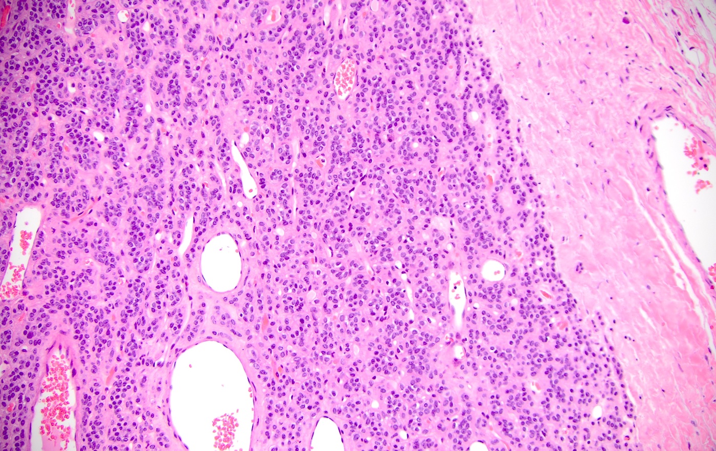
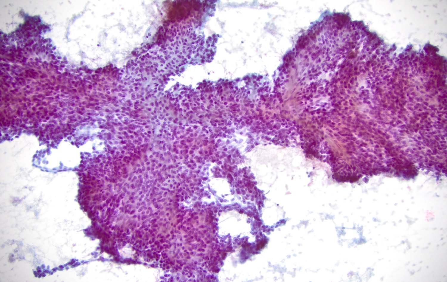
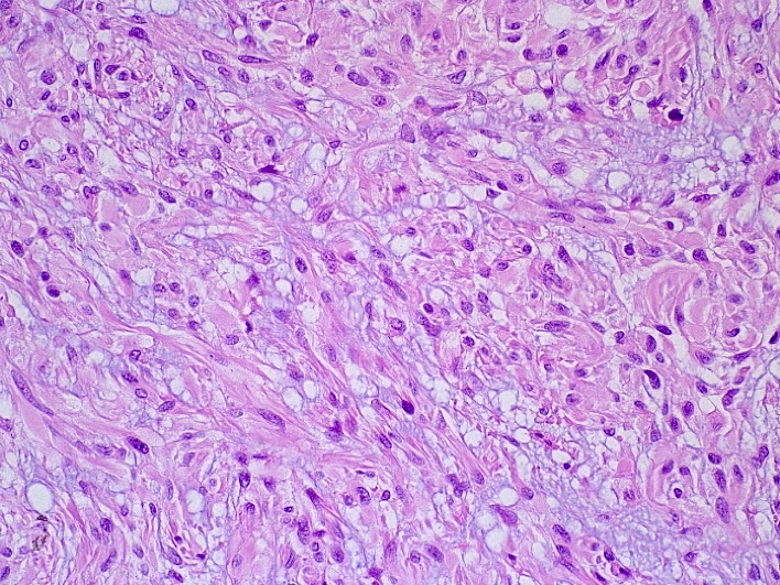
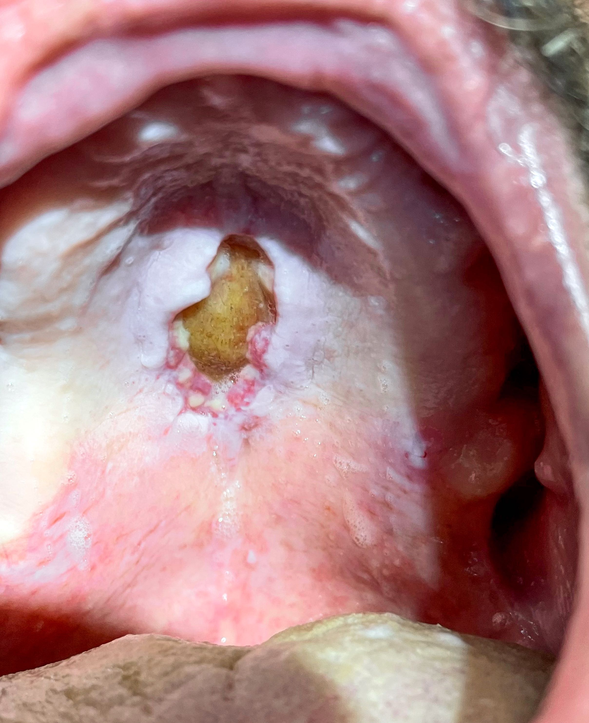
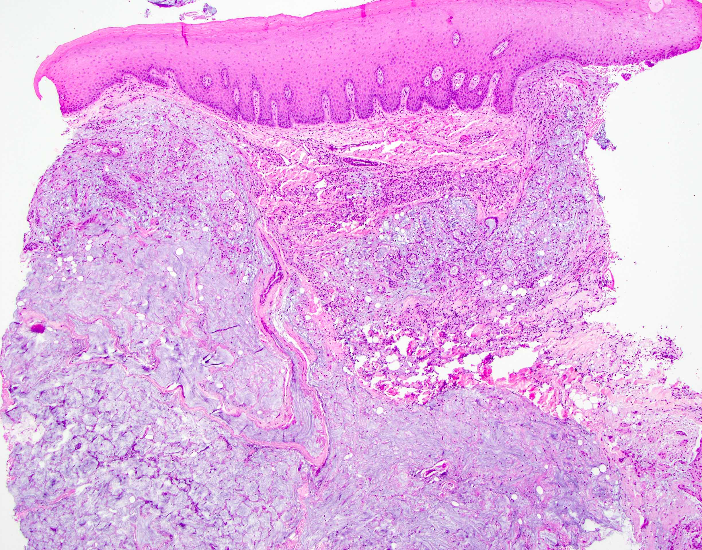
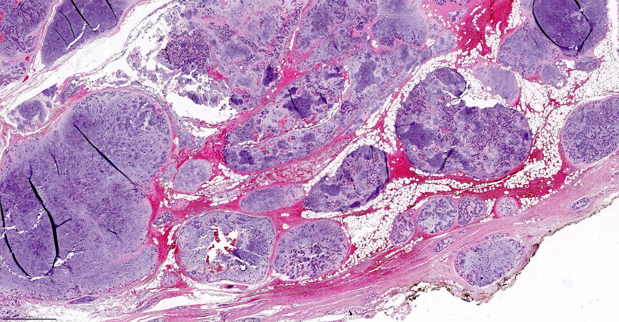
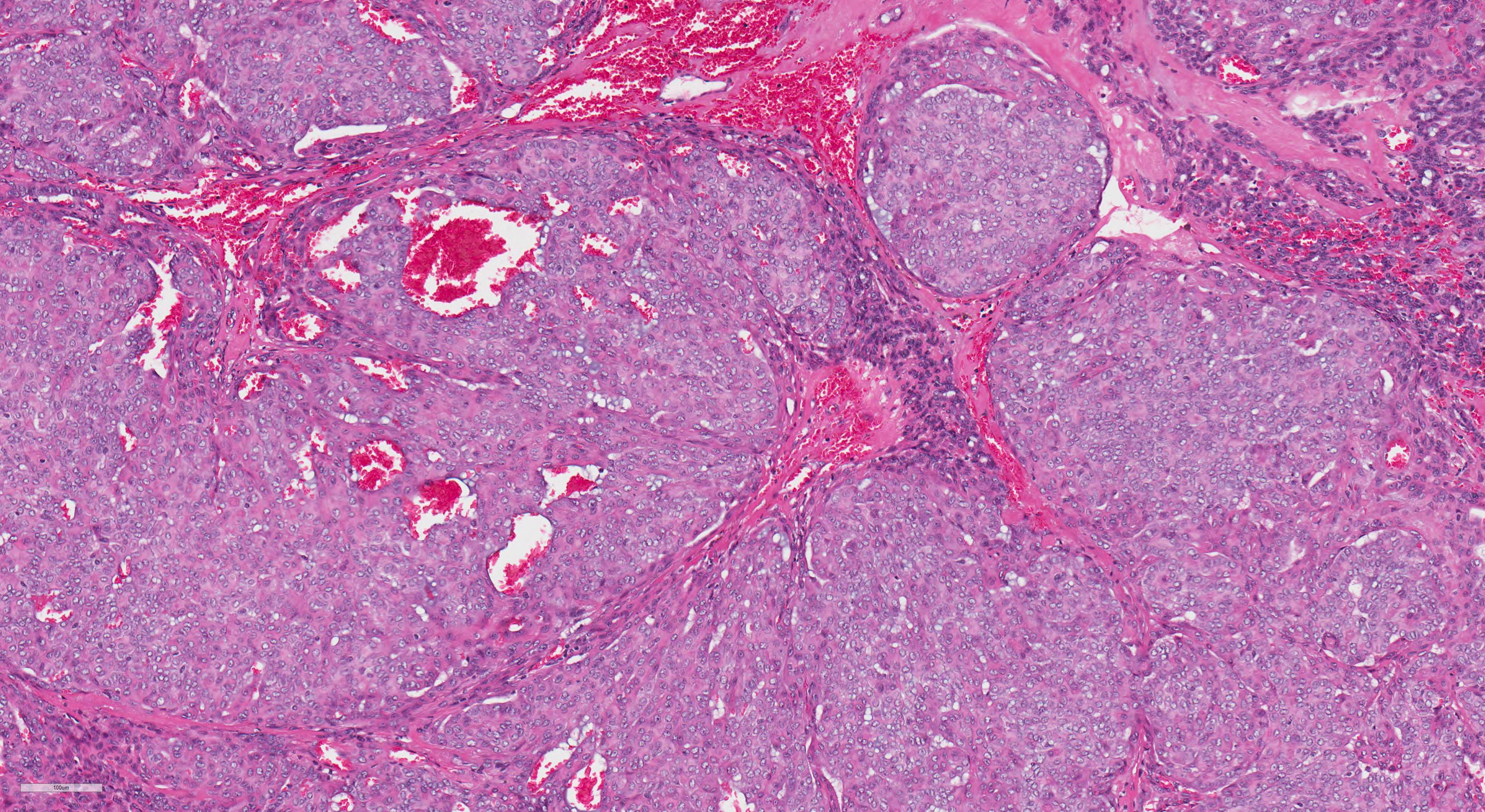
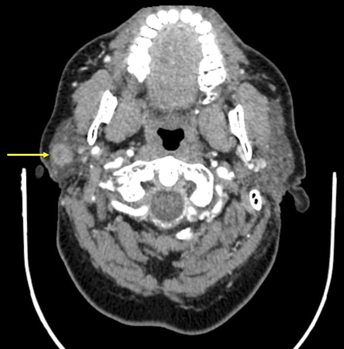
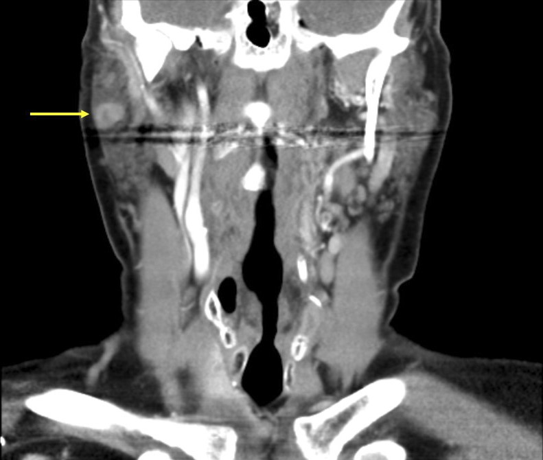
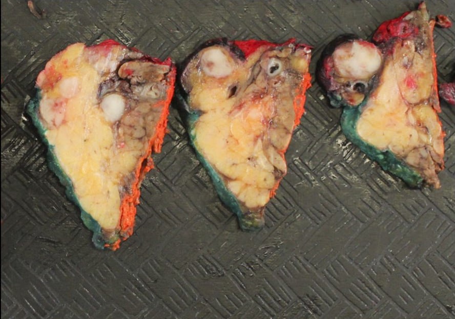
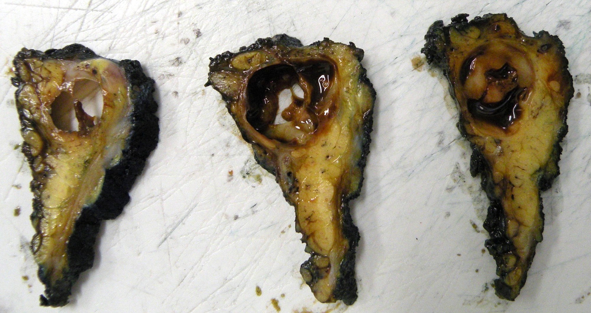
Superpage
Salivary glands


Copyright: 2003-2024, PathologyOutlines.com, Inc.

Head & Neck related: Jobs, Fellowships, Conferences, Cases, CME, Board Review

Editorial Board oversight: Marc Pusztaszeri, M.D. (last reviewed December 2022), Lisa Rooper, M.D. (last reviewed December 2021)
Page views in 2024 to date: 26

Acinic cell carcinoma
Definition / general
  • Malignant epithelial neoplasm of the salivary glands characterized by serous acinar cell differentiation (cytoplasmic zymogen granules) in at least some of the neoplastic cells
Essential features
  • ~6 - 8% of all salivary gland neoplasms and 10 - 17% of primary salivary gland malignancies
  • Parotid gland is the most common site
  • Solid, microcystic, papillary cystic or follicular growth of acinar cells, with granular to vacuolated cytoplasm and eccentric round nuclei with conspicuous nucleoli
  • Neoplastic cells positive for pankeratins, DOG1, SOX10 and PASD
  • Favorable prognosis, with low rates of local recurrence, distant metastasis and high grade transformation
Terminology
N/A
ICD coding
  • ICD-O: 8550/3 - acinic cell carcinoma
  • ICD-10: C08.9 - malignant neoplasm of salivary gland, unspecified
Epidemiology
Sites
  • Most common site: parotid gland (80%)
  • Submandibular, sublingual and minor salivary glands rarely involved (13 - 17%)
    • Most common sites in the oral cavity are the buccal mucosa and upper lip (Cancer 1983;52:542)
Pathophysiology
N/A
Etiology
Diagrams / tables
N/A
Clinical features
  • Typical clinical presentation is a slow growing mass
  • Pain, fixation to surrounding tissues and cranial nerve VII dysfunction are uncommon
  • Cervical lymph node metastasis uncommon (up to 10%)
  • Reference: Cancer 1991;67:172
Diagnosis
  • Radiology (ultrasound [US], computed tomography [CT] or magnetic resonance imaging [MRI]) demonstrates presence and extent of mass lesion
    • MRI is superior to CT in assessing invasion of adjacent structures
  • Fine needle aspiration cytology and core biopsy
    • Low sensitivity in parotid
      • High false negatives due to morphologic overlap with normal acini (Arch Pathol Lab Med 2005;129:26)
      • Diagnosis more challenging if tumor cells lack secretory granules
      • Diagnostic accuracy higher in cases with high grade transformation
  • Definitive diagnosis most often established on a surgical excision specimen (e.g., parotidectomy)
  • Positron emission tomography (PET) may be done postoperatively in cases of high grade transformation (Eur Arch Otorhinolaryngol 2016;273:3511)
Laboratory
N/A
Radiology description
  • Ultrasound: well demarcated, lobular, hypoechoic, heterogeneous and poorly vascularized
  • Computed tomography
    • Findings are nonspecific and there is an overlap with benign tumors
    • Solid to cystic mass
    • Focal low attenuating portions on CT may be present, corresponding to cystic degeneration or hemorrhage (J Comput Assist Tomogr 2005;29:121)
Radiology images

Images hosted on other servers:
MRI: cystic lesion of the upper lip

MRI: cystic lesion of the upper lip

CT and US:  right parotid nodule

CT and US: right parotid nodule

Prognostic factors
  • 5 year survival: 97%
  • May recur years or decades after initial diagnosis
  • Metastatic rate ~ 16%
  • Local recurrence rate is up to 33%; 12% in another large study (Cancer 1978;41:924, Cancer 1983;52:542)
  • Adverse prognostic factors include
    • Male gender
    • Age > 45 (JAMA Otolaryngol Head Neck Surg 2013;139:1195)
    • Tumor > 3 cm at presentation (Oral Oncol 2018;82:53)
    • Metastatic disease
    • Tumor necrosis
    • Nuclear pleomorphism
    • Mitosis (> 2/10 high power fields [HPF])
    • Ki67 index > 5%
    • High grade transformation
    • Vascular and perineural invasion
    • Extracapsular extension (Cancer 2009;115:2128)
    • Positive resection margins
    • Invasion of the skull base
    • Depletion of lymphocytes within the stroma
Case reports
Treatment
  • Complete surgical excision (J Oral Maxillofac Surg 2005;63:917)
  • Radiotherapy may be indicated for tumors that metastasize or cannot be completely excised
  • Systemic therapy may be indicated for high grade tumors, positive margins, advanced stage disease or metastatic tumors
Clinical images

Images hosted on other servers:
1.2 cm upper lip nodule

1.2 cm upper lip nodule

Gross description
  • Tumor dimensions range from < 1 cm to 13 cm (85% of the lesions are < 3 cm) (Cancer 1983;52:542)
  • Well circumscribed, soft rubbery, tan-yellow to pink mass with homogeneous cut surface
  • Cystic change and hemorrhage may occur
Gross images

Images hosted on other servers:
Well circumscribed multilobulated mass

Well circumscribed multilobulated mass

Frozen section description
N/A
Frozen section images
N/A
Microscopic (histologic) description
  • Multiple cell types are noted
    • Cell types include serous acinar, intercalated ductal, vacuolated, nonspecific glandular and clear cells
  • Acinar cells are large and polyhedral, with basophilic granular cytoplasm and eccentric nuclei
    • Cytoplasmic diastase resistant positive periodic acid-Schiff (PAS) reaction may be focal
  • Variety of morphologic patterns, including solid, microcystic, papillary cystic and follicular (Cancer 1983;52:542)
  • Prominent lymphoid infiltrate may be present (tumor associated lymphoid proliferation varies from randomly scattered, patchy collections of lymphocytes to diffuse, dense infiltrates with well formed follicles throughout the tumor) (Oral Surg Oral Med Oral Pathol 1994;77:19)
  • Mitosis, necrosis and significant pleomorphism usually absent
  • Proposed grading system (Am J Surg Pathol 2022;46:933)
    • Low to intermediate grade: mitotic count < 5/10 HPF and no necrosis
    • High grade: mitotic count ≥ 5/10 HPF or necrosis
Microscopic (histologic) images

Contributed by Rema A. Rao, M.D. and Arash H. Lahouti, M.D.
Well circumscribed tumor

Well circumscribed tumor

Finely vacuolated cytoplasm

Finely vacuolated cytoplasm

Tumor associated lymphoid proliferation

Tumor associated lymphoid proliferation

Cystic pattern and hemorrhage Cystic pattern and hemorrhage

Cystic pattern and hemorrhage


NR4A3 nuclear stain NR4A3 nuclear stain

NR4A3 nuclear stain

Virtual slides

Images hosted on other servers:
Acinic cell carcinoma, parotid

Acinic cell carcinoma, parotid

Acinic cell carcinoma, cystic change

Acinic cell carcinoma, cystic change

Cytology description
  • Cellular aspirates
  • Cytologic diagnosis based on identifying acinar-like cells
  • Tend to form small groups and loosely cohesive clusters as well as single cells (Diagn Histopath 2018;24:417)
  • Cells are large but morphologically resemble normal acinar cells
    • Round nuclei with tiny nucleoli
    • Abundant finely vacuolated to granular cytoplasm
  • Stripped or bare nuclei may be present in the background, can mimic lymphocytes
  • Lymphocytic infiltrate may be prominent
  • Laminated psammoma body-like calcification may be present
  • Pitfall: acinic cells may resemble oncocytes and in the presence of lymphocytic infiltrate, the tumor may be misdiagnosed as Warthin tumor
Cytology images

Contributed by Rema A. Rao, M.D. and Arash H. Lahouti, M.D.
Loosely cohesive groups

Loosely cohesive groups

Cellular smears

Cellular smears

Finely vacuolated to granular cytoplasm

Finely vacuolated to granular cytoplasm

Round eccentric nuclei

Round eccentric nuclei

Loosely cohesive clusters

Loosely cohesive clusters

PASD

PASD

Immunofluorescence description
N/A
Immunofluorescence images
N/A
Positive stains
Negative stains
Electron microscopy description
N/A
Electron microscopy images
N/A
Molecular / cytogenetics description
  • Recurrent (t[4;9][q13;q31]) genomic rearrangement (resulting in translocation of active enhancer regions from the secretory Ca binding phosphoprotein [SCPP] gene cluster to the region upstream of nuclear receptor subfamily 4 group A member 3 [NR4A3], leading to NR4A3 IHC overexpression) (Cancer Cytopathol 2021;129:104)
  • Loss of function mutations of CDKN2A / B gene are associated with high grade histology and distant metastasis (Mod Pathol 2023;36:100150)
Molecular / cytogenetics images

Images hosted on other servers:
FISH, NR4A3 break apart probe

FISH, NR4A3 break apart probe

Videos
N/A
Sample pathology report
  • Parotid gland, right, inferior, partial parotidectomy:
    • Acinic cell carcinoma, 1.7 cm (see synoptic report)
    • No lymphovascular invasion identified
    • No perineural invasion identified
    • Resection margins uninvolved by carcinoma
    • 2 lymph nodes, negative for carcinoma (0/2)
    • Tumor associated lymphoid proliferation present
Differential diagnosis
Board review style question #1
Which of the following statements is true regarding acinic cell carcinoma of salivary glands?

  1. Acinic cell carcinoma is a high grade malignant neoplasm
  2. Most commonly occurs in minor salivary glands
  3. Neoplastic cells express DOG1 and SOX10
  4. Younger age at presentation and presence of tumor associated lymphocytes are associated with a poor prognosis
Board review style answer #1
C. Neoplastic cells express DOG1 and SOX10. DOG1 and SOX10 are expressed in benign acinar cells and intercalated duct cells and other salivary gland neoplasms. However, strong diffuse expression of these markers can help distinguish acinic cell carcinoma from other salivary gland neoplasms. Answer A is incorrect because acinic cell carcinoma is considered low intermediate grade malignancy of salivary glands, although high grade transformation may occur. Answer B is incorrect because the majority of cases arise in the parotid gland. Answer D is incorrect because older age at the onset (> 45) and depletion of lymphocytes within the stroma are linked to worse outcomes.

Comment Here

Reference: Acinic cell carcinoma
Board review style question #2

A 51 year old woman presents with a left parotid mass. CT images show a well circumscribed cystic lesion measuring 3 cm. An excisional biopsy is performed (see image above). The neoplastic cells are positive for periodic acid-Schiff with diastase (PASD). Which of the following statements is true regarding this lesion?

  1. Cytology aspirates of this tumor can show predominance of extracellular mucin
  2. Mitosis, necrosis and significant pleomorphism are usually present
  3. MYB immunohistochemical staining in FNA specimens is useful in the diagnosis of this tumor
  4. Pain and facial nerve dysfunction are uncommon presenting symptoms
Board review style answer #2
D. The most common initial presentation in patients with acinic cell carcinoma is a painless mass without facial nerve involvement. Answer C is incorrect because MYB immunohistochemical staining is useful in the diagnosis of adenoid cystic carcinoma. Answer A is incorrect because the presence of extracellular mucin in aspirates is a feature of mucoepidermoid carcinoma. Answer B is incorrect because acinic cell carcinoma is usually a low grade malignant neoplasm without significant mitosis, necrosis or pleomorphic nuceli.

Comment Here

Reference: Acinic cell carcinoma

Acinic cell carcinoma
Definition / general
Essential features
  • Approximately 6 - 8% of all salivary gland neoplasms and 10 - 17% of primary salivary gland malignancies
  • Parotid gland is the most common site
  • Solid, microcystic, papillary cystic or follicular growth of acinar cells, with granular to vacuolated cytoplasm and eccentric round nuclei with conspicuous nucleoli
  • Neoplastic cells positive for pankeratins, DOG1, SOX10 and PAS-D
  • Favorable prognosis, with low rates of local recurrence, distant metastasis and high grade transformation
ICD coding
  • ICD-O: 8550/3 - acinic cell carcinoma
  • ICD-10: C08.9 - malignant neoplasm of salivary gland, unspecified
Epidemiology
Sites
  • Most common site: parotid gland (80%)
  • Submandibular, sublingual and minor salivary glands rarely involved (13 - 17%)
    • Most common sites in the oral cavity are the buccal mucosa and upper lip (Cancer 1983;52:542)
Etiology
Clinical features
  • Typical clinical presentation is a slow growing mass
  • Pain, fixation to surrounding tissues and cranial nerve VII dysfunction are uncommon
  • Cervical lymph node metastasis uncommon (up to 10%)
  • Reference: Cancer 1991;67:172
Diagnosis
  • Radiology (ultrasound, CT or MRI) demonstrates presence and extent of mass lesion
    • MRI superior to CT in assessing invasion of adjacent structures
  • Fine needle aspiration cytology and core biopsy
    • Low sensitivity in parotid:
      • High false negatives due to morphologic overlap with normal acini (Arch Pathol Lab Med 2005;129:26)
      • Diagnosis more challenging if tumor cells lack secretory granules
      • Diagnostic accuracy higher in cases with high grade transformation
  • Definitive diagnosis most often established on a surgical excision specimen (e.g., parotidectomy)
  • Positron emission tomography (PET) may be done postoperatively in cases of high grade transformation (Eur Arch Otorhinolaryngol 2016;273:3511)
Radiology description
  • Ultrasound: well demarcated, lobular, hypoechoic, heterogeneous and poorly vascularized
  • Computed tomography (CT):
    • Findings are nonspecific and there is an overlap with benign tumors
    • Solid to cystic mass
    • Focal low attenuating portions on CT may be present corresponding to cystic degeneration or hemorrhage (J Comput Assist Tomogr 2005;29:121)
Radiology images

Images hosted on other servers:

MRI: cystic lesion of the upper lip

CT and US: right parotid nodule

Prognostic factors
  • 5 year survival: 97%
  • May recur years or decades after initial diagnosis
  • Metastatic rate ~ 16%
  • Local recurrence rate is up to 33%; 12% in another large series (Cancer 1978;41:924, Cancer 1983;52:542)
  • Adverse prognostic factors include
    • Male gender
    • Age > 45 (JAMA Otolaryngol Head Neck Surg 2013;139:1195)
    • Tumor > 3 cm at presentation (Oral Oncol 2018;82:53)
    • Metastatic disease
    • Tumor necrosis
    • Nuclear pleomorphism
    • Mitosis (> 2/10 high power fields)
    • Ki67 index > 5%
    • High grade transformation
    • Vascular and perineural invasion
    • Extracapsular extension (Cancer 2009;115:2128)
    • Positive resection margins
    • Invasion of the skull base
    • Depletion of lymphocytes within the stroma
Case reports
Treatment
  • Complete surgical excision (J Oral Maxillofac Surg 2005;63:917)
  • Radiotherapy may be indicated for tumors that cannot be completely excised or metastatic disease
  • Systemic therapy may be indicated for high grade tumors, positive margins, advanced stage disease, or metastatic tumors
Clinical images

Images hosted on other servers:

1.2 cm upper lip nodule

Gross description
  • Tumor dimensions range from less than 1 cm to 13 cm (85% of the lesions less than 3 cm) (Cancer 1983;52:542)
  • Well circumscribed, soft rubbery, tan-yellow to pink mass with homogeneous cut surface
  • Cystic change and hemorrhage may occur
Gross images

Images hosted on other servers:

Well circumscribed multilobulated mass

Microscopic (histologic) description
  • Multiple cell types are noted
    • Cell types include serous acinar, intercalated ductal, vacuolated, nonspecific glandular and clear cells
  • Acinar cells are large and polyhedral, with basophilic granular cytoplasm and eccentric nuclei
    • Cytoplasmic diastase resistant positive periodic acid-Schiff (PAS) reaction may be focal
  • Variety of morphologic patterns, including solid, microcystic, papillary cystic and follicular (Cancer 1983;52:542)
  • Prominent lymphoid infiltrate may be present (tumor associated lymphoid proliferation varies from randomly scattered, patchy collections of lymphocytes to diffuse, dense infiltrates with well formed follicles throughout the tumor) (Oral Surg Oral Med Oral Pathol 1994;77:19)
  • Mitosis, necrosis and significant pleomorphism usually absent
  • No specific histologic grading system
Microscopic (histologic) images

Contributed by Rema A. Rao, M.D. and Arash H. Lahouti, M.D.
Well circumscribed tumor

Well circumscribed tumor

Finely vacuolated cytoplasm

Finely vacuolated cytoplasm

TALP

Tumor associated lymphoid proliferation

Cystic pattern and hemorrhage Cystic pattern and hemorrhage

Cystic pattern and hemorrhage


NR4A3 nuclear stain NR4A3 nuclear stain

NR4A3 nuclear stain

Virtual slides

Images hosted on other servers:

Acinic cell carcinoma, parotid

Acinic cell carcinoma, cystic change

Cytology description
  • Cellular aspirates
  • Cytologic diagnosis based on identifying acinar-like cells
  • Tend to form small groups and loosely cohesive clusters as well as single cells (Diagn Histopath 2018;24:417)
  • Cells are large but morphologically resemble normal acinar cells
    • Round nuclei with tiny nucleoli
    • Abundant finely vacuolated to granular cytoplasm
  • Stripped or bare nuclei may be present in the background, can mimic lymphocytes
  • Lymphocytic infiltrate may be prominent
  • Laminated psammoma body-like calcification may be present
  • Pitfall: acinic cells may resemble oncocytes and in the presence of lymphocytic infiltrate, the tumor may be misdiagnosed as Warthin tumor
Cytology images

Contributed by Rema A. Rao, M.D. and Arash H. Lahouti, M.D.
Loosely cohesive groups

Loosely cohesive groups

Cellular smears

Cellular smears

Finely vacuolated to granular cytoplasm

Finely vacuolated to granular cytoplasm

Round eccentric nuclei

Round eccentric nuclei

Loosely cohesive clusters

Loosely cohesive clusters

PASD

PASD

Positive stains
Negative stains
Molecular / cytogenetics description
  • Recurrent (t[4;9][q13;q31]) genomic rearrangement (resulting in translocation of active enhancer regions from the secretory Ca binding phosphoprotein [SCPP] gene cluster to the region upstream of nuclear receptor subfamily 4 group A member 3 [NR4A3], leading to NR4A3 IHC overexpression) (Cancer Cytopathol 2021;129:104)
Molecular / cytogenetics images

Images hosted on other servers:

FISH, NR4A3 break apart probe

Sample pathology report
  • Parotid gland, right, inferior, partial parotidectomy:
    • Acinic cell carcinoma, 1.7 cm (see synoptic report)
    • No lymphovascular invasion identified
    • No perineural invasion identified
    • Resection margins uninvolved by carcinoma
    • Two lymph nodes, negative for carcinoma (0/2)
    • Tumor associated lymphoid proliferation present
Differential diagnosis
Board review style question #1
Which of the following statements is true regarding acinic cell carcinoma of salivary glands?

  1. Acinic cell carcinoma is a high grade malignant neoplasm
  2. Most commonly occurs in minor salivary glands
  3. Younger age at presentation and presence of tumor associated lymphocytes are associated with a poor prognosis
  4. Neoplastic cells express DOG1 and SOX10
Board review style answer #1
D. Neoplastic cells express DOG1 and SOX10. DOG1 and SOX10 are expressed in benign acinar cells and intercalated duct cells and other salivary gland neoplasms. However, strong diffuse expression of these markers can help distinguish acinic cell carcinoma from other salivary gland neoplasms. Acinic cell carcinoma is considered low intermediate grade malignancy of salivary glands, although high grade transformation may occur (answer A). The majority of cases arise in the parotid gland (answer B). Older age at the onset (> 45) and depletion of lymphocytes within the stroma are linked to worse outcomes (answer C).

Comment Here

Reference: Acinic cell carcinoma
Board review style question #2

A 51 year old woman presents with a left parotid mass. CT images show a well circumscribed cystic lesion measuring 3 cm. An excisional biopsy is performed (see image above). The neoplastic cells are positive for periodic acid-Schiff with diastase (PASD). Which of the following statements is true regarding this lesion?

  1. Pain and facial nerve dysfunction are uncommon presenting symptoms
  2. Mitosis, necrosis and significant pleomorphism are usually present
  3. MYB immunohistochemical staining in FNA specimens is useful in diagnosis of this tumor
  4. Cytology aspirates of this tumor can show predominance of extracellular mucin
Board review style answer #2
A. Pain and facial nerve dysfunction are uncommon presenting symptoms

The most common initial presentation in patients with acinic cell carcinoma is painless mass. Clinical examination shows a firm, mobile nodule in the parotid gland. Facial nerve paralysis or paresis is an uncommon initial finding. MYB immunohistochemical staining is useful in diagnosis of adenoid cystic carcinoma. The presence of extracellular mucin in aspirates is a feature of mucoepidermoid carcinoma.

Comment Here

Reference: Acinic cell carcinoma

Adenocarcinoma, NOS
Definition / general
  • Invasive tumor, often aggressive, with glandular or ductal differentiation but no features characteristic of other specific types (Arch Pathol Lab Med 2004;128:1385)
  • Common, 5 - 10% of salivary gland tumors
  • 6 - 10% of salivary gland malignancies, 17% of parotid gland malignancies, 15% of minor salivary gland malignancies
Sites
  • Parotid gland, submandibular gland, palate, buccal mucosa
Clinical features
  • Mean age 58 years (median 67 years), range 10 - 93 years
  • Usually asymptomatic
  • Often fixed to skin or deep tissues
  • Palatal lesions often ulcerated and involve bone
  • Gender predominance debated, recent reports show male predominance
  • Cervical lymph node metastases in 23%, distant metastases in 37%
  • Diagnosis of exclusion (not metastatic, not another salivary gland carcinoma)
  • 5 year disease specific survivals is 57%
Case reports
Treatment
  • Complete surgical excision
Gross description
  • Poorly circumscribed with infiltrative borders
  • Solid tan cut surface with hemorrhage and necrosis
Microscopic (histologic) description
  • Invasive with glandular or ductal differentiation but no features characteristic of other specific types
  • Patterns include glandular spaces with cyst formation, papillary formation, solid sheets, comedonecrosis, hyalinized "shadow" nodules
  • Small clusters of cuboidal, round or ovoid cells with distinct borders and abundant cytoplasm
  • May have clear cell or oncocytic features
  • Low, intermediate or high grade based on cytomorphic features
  • In situ component in 68% (Virchows Arch 2006;449:159)
Microscopic (histologic) images

Contributed by Marino Leon, M.D.

Oncocytic carcinoma, H&E and p63

Positive stains
Negative stains
Molecular / cytogenetics description
  • EGFR gene amplification, increased EGFR gene copy number
  • HER2 amplification, high HER2 gene copy number
  • KRAS mutation rarely (Arch Pathol Lab Med 2000;124:836)
Differential diagnosis

Adenoid cystic carcinoma
Definition / general
  • A carcinoma of primary salivary gland or minor salivary gland of upper aerodigestive tract characterized by its biphasic ductal and myoepithelial differentiation, tubular / cribriform / solid architecture (Head Neck 2017;39:456)
  • May occur in major and minor salivary glands
    • Among major salivary glands, the most commonly affected site is the parotid gland
    • Minor salivary glands that may be affected include those of oral cavity, sinonasal tract, nasopharynx, oropharynx and trachea
Essential features
  • Grows in tubular, cribriform, and/or solid patterns: solid pattern is associated with adverse outcome
  • 60% - 90% carry MYB-NFIB or MYBL1-NFIB fusion
  • Biphasic tumor showing ductal and myoepithelial differentiation
  • Frequent perineural invasion
Epidemiology
Diagrams / tables

Images hosted on other servers:
Missing Image

Histologic impact on survival

Clinical features
Diagnosis
  • Diagnosis is based on tissue examination findings of typical histologic features or the presence of fusion involving MYB, MYBL1 or NFIB genes
Radiology images

Images hosted on other servers:
Missing Image

MRI parotid mass

Missing Image

Lung metastases

Prognostic factors
Case reports
Treatment
  • Complete surgical resection with appropriate margins is the mainstream treatment
  • Most patients receive adjuvant radiation therapy given the high rate of local recurrence and dismal long term survival
  • Chemotherapy for patients with distant metastasis
  • Multiple clinical trials are ongoing investigating the effects of tyrosine kinase inhibitors, e.g. axitinib (Ann Oncol 2016;27:1902) and dovitinib (Clin Cancer Res 2017;23:4138)
Clinical images

Images hosted on other servers:
Missing Image

Vocal cord mass

Gross description
  • Infiltrative ill defined firm mass
Gross images

Contributed by Bin Xu, M.D., Ph.D. and Kelly Magliocca, D.D.S., M.P.H.
Missing Image

Parotid mass

Cut section turbinate to palate

Cut section turbinate to palate

Frozen section description
  • Biphasic pattern of epithelial and myoepithelial cells and the typical tubular / cribriform / solid architecture are useful diagnostic cues
Microscopic (histologic) description
  • Biphasic salivary gland tumor, composed of ductal and myoepithelial cells
  • Myoepithelial cells have dark angulated nuclei and scanty cytoplasm, giving a basaloid appearance
  • Tubular, cribriform and solid architecture
  • Tubular pattern contains simple tubules composed of inner ductal and outer myoepithelial cells
  • Cribriform pattern is composed of predominantly myoepithelial cells with myxoid or hyalinized globules
  • Scattered ductal elements may be seen within the cribriform area
  • Solid pattern is solid nests composed of sheets of basaloid cells
  • Perineural invasion is frequent
  • High grade transformation can be seen in a small number of cases, defined as comedo type tumor necrosis, frequent mitoses (often > 10 per 10 high power fields) and marked nuclear atypia; high grade transformation is associated with high risk of lymph node metastasis, distant metastasis and disease related death (Am J Surg Pathol 2007;31:1683)
  • Other rare features that may present occasionally include:
Microscopic (histologic) images

Contributed by Bin Xu, M.D., Ph.D. and Kelly Magliocca, D.D.S., M.P.H.
Missing Image

Tubular pattern

Missing Image

Cribriform pattern

Missing Image

Solid pattern

Missing Image

High grade transformation

Missing Image Missing Image

Perineural invasion


Missing Image

Squamous metaplasia

Missing Image

Clear myoepithelial cells

Missing Image

p40

Missing Image

SMA

Missing Image

CAM5.2

Missing Image

MYB


H&E of turbinate to palate

H&E of turbinate to palate

Cytology description
  • Matrix containing and basaloid
  • Diff-Quik stained smears: metachromatic matrix spheres with sharply defined border (hyaline globules) surrounded by basaloid tumor cells
  • Uniform basaloid cells with scant cytoplasm, oval to angulated hyperchromatic nuclei and indistinct nucleoli
  • Various architecture patterns, including microcystic, tubular and syncytial clusters
  • Characteristic extracellular hyaline globules or cylinders of acellular metachromatic stroma with sharp borders best seen on Romanowsky type stain
  • Marked cytologic atypia and necrosis in cases with high grade transformation
Cytology images

Contributed by Bin Xu, M.D., Ph.D. and Jen-Fan Hang, M.D.
Missing Image Missing Image

Smear, Diff-Quik

Missing Image Missing Image

Smear, Papanicolaou


Basaloid cells

Microcystic pattern

Branching pattern

Positive stains
Molecular / cytogenetics description
  • Absence of high risk HPV bby DNA or RNA in situ hybridization
  • 60 - 90% have characteristic and diagnostic fusion involving MYB, MYBL1 or NFIB gene, the most common being t(6,9) MYB-NFIB fusion (Nat Genet 2013;45:791, Proc Natl Acad Sci U S A 2009;106:18740, Cancer Discov 2016;6:176)
  • Additional mutations involving FGF-IGF-PI3K pathway (in 30%) and chromatin state regulators (e.g. SMARCA2, CREBBP and KDM6A) are reported (Nat Genet 2013;45:791)
  • Although commonly overexpresses c-KIT by immunohistochemistry, it is not associated with c-KIT mutation
Sample pathology report
  • Left parotid, parotidectomy:
    • Adenoid cystic carcinoma, solid, cribriform and tubular pattern (see synoptic report)
    • Comment: Solid component accounts for 20% of total tumor volume.
Differential diagnosis
  • Epithelial myoepithelial carcinoma:
    • Also biphasic with ductal and myoepithelial differentiation
    • Typically contains compact branching tubules (adenoid cystic carcinoma contains simple tubules)
    • Generally lacks cribriform growth pattern: if present, cribriform pattern is often very focal
    • Papillocystic architecture may be seen (typically absent in adenoid cystic carcinoma)
    • Contains clear myoepithelial cells (adenoid cystic carcinoma contains myoepithelial cells with dark angulated nuclei and scanty cytoplasm)
    • Characterized by HRAS mutation; lacks fusions involving MYB, NFIB or MYBL1
  • Basal cell adenocarcinoma:
    • Also biphasic and basaloid
    • Trabecular, tubular and solid growth and usually lacks cribriform growth (adenoid cystic carcinoma lacks trabecular growth)
    • Prominent peripheral palisading and may contain areas with squamoid eddies
    • Characterized by mutation of Wnt / beta catenin pathway leading to abnormal nuclear accumulation of beta catenin by immunohistochemistry; lacks fusions involving MYB, NFIB or MYBL1
  • Polymorphous adenocarcinoma:
    • Also may show tubular, cribriform and solid growth
    • May have other growth patterns, e.g. single files, trabecular or papillary architecture
    • Shows cytologic monotony with only one type of tumor cells
      • Nuclei contain open chromatin resemble nuclei of papillary thyroid carcinoma
    • Typically p63 positive / p40 negative, whereas adenoid cystic carcinoma is typically positive for both p63 and p40 (Am J Surg Pathol 2014;38:257)
  • HPV related multiphenotypic sinonasal carcinoma:
    • Also can affect sinonasal tract and show similar morphology and expression
    • May have surface dysplasia
    • Positive for high risk HPV; negative for MYB-NFIB fusion
  • Squamous cell carcinoma:
    • Basaloid squamous cell carcinoma and HPV+ oropharyngeal squamous cell carcinoma may contain adenoid cystic carcinoma-like areas
    • Typically shows more pleomorphism, is negative for MYB-NFIB fusion, is diffusely positive for squamous markers (p40, p63 and CK5/6), is negative for myoepithelial markers (S100, calponin and SMA) and may be HPV+
Board review style question #1

    A parotid tumor is resected and the H&E is shown. Which of the following statements is true?

  1. It has an excellent prognosis with 5% risk of recurrence following complete resection of the primary tumor
  2. It is characterized by point mutation involving MYB gene
  3. Solid pattern is associated with adverse clinical outcome
  4. The most common site of this tumor is the parotid gland, followed by submandibular gland
Board review style answer #1
C. Solid pattern is associated with adverse clinical outcome

Comment Here

Reference: Adenoid cystic carcinoma
Board review style question #2
    What is the most common molecular alteration in adenoid cystic carcinoma?

  1. t(6,9) MYB-NFIB fusion
  2. t(11,19) MAML2-CRTC1 fusion
  3. t(12,15) ETV6-NTRK3 fusion
  4. t(12,22) ATF1-EWSR1 fusion
Board review style answer #2
A. t(6,9) MYB-NFIB fusion

Comment Here

Reference: Adenoid cystic carcinoma
Board review style question #3

Which histological diagnosis is most likely to correspond to this cytologic aspirate?

  1. Acinic cell carcinoma
  2. Adenoid cystic carcinoma
  3. Mucoepidermoid carcinoma
  4. Secretory carcinoma
Board review style answer #3
B. Adenoid cystic carcinoma

Basaloid tumor cells with prominent hyaline globules arranging in cribriform pattern are characteristic for an adenoid cystic carcinoma.

Comment Here

Reference: Adenoid cystic carcinoma

Adenomatoid hyperplasia and sialadenosis
Definition / general
  • Presents as submucosal nodule or mass; may be hyperplastic or hamartomatous in nature
  • Usually hard palate, also retromolar but may occur anywhere in the oral cavity
  • Called sialadenosis in major salivary glands
  • Etiology unknown; in major salivary glands, sialadenosis is associated with metabolic conditions
Microscopic (histologic) description
  • Coalescing lobules of normal mucinous acini

Anatomy & histology
Definition / general
  • 3 paired major salivary glands:
    • Parotid glands
    • Submandibular glands
    • Sublingual glands
  • In addition, there are numerous (600 - 1000) minor salivary glands distributed throughout the submucosa of the upper aerodigestive tract
Essential features
  • Functional unit of all salivary glands consists of acini and ducts
  • Acini can be composed predominantly of serous or mucous cells or can be mixed seromucous type
  • 3 main types of ducts are intercalated, striated or interlobular
  • Myoepithelial cells embrace the secretory unit and are critical for the expulsion of saliva
  • Saliva has a crucial protective and digestive function within the upper aerodigestive cavity, providing mucosal lubrication, alkaline environment for dental protection, facilitating taste, initiating enzymatic digestion of carbohydrates and contributing to oral immunity
  • Unique to the parotid gland is the presence of sebaceous glands and intraparenchymal lymph nodes
Embryology
  • Salivary glands are of ectodermal origin
  • Developmental process begins when preacinar buds invaginate the adjacent mesenchyme, proliferating through cords, which eventually form clefts and produce extensive branching; end result is terminal bulbs, which form the secretory unit (Eur J Mol Clin Med 2020;764)
  • Parotid anlage appears in the fifth week, followed by the submandibular and sublingual primordia in the sixth and eighth weeks, respectively
  • Minor salivary glands develop during ninth to twelfth weeks
  • Connective tissue component of the developing glands progressively lessens with maturation
  • Capsule of each gland is formed by the surrounding mesenchyme, a process which is delayed for the parotid gland, therefore allowing the integration of parotid buds and lymphoid tissue:
    • Results in the presence of both intraparotid lymph nodes as well as lymph nodes containing glandular and ductal inclusions
  • As the parotid gland forms, its embryologic branches intertwine with branches of the facial nerve
  • Minor salivary glands develop in a similar fashion to the major glands; however, the branching is less extensive and remains located within the submucosa
Anatomy
  • Parotid gland:
    • Largest of the major salivary glands (weight 15 - 30 g)
    • Pyramidal shaped and encased in robust parotid fascia, which is a continuation of deep cervical fascia as it travels superiorly
    • Located in a shallow, preauricular compartment defined by:
      • Superficially: the superficial musculoaponeurotic system (SMAS) forming parotid fascia and platysma
      • Inferiorly: the sternocleidomastoid muscle
      • Medially: the parapharyngeal space
      • Anteromedially: the mandibular ramus covered by the masseter muscle
      • Posteriorly: the cartilage of the ear canal and temporal bone
    • Extracranial trunk of the facial nerve extends from the stylomastoid foramen into the parotid gland and divides into upper and lower division
      • Branches of the facial nerve traverse the body of the parotid gland to artificially divide the gland into superficial and deep lobes and exit in the form of 5 terminal branches from its anteromedial surface to supply 22 muscles of facial expression
      • Parotidectomy requires precise dissection of the parotid gland to identify and preserve the integrity of the facial nerve, preventing ipsilateral hemifacial paralysis
    • Stensen duct (~7 cm) is the main excretory duct of the parotid gland and emerges from the anterior border of the gland to travel horizontally along the superficial surface of the masseter muscle before turning medially to pierce the buccinator muscle and enter the oral cavity adjacent to the second maxillary molar
    • Accessory parotid tissue, usually adjacent to Stensen duct, can be identified in approximately 20% of individuals (Otolaryngol Clin North Am 2016;49:273)
      • Clinically important as salivary gland tumors can arise in this location
  • Submandibular gland:
    • Second largest of the major salivary glands (weight 7 - 15 g)
    • Hook shaped around the mylohyoid muscle and enveloped by a capsule continuous with the superficial layer of the deep cervical fascia
    • Divided into superficial and deep arms
      • Superficial (largest) portion lies outside the oral cavity below the mylohyoid muscle in the submandibular triangle bound by:
        • Superficial: platysma
        • Superior: mandible
        • Inferior: digastric muscle
      • Deep portion is formed as the gland curves around the posterior border of the mylohyoid muscle to enter the oral cavity
    • Wharton duct (~5 cm) is the main excretory duct of the submandibular gland, arises from the deep part of the gland, courses anteriorly and opens onto the sublingual caruncle adjacent to the frenulum of the tongue
    • Lingual nerve crosses Wharton duct twice lateral to medial and the hypoglossal nerve (CN XII) travels inferior to the duct (Folia Morphol (Warsz) 2018;77:521)
    • Vertical posterior edge of the submandibular gland and the digastric muscle can be used as a boundary between levels I and II of the neck (Arch Otolaryngol Head Neck Surg 2008;134:536)
  • Sublingual gland:
    • Smallest of the major salivary glands (weight 2 - 4 g)
    • Almond shaped and enveloped by loose connective tissue (no discrete capsule)
    • Located above the mylohyoid muscle lateral to the submandibular duct and lingual nerve
    • Fills the medial groove or sublingual fossa of the mandible
    • Superiorly, the gland creates a mucosal elevation (sublingual fold) on the floor of the oral cavity
    • There may be multiple minor sublingual ducts, also known as ducts of Rivinus, opening directly onto the crest of the sublingual fold; these may be replaced anteriorly by the major sublingual duct (Bartholin duct), which may connect with the submandibular duct at the sublingual caruncle
  • Minor salivary glands:
    • Scattered throughout the oral submucosa, with the exception of the gingiva and anterior hard palate
    • Not encapsulated and each gland possesses 1 duct draining directly into the oral cavity
    • Predominantly mucinous apart from the purely serous von Ebner glands of the dorsal tongue that surround the circumvallate papillae
  • Vasculature:
    • Arterial supply
      • External carotid artery supplies the major salivary glands, coursing superiorly from the carotid bifurcation and providing the facial and lingual arteries that supply the submandibular and sublingual glands
        • Facial artery is intimately associated with the submandibular gland and is routinely divided above and below the submandibular gland during its removal as there are numerous branches
      • External carotid artery arrives at the posteromedial surface of the parotid gland, dividing into 2 terminal branches, the superficial temporal artery and maxillary artery, which exit from superior and anteromedial surfaces, respectively
      • Superficial temporal artery provides the transverse facial artery, which travels anteriorly to supply the parotid gland and parotid duct
    • Venous outflow
      • Retromandibular vein is formed in the body of the parotid gland by the union of the superficial temporal and maxillary veins and provides venous return for the gland, descending superficial to the external carotid artery and deep to the facial nerve before eventually joining the external jugular vein
        • Retromandibular vein is intimately associated with the facial nerve and is used as a surrogate anatomic landmark to determine whether tumors are in the deep or superficial lobe of the parotid gland
      • Lingual and facial veins drain the submandibular and sublingual glands and flow into the internal jugular vein
        • Facial vein lies over the submandibular gland and needs to be ligated to access the gland during surgery
      • Venous anatomy can be variable
    • Lymphatic drainage
      • Lymphatic drainage of the parotid gland follows the venous system, draining to a superficial (fascial) and deep (intraparenchymal) group of lymph nodes before flowing into the superior deep cervical nodes
      • Lymphatic drainage of the submandibular and sublingual glands is received predominantly by the submandibular lymph nodes before progressing to the jugular chain
  • Innervation:
    • Both parasympathetic and sympathetic fibers innervate the salivary glands
      • Parasympathetic syndromes cause salivation
      • Sympathetic syndromes, anxiety cause dry mouth
    • Within the oral cavity, parasympathetic innervation to salivary glands is by the facial nerve (VII), branches of which join the maxillary (V2) and mandibular (V3) nerves
      • Parasympathetic fibers conveyed by the chorda tympani branch of the facial nerve (VII) innervate the submandibular and sublingual glands via the lingual nerve
      • Chorda tympani nerve unites with the lingual nerve of V3 in the infratemporal fossa before entering the oral cavity to synapse with postganglionic fibers in the submandibular ganglion
      • Submandibular ganglion is suspended from the lingual nerve and located inferior to the submandibular duct, supplying postganglionic parasympathetic fibers that pass directly to the submandibular and sublingual glands
        • Branches from lingual nerve to the submandibular gland are ligated during gland excision
    • Beyond the oral cavity, the parotid gland receives parasympathetic innervation by the auriculotemporal nerve (branch of the mandibular nerve [V3]), a nerve which derives its fibers from the glossopharyngeal nerve (IX) in the infratemporal fossa
      • Delayed complication of parotid surgery is Frey syndrome, characterized by gustatory sweating
    • Postganglionic sympathetic fibers branch from the carotid plexus of the external carotid artery and innervate the salivary glands along blood vessels
Physiology
  • Saliva has a crucial protective and digestive function within the upper aerodigestive cavity, providing mucosal lubrication and dental protection, facilitating taste, initiating enzymatic digestion of carbohydrates and contributing to oral immunity
  • Saliva synthesizing acinar cells are capable of producing a volume of up to 1.5 L daily, secreted in response to parasympathetic and sympathetic stimulation
    • Parasympathetic activity favors the production of serous or watery solution
    • Sympathetic activity triggers protein secretion
  • Parotid glands are responsible for up to 50% of saliva production upon stimulation (J Med Life 2009;2:303)
  • Saliva primarily contains the enzymes amylase and lysozyme, IgA and IgG antibodies, inorganic ions and a variable quantity of mucous
  • Serous and mucous cells of the salivary glands contribute to the composition of saliva and the striated ducts perform critical modification processes, specifically electrolyte reabsorption and secretion
    • Initially isotonic, saliva becomes a hypotonic, bicarbonate rich solution as it passes through the mitochondria packed striated ducts that facilitate ion transport
Diagrams / tables

Contributed by Theoni Haralabopoulos, M.D. and AFIP

3 major salivary glands (left side)

Schematic representation of acinus and ductal system

Missing Image

Embryological development of oral cavity

Missing Image

Parotid gland lymph nodes

Missing Image

Horizontal section through parotid gland


Images hosted on other servers:

Overview of major salivary gland anatomy

Clinical implications
  • Each cellular component can give rise to benign and malignant lesions that may present as swellings, with or without associated discomfort or pain
  • Salivary gland inflammation (sialadenitis) manifests as lymphoid infiltration and destruction of serous and mucinous acini, leading to pain, swelling and dry mouth; this can be secondary to
    • Bacterial or viral infection
    • Autoimmune (e.g., Sjögren syndrome and IgG4 related disease)
    • Impaction of calculi (sialolithiasis)
    • Trauma
    • Irradiation
  • Dilatation of salivary gland ducts (sialectasis) may be associated with recurrent sialadenitis and typically presents as painful, diffuse parotid gland swelling
  • Xerostomia following irradiation of the major or minor salivary gland tissues
  • Neoplasms:
    • Most (80%) of the salivary gland neoplasms arise in the parotid gland
      • 80% of these are benign; on the other hand, most neoplasms of the sublingual gland are malignant
    • Primary epithelial
      • Pleomorphic adenoma, a benign, morphologically diverse tumor, displays both epithelial and myoepithelial derivation
        • Most common (80%) salivary gland tumor
        • Typically, the lesion presents as a slow growing, painless and mobile parotid mass (Wiad Lek 2020;73:2339)
      • Warthin tumor, the second most frequent benign epithelial neoplasm, originates from heterotrophic salivary ducts present in intra or periparotid lymph nodes
        • Dual morphological components of a lymphoid stroma and a bilayered, oncocytic epithelium that lines papillary and cystic structures
        • Oncocytic epithelium rich in mitochondria is derived from the striated ducts
        • This entity displays male predominance and usually presents as a symptomless, fluctuant parotid lump (J Clin Diagn Res 2014;8:ZD37)
        • Mitochondria rich oncocytic epithelium can often appear PET avid
      • Interlobular and intralobular salivary duct epithelium gives rise to the most common salivary gland malignancy, mucoepidermoid carcinoma, comprising mucinous, squamoid and intermediate cells arranged in a solid and cystic architecture
        • Mucoepidermoid carcinoma predominantly presents as a solitary asymptomatic swelling within the parotid gland; pain and facial paralysis may occasionally occur (Adv Anat Pathol 2006;13:293)
      • WHO classification system of salivary gland tumors includes nearly 30 other subtypes including adenoid cystic carcinoma, secretory carcinoma, epithelial myoepithelial carcinoma, salivary duct carcinoma and carcinoma ex pleomorphic adenoma
    • Other neoplasms include lymphomas and metastatic malignancies from the skin and soft tissues of the scalp (squamous cell carcinoma, melanoma, angiosarcoma, pleomorphic dermal sarcoma); these are more likely to occur in the parotid gland due to the presence of native lymph nodes
    • Malignancies of the parotid gland can involve the facial nerve as the nerve passes through the gland
    • Minor salivary gland neoplasms generally manifest as submucosal nodules
Gross description
Gross images

Contributed by Ruta Gupta, M.B.B.S., M.D. and Theoni Haralabopoulos, M.D.

Parotid gland

Cut surface of parotid gland

Submandibular gland

Cut surface of submandibular gland

Sublingual gland

Cut surface of sublingual gland

Microscopic (histologic) description
  • Functional unit of all salivary glands consists of acini and ducts
  • Within the major salivary glands, these functional secretory units are arranged in lobules
  • Interlobular regions include large excretory ducts and neurovascular bundles (Anat Rec A Discov Mol Cell Evol Biol 2006;288:498)
  • Acini can be composed predominantly of serous or mucous cells or can be mixed seromucous type
    • Serous acini
      • Dominant component in the parotid gland and von Ebner glands of the tongue
      • Polygonal shaped cells with dense, intracytoplasmic, basophilic zymogen enzyme containing granules
      • Cells are periodic acid-Schiff (PAS) reagent positive
    • Mucous acini
      • Dominant component in the minor salivary glands of the palate and base of tongue
      • Round cells filled with mucin and containing a basally located, condensed nucleus
      • Positive for mucicarmine, alcian blue and PAS with diastase resistance (DPAS)
    • Mixed seromucous acini
      • Dominant component in the submandibular (serous predominance) and sublingual (mucous predominance) glands, minor salivary glands of the lip, cheek and anterior tongue
      • Mucous acini with crescent shaped caps (demilunes) of serous cells
  • 3 main types of ducts are intercalated, striated or interlobular:
    • Intercalated ducts
      • Smallest caliber
      • Emerge from the acinar unit
      • Lined by a single layer of cuboidal epithelium
      • Contain reserve cells that regenerate the terminal secretory unit
    • Striated ducts
      • Proceed from the intercalated ducts
      • Intralobular
      • Intermediate caliber between intercalated and interlobular ducts
      • Lined by columnar epithelium containing basal cytoplasmic striations, formed by rows of mitochondria alternating with folded cell membrane (Anat Rec 2001;264:121)
      • Contribute to the ionic composition of the saliva
      • Drain into interlobular ducts
    • Interlobular ducts
      • Largest caliber
      • Located within interlobular septa and lined by pseudostratified columnar epithelium
      • Merge to form the major excretory duct outside the gland, lined distally by nonkeratinized stratified squamous epithelium
  • Myoepithelial cells are critical for the expulsion of saliva
    • Contractile myoepithelial cells surround acini, intercalated ducts and striated ducts
    • Located between epithelial cells and the basement membrane, embracing the secretory unit with long cytoplasmic processes
    • Generally spindle shaped with blunt ended nuclei and elongated cytoplasmic processes (Microsc Res Tech 1993;26:5)
  • Normal inclusions within the salivary glands:
    • Presence of sebaceous glands within the parotid gland is common and are usually found associated with interlobular ducts or may exist as isolated cells within the wall of intercalated or striated ducts (Am J Surg Pathol 1989;13:879)
    • Unique to the parotid gland is the presence of intraparotid lymph nodes, most of which are located superficial to the facial nerve (Br J Plast Surg 1985;38:1)
      • Often the lymph nodes may lack a well defined capsule and merge with the adjacent salivary gland acini
    • Fatty infiltration of the major salivary glands is an age induced variation (Am J Surg Pathol 1989;13:879)
Microscopic (histologic) images

Contributed by Ruta Gupta, M.B.B.S., M.D. and Theoni Haralabopoulos, M.D.

Major salivary glands:

Parotid gland

Serous acini of Parotid gland

Striated ducts of parotid gland

Interlobular duct of parotid gland

Intraparotid lymph node

Intraparotid lymph node with glandular inclusions


Sebaceous cells in parotid gland

Submandibular gland

Mucous acinar cells of submandibular gland

Sublingual gland

Seromucous acini of sublingual gland


Minor salivary glands:

Minor salivary glands

Minor salivary glands are unencapsulated


Special stains and immunohistochemistry:

Mucicarmine stain of sublingual gland

PAS stain of sublingual gland

SMA positivity of myoepithelial cells

EMA positivity of ductal cells

AE1 / AE3 positivity of all cell types

p63 positivity of myoepithelial cells

Positive stains
Videos

Shotgun histology parotid gland

Shotgun histology submandibular gland

Shotgun histology sublingual gland

Board review style question #1

Which type of salivary duct, pictured above, is responsible for the ion transport that alters the isotonic secretion produced by acinar cells to hypotonic saliva?

  1. Intercalated duct
  2. Interlobular duct
  3. Major excretory duct
  4. Striated duct
Board review style answer #1
D. The striated ducts contain abundant mitochondria to provide the energy for ion transport

Comment Here

Reference: Salivary glands - Anatomy & histology
Board review style question #2
Which of the following is true about the parotid gland?

  1. The acini are purely of mucous type
  2. Intraparenchymal lymph nodes may contain glandular inclusions
  3. Wharton duct, the main excretory duct, pierces the masseter muscle to enter the oral cavity
  4. Sebaceous cells may be found within fatty tissue
  5. The facial nerve traverses the superficial surface of the gland
Board review style answer #2
B. Intraparenchymal lymph nodes may contain glandular inclusions. A is incorrect because the acini are purely of serous type in the parotid gland. C is incorrect since the main excretory duct of the parotid gland is Stensen duct, which emerges from the anterior border of the gland, courses horizontally along the masseter muscle and turns medially to pierce the buccinator muscle. D is incorrect because sebaceous cells may exist within the wall of intercalated or striated ducts. E is incorrect because the facial nerve traverses the body of the gland to divide it into superficial and deep lobes.

Comment Here

Reference: Salivary glands - Anatomy & histology

Atypia of undetermined significance (AUS)
Definition / general
Essential features
  • Category is heterogeneous in nature
  • Differential includes nonneoplastic and neoplastic entities
  • Risk of malignancy is ~20%; the implied risk of malignancy was estimated to be ~20% in the 1st edition of the Milan system but was found to be higher (~30% on average) in subsequent studies with surgical follow up
  • It is recommended that no more than 10% of FNAs should be interpreted as atypia of undetermined significance
CPT coding
  • 88172 - determination of adequacy of specimen, first pass
  • 88177 - determination of adequacy of specimen, subsequent pass
  • 88173 - FNA interpretation
  • 88305 - cell block
Sites
  • Major salivary glands (parotid, submandibular, sublingual), less frequent in minor salivary glands
Case reports
Treatment
Cytology description
  • Atypia in a scant cellularity specimen which may represent reactive or reparative atypia but is indeterminate for a neoplastic process
  • Epithelial cells which have squamous, oncocytic or metaplastic changes but have limited cellularity and are indeterminate for a neoplastic process
  • Scant cellularity specimens which are suggestive of but not diagnostic of a neoplasm
  • Poorly preserved specimens which preclude determination of a neoplastic versus nonneoplastic diagnosis
  • A predominantly cystic lesion with a mucinous background with rare or absent epithelial cells
  • A lymphoid predominant aspirate in which a hematolymphoid neoplasm cannot be excluded
  • Rare atypical cells present when the differential includes a basaloid neoplasm
  • Reference: Faquin: The Milan System for Reporting Salivary Gland Cytopathology, 1st Edition, 2018
Cytology images

Contributed by Umamaheshwari Golconda, M.D.
Rare atypical cells

Rare atypical cells

Lymphoid rich aspirate

Lymphoid rich aspirate

Abundant extracellular mucin

Abundant extracellular mucin

Sample pathology report
  • Parotid, right, fine needle aspiration smears:
    • Atypia of undetermined significance
    • Scant histiocytes in a background of abundant mucin (see comment)
    • Comment: The specimen is limited by scant cellularity. The differential includes a mucinous cyst, though a low grade mucoepidermoid carcinoma is not entirely excluded. Clinical and radiologic correlation is suggested.

  • Submandibular, left, fine needle aspiration smears:
    • Atypia of undetermined significance
    • Heterogenous lymphoid elements (see comment)
    • Comment: Insufficient material was available for flow cytometry. Additional sampling with dedicated material for flow cytometry is suggested if clinical suspicion for a lymphoproliferative disorder persists.

  • Parotid, right, fine needle aspiration smears:
    • Atypia of undetermined significance
    • Rare atypical cells (see comment)
    • Comment: The specimen is limited by scant cellularity. The differential includes reactive atypia, though a neoplastic process cannot be entirely excluded. Clinical and radiologic correlation is suggested.
Differential diagnosis
  • Differential diagnosis of lymphocyte rich aspirates includes:
    • Nonneoplastic:
      • Chronic sialadenitis:
        • Hypocellular aspirate with basaloid (atrophic) or metaplastic ductal cells and few / absent acinar cells
        • Chronic inflammation (lymphocytes and plasma cells)
      • Lymphoepithelial sialadenitis:
        • More common in parotid gland (~90% of cases), in women and associated with autoimmune conditions like Sjögren syndrome
        • Cellular aspirate with large sheets of reactive ductal cells with or without squamous metaplasia and percolating mature lymphocytes
      • Lymphoepithelial cyst (HIV associated) cyst:
        • Associated with autoimmune disease (e.g., Sjögren syndrome) or young HIV+ individuals (usually multiple and bilateral) (Diagn Cytopathol 2012;40:684)
        • Proteinaceous and keratin debris with squamous cells (without significant atypia), lymphocytes and lymphohistiocytic aggregates
    • Neoplastic:
      • Warthin tumor:
        • FNA with predominant dirty granular background material and scant epithelial component can be falsely interpreted as necrosis
      • False negative rate: 1.3% (4/304) (Cancer Cytopathol 2021;129:43)
      • Mucoepidermoid carcinoma:
        • Predominantly mucus cells floating in extracellular mucin (low grade)
          • High grade nuclear features with pleomorphic nuclei, prominent nucleoli, intermediate cells and mucous cells are rare
      • Acinic cell carcinoma:
        • Cells are large but morphologically resemble normal acinar cells
          • Round nuclei with small nucleoli
          • Abundant finely vacuolated to granular cytoplasm
      • Malignant lymphoma:
        • Lymphomas originating from the salivary glands are rare, accounting for ~3% of all malignant tumors which more commonly affect the parotid glands
  • Differential diagnosis of lesions that can yield cystic fluid includes:
    • Nonneoplastic:
      • Obstructive sialoadenopathy
      • Neoplastic:
        • Warthin tumor:
          • Sheets of oncocytes and mixed population of lymphocytes in a characteristic dirty proteinaceous background
        • Pleomorphic adenoma:
          • Mixture of myoepithelial cells, ductal cells and extracellular stroma
        • Mucoepidermoid carcinoma:
          • Predominantly mucus cells floating in extracellular mucin (low grade)
            • High grade nuclear features with pleomorphic nuclei, prominent nucleoli, intermediate cells and mucous cells are rare
        • Acinic cell carcinoma:
          • Cells are large but morphologically resemble normal acinar cells
            • Round nuclei with small nucleoli
            • Abundant finely vacuolated to granular cytoplasm
          Cystadenoma:
          • Cyst wall with simple lining of glandular cells, oncocytic cells or squamous cells
        • Secretory carcinoma:
          • Cohesive clusters or sheets of epithelial cells with variable eosinophilic, granular to vacuolated cytoplasm and uniform nuclei with single nucleoli
Board review style question #1

This aspirate smear contains abundant extracellular mucin without epithelial cells. Under which category should this be placed according to the Milan system for reporting salivary gland cytopathology?

  1. Atypia of undetermined significance
  2. Nondiagnostic
  3. Salivary gland neoplasm of uncertain malignant potential
  4. Suspicious for malignancy
Board review style answer #1
A. Atypia of undetermined significance. The image shows abundant extracellular mucin. The differential diagnosis of mucin containing cysts includes mucocele, mucus retention cysts and low grade mucoepidermoid carcinoma. Therefore, atypia of undetermined significance is the correct interpretation on FNA.

Comment Here

Reference: Atypia of undetermined significance (AUS)
Board review style question #2
What is the implied risk of malignancy on excision if a diagnosis of atypia of undetermined significance is rendered on a fine needle aspiration (FNA) of the parotid?

  1. < 5%
  2. 20%
  3. 45%
  4. 90%
Board review style answer #2
B. 20%. The implied risk of malignancy is ~20% as per the Milan system for reporting salivary gland cytopathology (1st edition 2018), based on a limited number of studies. However, subsequent studies using the Milan system criteria have found the risk of malignancy for a diagnosis of atypia of undetermined significance with surgical follow up to be significantly higher (30% on average) with wide variations across institutions that reflect different types of practices.

Comment Here

Reference: Atypia of undetermined significance (AUS)

Basal cell adenocarcinoma
Definition / general
Essential features
  • Biphasic, low grade malignancy of salivary gland
  • Parotid is the most common location; rare in other salivary glands and minor salivary gland tissue
  • Histologic features similar to basal cell adenoma but with evidence of invasive growth pattern
  • Diagnosis based on demonstration of a dual cell population of centrally located ductal cells and abluminal basal cells with peripheral palisading of nuclei
  • Squamous and sebaceous differentiation may be seen
  • Surgery with clear margins is the mainstay of treatment
Terminology
  • Basal cell adenocarcinoma (BCAC)
  • Use of other terminology is not recommended
ICD coding
  • ICD-O: 8147/3 - basal cell adenocarcinoma
Epidemiology
  • Rare primary salivary gland tumor, accounting for < 1 - 2% of salivary gland malignancies
  • Wide age range (40 - 90)
  • Most frequent in adults in seventh to eighth decade; exceedingly rare in children
  • Slight female predominance (F:M = 1.2:1) (Surg Pathol Clin 2021;14:25)
Sites
  • Parotid (75 - 90%), submandibular, minor salivary gland of oral cavity
Etiology
  • Arises from pluripotential, ductal and myoepithelial cells
  • Majority arise de novo; approximately 25% of cases arise from basal cell adenoma (Cancer 1996;78:2471)
  • Rare examples may occur in the context of familial cylindromatosis syndromes, such as Brooke-Spiegler syndrome (Surg Pathol Clin 2021;14:25)
Clinical features
Diagnosis
  • Imaging modalities for workup of salivary gland neoplasms include ultrasonography, CT and MRI (Otolaryngol Head Neck Surg 2021;164:27)
  • Cytologic diagnosis exceedingly difficult due to morphologic overlap with other more common biphasic salivary gland neoplasms, including pleomorphic adenoma and adenoid cystic carcinoma (Diagn Pathol 2013;8:171)
Radiology description
  • No specific radiological findings
Prognostic factors
Case reports
Treatment
Gross description
Gross images

Contributed by Natasha Prosser, B.Sc., M.B.B.S.

Parotid gland tumor


Images hosted on other servers:

Bisected specimen with focal hemorrhage

Frozen section description
  • Should not be used; diagnosis may not be possible on frozen section
Microscopic (histologic) description
Microscopic (histologic) images

Contributed by Natasha Prosser, B.Sc., M.B.B.S. and Ruta Gupta, M.D.

Peripheral palisading

Biphasic tumor composition

Solid pattern

Tubular and trabecular growth patterns

Membranous pattern


Glandular elements

Squamous morules

Invasion


Perineural invasion

DPAS special stain

Beta catenin IHC

Cytokeratin 7 IHC

p63 IHC

Ki67 IHC

Cytology description
Positive stains
Electron microscopy description
  • Not used in routine practice
  • Ultrastructural features identical for basal cell adenoma and BCAC
  • Cells show basal, myoepithelial and ductal differentiation
  • Excess basal lamina, marginally and intercellularly seen around nonluminal cells
  • Reference: Oral Surg Oral Med Oral Pathol Oral Radiol Endod 1999;87:485
Molecular / cytogenetics description
Sample pathology report
  • Left superficial parotid, excision:
    • Basal cell adenocarcinoma (see comment)
    • Comment: The sections show a partly encapsulated tumor composed of anastomosing tubules and ductules with a prominent hyaline stroma. In most areas, there appears to be a double cell layer, with CK7 positive pale cells surrounded by SMA positive darker cells abutting the hyaline stroma. There is invasion of adipose tissue with an extension close to the parotid gland. No perineural invasion was found. The tumor is 0.8 mm from the nearest inked margin of excision. Mitoses are sparse.
Differential diagnosis
  • Basal cell adenoma:
    • Well circumscribed, no invasion / infiltration into adjacent tissues
    • Multifocal or membranous type can simulate invasion
    • Nuclear beta catenin+
  • Pleomorphic adenoma:
    • Presence of myxoid / chondromyxoid matrix
    • Plasmacytoid and spindled cells frequent
    • Epithelial / myoepithelial cells blend into stroma; BCAC has abrupt border with matrix
    • Multinodular growth may simulate invasion
  • Canalicular adenoma:
    • May be multifocal but nearly always involves upper lip / minor salivary gland
    • Beaded chains of short columnar cells
    • No basement membrane material
    • S100+, SOX10+, GFAP+ (only at periphery)
    • p63-, actin-
  • Adenoid cystic carcinoma:
    • Cribriform patterns and punched out spaces surrounded by small cells with minimal cytoplasm
    • Palisading not prominent
    • Lacks large pale and small dark cells common in BCAC
    • Coarse nuclear chromatin, high mitotic rate
    • Of note: basal cell adenocarcinoma may occasionally show MYB overexpression (J Oral Pathol Med 2017;46:798)
  • Basaloid squamous cell carcinoma:
    • High grade neoplasm, usually oral or sinonasal tract rather than salivary glands
    • Basaloid, comedonecrosis and hyaline type material seen in both
    • Abrupt squamous differentiation
  • Polymorphous adenocarcinoma:
    • Occurs in minor salivary glands only
    • Polymorphous architectural patterns
    • Monotonous population of single cell type neoplastic proliferation
    • Cells have open, vesicular nuclear chromatin with small nucleoli (not basaloid cells)
    • Perineural invasion
    • Immunohistochemistry highlights only single cell population (not biphasic)
  • Skin basal cell carcinoma:
    • Skin tumors may metastasize to intraparotid lymph nodes or directly invade parotid
    • Palisading, basaloid architecture, necrosis and increased mitotic figures
    • Lacks biphasic appearance and myoepithelial cells
    • BerEP4+
Board review style question #1

A 70 year old man presents with a slow growing, painless lesion in the parotid. On gross examination, the lesion is well circumscribed, with a cream, partly cystic lesion. Which of the following is correct?

  1. Cytology is helpful in the diagnosis of this lesion
  2. Invasion distinguishes this lesion from its benign counterpart
  3. Molecular features are often used to differentiate this tumor from mimics
  4. This low grade neoplasm frequently metastasizes to the lung
Board review style answer #1
B. Invasion distinguishes this lesion from its benign counterpart. The tumor is a basal cell adenocarcinoma, a rare, indolent malignant counterpart to basal cell adenoma. Evidence of invasion is essential for diagnosis.

Comment Here

Reference: Basal cell adenocarcinoma

Basal cell adenoma
Definition / general
  • Benign salivary tumor composed of nests and cords of small basaloid cells with inner ductal epithelial cells
  • First described by Kleinasser and Klein in 1967 (Arch Klin Exp Ohren Nasen Kehlkopfheilkd 1967;189:302)
  • Initially included in the group of monomorphic adenoma (unfavored term) along with canalicular adenoma; in 1991, recognized as an independent entity in the second edition of the Salivary Gland Tumors Classification by the WHO
Essential features
  • Rare benign epithelial tumor of the salivary gland, displaying monomorphic basaloid cells without a myxochondroid component
Terminology
  • Also called monomorphic adenoma, basaloid salivary gland adenoma
  • Membranous adenoma also called dermal analogue tumor
ICD coding
  • ICD-O: 8147/0 - basal cell adenoma
  • ICD-10: D11.9 - benign neoplasm of major salivary gland, unspecified
Epidemiology
Sites
  • > 80% arise in the major salivary glands, mostly parotid gland (mainly in the superficial lobe), followed by the submandibular gland (5%) and 6% in intraoral location with upper lip being the most common site, followed by the buccal mucosa
Pathophysiology
  • Intercalated duct lineage (IDL) could be precursor; IDL, tubular basal cell adenoma (BCA) and nontubular BCA form a continuum of lesions in which IDLs are related closely to tubular BCA (Histopathology 2014;64:880)
  • According to some studies, myoepithelial or intercalated duct lineage might be the origin of BCA (J Maxillofac Oral Surg 2010;9:289)
  • Epithelial tumor cells of pleomorphic adenoma (PA) may form BCA through certain differentiation mechanisms; basal cells of the epithelium of PA possess reserve cell functions, through epithelial - mesenchymal transdifferentiation, forming the predominant basaloid cell population of basal cell adenoma (Auris Nasus Larynx 2006;33:97)
Etiology
  • Etiology and risk factors for BCA are unknown
  • Membranous type basal cell adenomas can associate with Brooke-Spiegler syndrome (multiple familial trichoepithelioma) caused by CYLD mutation (Eur J Dermatol 2004;14:139)
Clinical features
  • Rare, 1 - 2% of epithelial tumors of salivary glands; 2% of benign salivary gland tumors
  • Solitary or part of turban tumor, Brooke-Spiegler syndrome
  • Usually adults, 67% female, mean age 58 years
  • Rarely is congenital and resembles embryoma
  • Almost all arise in parotid gland, with submandibular gland being a distant second
  • Usually appears as a firm and mobile, slow growing, asymptomatic mass
  • Low recurrence rate except for membranous type
  • Rarely transforms into malignancy; more likely if membranous type (4% malignant transformation)
  • Usually some myoepithelial differentiation using immunostains (Arch Pathol Lab Med 2000;124:401)
  • Membranous type may present as multiple nodules; may coexist with dermal cylindromas or trichoepitheliomas
Diagnosis
  • Diagnosis is established by the histological study; generally, biopsy is accepted as the most accurate method to obtain the diagnosis, although some authors advocate for fine needle aspiration if physical access to the tumor is available
Radiology description
Radiology images

Images hosted on other servers:

Basal cell adenoma in left parotid gland

Basal cell adenoma versus pleomorphic adenoma of the parotid gland

Prognostic factors
  • Recurrence rate ~ 2% on average; almost nonexistent for the solid and trabecular - tubular variants
  • Membranous type most commonly associated with recurrence (25 - 37%), likely due to the multicentricity rather than because of true recurrences (Acta Otolaryngol 1998;118:588)
  • Malignant transformation is more common in the membranous type (4%) than in the other types; total parotidectomy rather than superficial parotidectomy is proposed in the membranous type of BCA (J Oral Maxillofac Pathol 2016;20:142, Acta Otolaryngol 1998;118:588)
Case reports
Treatment
  • Excision: surgical resection with a cuff of normal salivary tissue as the main treatment
Gross description
Frozen section description
  • Adequate sectioning including the capsule should be made on frozen section
Microscopic (histologic) description
  • 2 types of tumor cells:
    • Basaloid cells with basal cells or myoepithelial phenotype
    • Luminal cells
  • Fibrocellular stroma can be present
  • 4 histologic patterns: mixture of at least 2 patterns is common (Ellis: Surgical Pathology of the Salivary Glands, 1st Edition, 1991)
    • Solid pattern: most common, basaloid cells are present in the form of solid masses which may show peripheral palisading
    • Trabecular pattern: narrow or broad trabeculae which may get interconnected with each other creating a reticular pattern
    • Tubular pattern: numerous tubules consisting of a central lumen and an outer single layer or double layered lining of cuboidal and basaloid cells is seen; the lumen often contains an eosinophilic secretion / mucin which is PAS positive
    • Membranous types: histologically identical to dermal cylindroma, eosinophilic hyaline membranes separating cell nests; BCA with greater than 50% membranous pattern should be designated as membranous type
  • Cyst formation is common and can be a main histopathologic feature of basal cell adenoma
  • Occasionally has acinar cells, squamous whorls or keratinization
  • No invasion, no perineurial invasion, no chondromyxoid matrix
Microscopic (histologic) images

Contributed by Shuanzeng Wei, M.D., Ph.D.

Solid pattern

Trabecular pattern

Tubular pattern

Membranous type

Virtual slides

Images hosted on other servers:

Basal cell adenoma with oncocytic features

Cytology description
  • Monomorphic basaloid cels with round nuclei and scant cytoplasm (Diagn Cytopathol 2007;35:85)
  • Irregular nests and trabecula, tubular or peripheral palisading architecture
  • Benign mimickers: cellular pleomorphic adenoma (with chondromyxoid stroma) and myoepithelioma
  • Malignant mimickers: basal cell adenocarcinoma and basaloid squamous cell carcinoma (with significant cytological atypia, mitosis and necrosis) (Am J Clin Pathol 2002;118:S100)
  • Membranous basal cell adenoma simulates adenoid cystic carcinoma (J Cytol 2018;35:55)
Cytology images

Contributed by Shuanzeng Wei, M.D., Ph.D.

Diff-Quik stain

Pap stain

Membranous type Pap stain

Positive stains
Electron microscopy description
Molecular / cytogenetics description
Sample pathology report
  • Parotid, left, parotidectomy:
    • Basal cell adenoma, 1.6 cm, membranous type, margins negative for tumor
Differential diagnosis
Board review style question #1

A 56 year old woman presents with a nodule close to the left ear. Imaging reveals a well circumscribed 1.6 cm nodule in parotid gland. Biopsy findings are shown in the picture above. Which of the following statements is true about this entity?

  1. Cystic change is uncommon
  2. Due to the high recurrence rate, total excision is required for all of these neoplasms
  3. It is one of the most common neoplasms in the salivary glands
  4. It occurs more often in younger patients
  5. It can be associated with Brooke-Spiegler syndrome
Board review style answer #1
E. It can be associated with Brooke-Spiegler syndrome. Brooke-Spiegler syndrome is a rare genetic disease characterized as an inherited skin tumor predisposition syndrome presenting with skin appendage tumors (cylindromas, spiradenomas and trichoepitheliomas) and salivary gland tumor. Some patients can have a membranous basal cell adenoma of the salivary gland. It is caused by germline mutations in the CYLD gene (16q12-q13).

Comment Here

Reference: Basal cell adenoma
Board review style question #2
Which of the following markers is most useful to differentiate tubular / trabecular predominant basal cell adenoma from pleomorphic adenoma?

  1. Beta catenin
  2. Calponin
  3. CMA
  4. p63
  5. S100
Board review style answer #2
A. Beta catenin. Jo et al. reported that nuclear beta catenin staining can be present in 83% of basal cell adenomas. All adenoid cystic carcinomas (0/20) and pleomorphic adenomas (0/20) were negative. 4 of 5 basal cell adenomas had exon 3 CTNNB1 mutations (Am J Surg Pathol 2016;40:1143).

Comment Here

Reference: Basal cell adenoma

Canalicular adenoma
Definition / general
Essential features
  • Monotonous, benign epithelial neoplasm; no basal / myoepithelial layer
  • Most frequently located in the minor salivary glands
  • May be multifocal
  • Treatment by surgical resection
Terminology
  • Retired terminology: canalicular tumor; canalicular mixed tumor; monomorphic adenoma, canalicular type; cystic adenoma; adenomatosis of accessory salivary glands
ICD coding
  • ICD-O: 8149/0 - canalicular adenoma
Epidemiology
Sites
  • Upper lip (80%), followed by buccal mucosa, lower lip, hard and soft palate (Head Neck Pathol 2015;9:181)
  • Extremely rare in major salivary glands (parotid); consider HMGA2-WIF1 fusion pleomorphic adenoma in differential diagnosis
Pathophysiology
Etiology
  • Unknown
Clinical features
Diagnosis
  • Clinical examination: movable, well circumscribed nodule (0.5 - 2 cm); unique or multiple nodules; possibly bilateral; exophytic mass or swelling; bluish or ulcerated mucosa (J Craniomaxillofac Surg 2017;45:1754)
  • Clinical differential diagnosis: mucocele, thrombosed vessel, lipoma, salivary gland tumor (Head Neck Pathol 2015;9:181)
Laboratory
  • Lack of specific laboratory tests
Radiology description
Radiology images

Images hosted on other servers:
Ultrasound examination image

Ultrasound examination (1B) and MRI (1C)

Prognostic factors
  • Favorable prognosis: complete resection
  • Persistence due to multifocality may be indistinguishable from recurrence (Head Neck Pathol 2015;9:181)
  • Recurrence rates: 3% recurrence, 5% recurrence after surgery
Case reports
Treatment
Clinical images

Images hosted on other servers:
Oral nodule Oral nodule

Oral nodule

Oral swelling / tumefaction / lesion

Oral swelling / tumefaction / lesion

Oral lesion after resection, postresection zone

Oral lesion after resection, postresection zone

Oral nodule

Oral nodule

Nodules (lip)

Nodules (lip)


Oral nodules / lesions

Oral nodules / lesions

Gross description
  • Often encapsulated; may be multifocal
  • Tumor may be resected as 1 or fragmented specimens
  • On cut surface: homogeneous or cystic spaces
  • Reference: Head Neck Pathol 2015;9:181
Gross images

Images hosted on other servers:
2 resected nodules

2 resected nodules

Resected specimen

Resected specimen

Resected specimen / nodule

Resected specimen / nodule

Resected lesion

Resected lesion

Resected nodule

Resected nodule

Frozen section description
Microscopic (histologic) description
  • Tumors may be single or multifocal
  • Bilayered strands or ribbons or anastomosing cords or branching tubules
  • Canalicular to cystic spaces between the cell strands; trabecular features
  • Lack of an outer layer of myoepithelial cells
  • Beading pattern, club ended cords
  • May infiltrate capsule and show extracapsular tumor islands (including in the normal salivary gland / multiple tumors)
  • Often cystic change
  • Tumoral columnar or cuboidal cells, foci of basaloid cells
  • Amphophil to eosinophilic cytoplasm, apocrine / oncocytic
  • Round to elliptical, uniform nuclei, focally nucleoli, basophilic chromatin, rare to absent mitoses
  • Mucous / mucinous metaplasia (Head Neck Pathol 2015;9:181)
  • Pigmented cells
  • Lacks or has exceptional necrosis (Head Neck Pathol 2015;9:181)
  • Microliths, tyrosine crystals, morules and squamous balls (intraluminal) (J Craniomaxillofac Surg 2017;45:1754, Head Neck Pathol 2015;9:181, Histopathology 1999;35:502)
  • Well vascularized, loose stroma; possibly sclerotic; perivascular eosinophil cuffs
  • Luminal or stromal histiocytes (foamy, lipofuscin, hemosiderin), luminal hemorrhage, degenerated / infarcted stroma (Head Neck Pathol 2015;9:181)
  • Malignant transformation not reported; lack of atypical figures (Head Neck Pathol 2015;9:181)
  • Occasionally reported as collision tumors or hybrid tumors (Eur J Cancer B Oral Oncol 1996;32B:251)
Microscopic (histologic) images

Contributed by Adriana Handra-Luca, M.D., Ph.D. (slides courtesy of Emmanuelle Vaz, M.D.)
Cystic tumor zone Cystic tumor zone

Cystic tumor zone

Solid tumor zone

Solid tumor zone

Microscopy

Microscopy

Canalicular pattern

Canalicular pattern


Alcian blue stain

Alcian blue stain

BCL2 expression

BCL2 expression

Virtual slides

Images hosted on other servers:

Canalicular adenoma

Cytology description
  • FNA smears (hematoxylin, eosin and May-Grünwald-Giemsa stains) pseudopapillary clusters, oval, spindle basaloid cells, scant cytoplasm or eosinophilic, isolated cells, bland nuclear chromatin, no visible nucleoli, no chondroid or myxoid substance (Head Neck Oncol 2014;6:32)
Cytology images

Images hosted on other servers:
Cytology of parotid case

Cytology of parotid case

Slides with cytology material

Slides with cytology material

Positive stains
Electron microscopy description
  • 2 rows of cells (tall columnar cells surrounding the canalicular lumina and conical cells situated between columnar cells, with direct contact with stromal connective tissue); interdigitating cells
  • Canalicular lumen: minimum amount of cell debris; no mucoid material
  • Small number of desmosomes
  • Ovoid nuclei, moderately prominent nucleoli, patchy condensation of chromatin
  • Reference: Cancer 1980;46:552
Electron microscopy images

Images hosted on other servers:
Apical zone of columnar cells

Apical zone of columnar cells

Basal zone of canalicular cells

Basal zone of canalicular cells

Zone of 2 parallel rows of tumor cells

Zone of 2 parallel rows of tumor cells

Basal zone of tumor cells

Basal zone of tumor cells

Molecular / cytogenetics description
Sample pathology report
  • Minor salivary gland, nodule resection:
    • Canalicular adenoma (1.5 cm); excised
Differential diagnosis
Board review style question #1
Which of the following is true about salivary gland canalicular adenoma?

  1. Cannot be multifocal
  2. Is a benign tumor
  3. Is rare in minor salivary glands
  4. Lacks stromal tissue
Board review style answer #1
B. Is a benign tumor

Comment Here

Reference: Canalicular adenoma
Board review style question #2
Which of the following is true about salivary gland canalicular adenoma?

  1. Is a type of adenocarcinoma
  2. Is often associated with lymph node metastases
  3. May contain squamous balls
  4. May show vascular emboli
Board review style answer #2
C. May contain squamous balls

Comment Here

Reference: Canalicular adenoma

Carcinoma ex pleomorphic adenoma
Definition / general
  • Epithelial or myoepithelial malignancy developing from primary or recurrent pleomorphic adenoma
Essential features
  • Identification of both malignant and benign components is essential for diagnosis; in cases where the benign component has been obliterated by carcinoma, a history of pleomorphic adenoma at the site must be documented
  • 5% of pleomorphic adenomas undergo malignant transformation, with the highest risk associated with recurrent or longstanding pleomorphic adenoma
  • Typical clinical presentation is a longstanding palpable mass in the parotid region that has undergone recent rapid growth
  • Salivary duct carcinoma and adenocarcinoma NOS are the most frequently encountered malignant components; other malignant components can be myoepithelial carcinoma or epithelial myoepithelial carcinoma
  • Main predictors of outcome are patient age, tumor diameter, degree of capsule invasion, histological subtype of malignant component and lymph node or distant metastases
Terminology
  • Malignant mixed tumor (not recommended due to possible confusion with carcinosarcoma)
  • Carcinoma ex mixed tumor
ICD coding
  • ICD-O: 8941/3 - carcinoma in pleomorphic adenoma
Epidemiology
Sites
Pathophysiology
Etiology
  • Unknown
Clinical features
  • Most common presentation is palpable mass in parotid region (Ear Nose Throat J 2019;98:504)
  • May present as a sudden increase in size of a preexisting lump, pain or facial paresis / paralysis
  • Long history of a mass prior to sudden increase in size is common, suggesting that long delays in treatment of pleomorphic adenoma increase the likelihood of malignant transformation (Acta Oncol 2009;48:132)
  • ≤ 25% of patients have a history of previously treated pleomorphic adenoma (Plast Reconstr Surg 2005;116:1206)
  • Advanced cases may present with skin ulceration or dysphagia (Head Neck 2001;23:705)
Diagnosis
  • Definitive diagnosis rests on microscopic analysis with documentation of both benign pleomorphic adenoma and malignant components
  • Sudden increase in size of a longstanding pleomorphic adenoma can raise clinical suspicion of carcinoma ex pleomorphic adenoma
Radiology description
  • MRI is superior to CT as it is most sensitive in detecting malignancy and allows better delineation of tissue planes and infiltrative edges (Head Neck Pathol 2012;6:1)
  • Characteristic MRI finding is the presence of both encapsulated and invasive components
  • Encapsulated (pleomorphic adenoma) component:
  • Invasive component:
    • Hemorrhage, necrosis, irregular margin or infiltration of surrounding tissues
    • Low to intermediate signal intensities on T2 weighted images (Acta Radiol 2012;53:303)
  • Diffusion weighted images may help differentiate between benign and malignant components (AJNR Am J Neuroradiol 2008;29:865)
  • PET signal avidity is inconsistent in salivary gland tumors and therefore not recommended (Head Neck 2019;41:269)
Radiology images

Images hosted on other servers:

Axial T2 weighted and ADC mapping

Salivary duct carcinoma ex PA, axial T2 weighted

Prognostic factors
Case reports
Treatment
  • Successful treatment requires input from a multidisciplinary team (Head Neck 2019;41:269)
  • Surgical excision with negative surgical margins is the mainstay of treatment
  • Extent of surgery depends on the size and location of the tumor, status of the adjacent cervical nodal basins and relationship to facial nerve, mandible, ear canal and lateral temporal bone
  • Adjuvant radiotherapy should be considered in some cases, including (Int J Radiat Oncol Biol Phys 2005;61:103, Otolaryngol Head Neck Surg 2019;160:1048):
    • Positive surgical margins
    • Advanced stage
    • Bone, perineural or lymphovascular invasion
    • Involved cervical lymph nodes
  • Adjuvant chemotherapy may be required in cases with distant metastases
Gross description
  • Gross appearance depends on the proportion of pleomorphic adenoma to carcinoma (Hum Pathol 2001;32:596, Head Neck 2001;23:705)
  • If pleomorphic adenoma is the dominant component, it may appear circumscribed and gray, glistening or white
  • If pleomorphic adenoma is not apparent, it may be a poorly circumscribed irregular infiltrative mass involving adjacent structures
  • Malignant component can show necrosis or hemorrhage
Gross images

Contributed by Ruta Gupta, M.B.B.S., M.D.
Parotid mass

Parotid mass

2 distinct tumor components

2 distinct tumor components

Parotid mass involving skin

Parotid mass involving skin

Frozen section description
  • Routine use of frozen section for salivary gland tumors is not appropriate (Head Neck 2019;41:269)
  • May be useful in some settings (e.g. in cases of facial nerve sacrifice to ensure negative nerve stump margin) (Head Neck 2019;41:269)
Microscopic (histologic) description
  • Must have either:
    • Microscopic confirmation of both benign and malignant components or
    • Microscopic diagnosis of carcinoma (see below) and previously documented pleomorphic adenoma at the site
  • Stratification based on extent of capsule invasion:
    • Intracapsular: good prognosis
    • Minimally invasive: generally considered as < 1.5 mm
      • Poorly defined category
      • Extent of invasion may be less relevant in intrinsically aggressive carcinomas, such as salivary duct carcinoma (Histopathology 2014;65:854)
      • Requires histologic evaluation of the entire lesion and possible examination through multiple deeper levels / sections
    • Widely invasive: ≥ 1.5 mm
  • Carcinomatous component may be salivary duct carcinoma, myoepithelial carcinoma or epithelial myoepithelial carcinoma
  • If adenocarcinoma:
    • Often high grade
    • Salivary duct carcinoma and adenocarcinoma NOS are most common
    • Other types of differentiation may be seen, including squamous, mucoepidermoid and polymorphous adenocarcinoma
  • If myoepithelial carcinoma, it is often low grade
  • Mixture of subtypes may also occur (Int J Oral Maxillofac Surg 2009;38:1116)
Microscopic (histologic) images

Contributed by Ruta Gupta, M.B.B.S., M.D.
2 distinct microscopic components

2 distinct microscopic components

Interface of distinct components

Interface of distinct components

Infiltrative component

Infiltrative component

DCIS-like morphology

DCIS-like morphology

HER2 IHC

HER2 IHC

Androgen receptor IHC

Androgen receptor IHC

Cytology description
  • Pleomorphic adenoma component:
    • Sheets or cohesive groups of ductal cells
    • Myoepithelial cells individually dispersed or in loose clusters
    • Dense fibrillary metachromatic matrix is the most characteristic finding: appears magenta in Romanowsky type stains
  • Carcinomatous component:
    • Pleomorphic, hyperchromatic cells, clumped chromatin, high N/C ratio (Cytojournal 2015;12:7)
    • Necrotic background
    • Features specific to the type of carcinoma may be seen (e.g. mucous cells, squamoid cells)
  • Fine needle aspiration has low sensitivity in the diagnosis of carcinoma ex pleomorphic adenoma, likely due to sampling error (Plast Reconstr Surg 2005;116:1206)
  • Even when a diagnosis is possible, cannot differentiate between noninvasive and invasive tumors (Cytojournal 2015;12:7)
  • Carcinomatous component is more likely to be identified on FNA if it is high grade and extensive (Diagn Cytopathol 2020;48:149)
Positive stains
Negative stains
Molecular / cytogenetics description
Sample pathology report
  • Carcinoma ex pleomorphic adenoma (see comment)
    • Comment: In large areas, the tumor shows a sclerotic nodule. Several infiltrative glands and ducts are seen extending from the periphery of this sclerotic nodule into the adjacent residual salivary gland and attached skeletal muscle. Occasional glands show internal cribriform architecture with Roman bridges and arches and resemble ductal carcinoma in situ of the breast. The glands are lined by cells with an apocrine appearance with apical snouts, moderate eosinophilic cytoplasm and enlarged nuclei with distinctive nucleoli. Mitoses are present. Multiple foci of lymphovascular involvement are present. Perineural involvement is seen. The tumor is present at the margin of resection. In addition, 5/25 lymph nodes in the accompanying neck dissection show metastatic carcinoma. History of a longstanding nodule in the region of the parotid gland with sudden increase in size and new onset facial palsy is noted. The clinical history and the morphologic features support a diagnosis of a carcinoma ex pleomorphic adenoma.
Differential diagnosis
  • Recurrent pleomorphic adenoma:
    • Commonly multinodular; may show numerous disconnected nodules with no obvious capsule, simulating invasion (Laryngoscope 2004;114:158)
    • History of incompletely resected or previously recurrent pleomorphic adenoma is a clue to the diagnosis
  • Pleomorphic adenoma with pseudopodia:
    • Small protrusions of tumor beyond the tumor capsule, found in up to a quarter of cases and without clinical consequence unless incompletely excised (Laryngoscope 2001;111:2195, Cancer 1998;82:617)
    • Must not be confused with malignant transformation
  • De novo carcinoma:
    • Identification of residual pleomorphic adenoma (or a history of recurrent / incompletely excised pleomorphic adenoma) is essential for diagnosis of carcinoma ex pleomorphic adenoma
    • PLAG1 and HMGA2 genetic aberrations are seen in carcinoma ex pleomorphic adenoma but not its malignant de novo counterparts (Hum Pathol 2015;46:26)
  • Metastatic carcinoma:
    • History of other primary head and neck malignancy
    • Absence of pleomorphic adenoma component
Board review style question #1
Regarding carcinoma ex pleomorphic adenoma, which of the following statements is true?

  1. Fine needle aspiration is a highly sensitive technique for the diagnosis of this entity
  2. Malignant transformation is equally likely to occur in a primary pleomorphic adenoma as it is in recurrent pleomorphic adenoma
  3. Not always possible to identify the preexisting pleomorphic adenoma macroscopically
  4. Submandibular gland is the most common site of origin
Board review style answer #1
C. It is sometimes not possible to identify the pleomorphic adenoma macroscopically or even microscopically; this is one of the reasons why thorough sampling is required. This is because the malignant component may overgrow and subsume the benign component. In such circumstances, a history of pleomorphic adenoma, particularly if longstanding or recurrent, is vital for correct diagnosis. Like pleomorphic adenoma, carcinoma ex pleomorphic adenoma arises most commonly in the parotid gland. Recurrent pleomorphic adenoma has a higher risk of malignant transformation than primary pleomorphic adenoma. Fine needle aspiration has low sensitivity in the diagnosis of carcinoma ex pleomorphic adenoma, largely due to sampling error.

Comment Here

Reference: Carcinoma ex pleomorphic adenoma
Board review style question #2

A 57 year old man presents with right facial swelling and paralysis. Following appropriate workup, he undergoes surgical resection of the right facial mass. A representative histological image is provided above. Regarding his diagnosis, which statement is true?

  1. Component on the left is commonly encountered in this entity
  2. Molecular features of the cellular component are completely distinct from those of the paucicellular component
  3. Paucicellular component on the right is irrelevant to the final diagnosis
  4. Presence or absence of a complete capsule around the cellular component does not alter the patient’s prognosis
Board review style answer #2
A. The correct diagnosis is carcinoma ex pleomorphic adenoma. The component on the left is morphologically identical to salivary duct carcinoma, the most common malignant component of carcinoma ex pleomorphic adenoma other than adenocarcinoma NOS. The component on the right is the preexisting pleomorphic adenoma, which is a requirement for diagnosis. PLAG1 and HMGA2 molecular aberrations are encountered in both pleomorphic adenoma and carcinoma ex pleomorphic adenoma. The presence of a tumor capsule is of paramount prognostic significance, as noninvasive or minimally invasive carcinoma ex pleomorphic adenoma is associated with excellent outcomes.

Comment Here

Reference: Carcinoma ex pleomorphic adenoma

Carcinosarcoma
Definition / general
  • First described by Kirklin in 1951 (Surg Gynecol Obstet 1951;92:721)
  • Characterized by a variable combination of malignant epithelial and sarcomatous tumor components
Essential features
  • High grade, aggressive salivary gland malignancy
  • Combined salivary tumor with malignant epithelial and mesenchymal component
  • May arise from pre-existing pleomorphic adenoma (PA)
  • Necrosis and hemorrhage are frequently seen
  • Poor prognosis
Terminology
  • Also known as true mixed malignant tumor
  • Sometimes named carcinosarcoma ex pleomorphic adenoma when tumor arises from longstanding or recurrent pleomorphic adenoma
ICD coding
  • ICD-O: 8980/3 - carcinosarcoma, NOS
  • ICD-11
    • 2B67.Y & XH2W45 - other specified malignant neoplasms of parotid gland & carcinosarcoma, NOS
    • 2B68.2 & XH2W45 - other specified malignant neoplasms of submandibular or sublingual glands & carcinosarcoma, NOS
Epidemiology
Sites
  • Most commonly affects the parotid glands (70.3%), followed by submandibular gland (18.9%)
  • Rarely affects the minor salivary glands (11.8%), with palate most commonly involved (Histopathology 2023;82:576)
Pathophysiology
Etiology
Clinical features
  • Large, rapidly growing infiltrative mass
  • May have a longstanding mass or history of recurrent mass that represents a pleomorphic adenoma (Laryngoscope 2020;130:E335)
  • Facial pain and paresis (facial nerve palsy)
  • Otalgia
  • Dysphagia
  • Skin ulceration
  • May have a history of radiation therapy for pleomorphic adenoma
  • Advanced stage of presentation with lymph node metastasis and distant hematogenous spread (17.2%), mostly to lung and liver; there is a high frequency of local recurrence (Laryngoscope 2020;130:E335, Histopathology 2023;82:576)
Diagnosis
  • Neck mass that typically leads to imaging studies (computed tomography [CT] / magnetic resonance imaging [MRI]) and fine needle aspiration (FNA)
  • Definite diagnosis only rendered after surgical resection
Radiology description
Radiology images

Images hosted on other servers:
CT of head and neck

CT of head and neck

CT showing cystic necrotic areas

CT showing cystic necrotic areas

MRI of parotid lesion

MRI of parotid lesion

MRI of heterogeneous mass lesion

MRI of heterogeneous mass lesion

Prognostic factors
Case reports
Treatment
  • Typically, surgical resection followed by adjuvant radiotherapy or chemotherapy
Clinical images

Images hosted on other servers:
Swelling of right parotid

Swelling of right parotid

Gross description
  • Often large (> 6 cm, mean size: 4 cm; range: 2 - 9 cm), infiltrating tumor (Laryngoscope 2020;130:E335)
  • Noncapsulated to poorly circumscribed
  • Can be a solid or solid cystic lesion
  • Gray-white to yellow appearance in solid area
  • Necrosis and hemorrhage are frequently seen
  • Calcifications may be seen (World J Surg Oncol 2018;16:103)
  • Smaller, often sclerotic nodule may be present that represents a pre-existing pleomorphic adenoma (Histopathology 2023;82:576)
Gross images

Contributed by Alexander Tang, M.B.B.S.
cut section of tan, fleshy carcinosarcoma

Cut section of tan, fleshy carcinosarcoma

Frozen section description
  • Diagnosis of high grade carcinoma or raising a possibility of sarcoma component is required from a representative section of the tumor for appropriate surgical management, including defining the extent of resection and the levels of neck dissection required
Microscopic (histologic) description
Microscopic (histologic) images

Contributed by Manish Mahadeorao Bundele, M.B.B.S., M.D.
Islands of carcinomatous and intervening sarcomatous components

Carcinomatous and intervening sarcomatous components

Carcinosarcoma with residual PA

Carcinosarcoma with residual PA

Sarcomatoid component with storiform pattern

Sarcomatoid component with storiform pattern

Undifferentiated carcinoma with necrosis

Undifferentiated carcinoma with necrosis

Undifferentiated carcinoma / pleomorphic sarcoma

Undifferentiated carcinoma / pleomorphic sarcoma

Focal squamoid component

Focal squamoid component


Focal squamoid component

Focal squamoid component

p40+ / SOX10- squamoid component

p40+ / SOX10- squamoid component

p40+ squamoid component

p40+ squamoid component

Focal SOX10 staining

Focal SOX10 staining

Nonspecific CD117 staining

Nonspecific CD117 staining

High proliferative index

High proliferative index


Diffuse positive p53

Diffuse positive p53

AE1 / AE3 + sarcomatoid component

AE1 / AE3 + sarcomatoid component

Calponin weak positive

Calponin weak positive

Variably increased p53 staining

Variably increased p53 staining

Myoepithelial cells in PA

Myoepithelial cells in PA

Cytology description
  • Scattered fragments of large epithelial cells
  • Epithelial cells have abundant cytoplasm; enlarged, pleomorphic, hyperchromatic nuclei and prominent nucleoli
  • Epithelial component can show squamoid features
  • Epithelial cells arranged in pseudoacinar pattern entrapped in myxoid materials have been reported (Diagn Cytopathol 2009;37:680)
  • Often show features of a high grade carcinoma without sarcomatous component
  • When sarcomatous component is present, it may appear as spindle cells showing atypical nuclei and giant cells in a background of necrosis (J Pathol Transl Med 2018;52:136, Acta Cytol 1998;42:1027)
Cytology images

Contributed by Manish Mahadeorao Bundele, M.B.B.S., M.D.
Cell block showing sheets of carcinoma cells

Cell block showing sheets of carcinoma cells

Cell block

Cell block

Parotid FNA (high grade carcinoma) Parotid FNA (high grade carcinoma)

Parotid FNA (high grade carcinoma)

Parotid FNA (high grade carcinoma) Parotid FNA (high grade carcinoma)

Parotid FNA (high grade carcinoma)


Pleural fluid metastasis Pleural fluid metastasis

Pleural fluid metastasis

Cytokeratin positive

Cytokeratin positive

CK7

CK7

Nonspecific CD117 positive Nonspecific CD117

Nonspecific CD117 positive

Positive stains
Negative stains
  • Carcinomatous component
    • Adenocarcinoma, NOS: AR and SOX10 negative
    • Salivary duct carcinoma: SOX10 negative
    • Squamous cell carcinoma: myoepithelial markers negative
Molecular / cytogenetics description
Sample pathology report
  • Left parotid, parotidectomy:
    • Carcinosarcoma (see comment)
    • Comment: The biphasic tumor features both malignant epithelial (undifferentiated carcinoma) and sarcomatous (chondrosarcoma) components. Focally, there is a sclerotic area with myoepithelial cells, which could be suggestive of residual pleomorphic adenoma component.
Differential diagnosis
  • Carcinoma ex pleomorphic adenoma:
  • Pleomorphic adenoma:
    • Lack of invasive features, completely encapsulated
    • Absence of carcinomatous and sarcomatous areas
  • Salivary carcinoma with sarcomatoid differentiation:
    • Generally smaller in size (< 30 mm) (Histopathology 2023;82:576)
    • Very rare heterologous differentiation
    • No pleomorphic adenoma component
  • Spindle cell squamous cell carcinoma:
    • Absence of heterologous differentiation
    • Absence of residual / sclerotic pleomorphic adenoma
  • Biphasic synovial sarcoma:
    • Most cases in adolescents and young adults
    • Monomorphic blue spindle cell sarcoma showing variable epithelial differentiation
    • Diffuse and strong nuclear immunostaining for TLE1
    • Demonstration of specific SS18::SSX1 / SS18::SSX2 / SS18::SSX4 gene fusion
  • Sarcoma, primary or metastatic:
    • Absence of carcinomatous and pleomorphic adenoma component
    • Clinicopathological correlation
Board review style question #1

A 68 year old man who has a history of longstanding right parotid mass presented with a rapidly enlarging mass of the right parotid. A radical resection is performed. What is the diagnosis?

  1. Carcinoma ex pleomorphic adenoma
  2. Carcinosarcoma
  3. Pleomorphic adenoma
  4. Spindle cell squamous carcinoma
Board review style answer #1
B. Carcinosarcoma. The slide shows a mixture of undifferentiated carcinoma and spindle / pleomorphic sarcoma components. Along with the history of longstanding right parotid mass, which is typically associated with pleomorphic adenoma, the histological features are consistent with carcinosarcoma. Answer C is incorrect because pleomorphic adenoma does not show carcinoma and sarcoma components. Answer A is incorrect because a sarcomatous component is not present in carcinoma ex pleomorphic adenoma. Answer D is incorrect because although spindle cell squamous carcinoma can exhibit a sarcomatoid appearance, there is a conventional squamous cell component in the form of intraepithelial dysplasia, carcinoma in situ or invasive squamous cell carcinoma. It is also typically not associated with a longstanding parotid mass.

Comment Here

Reference: Carcinosarcoma
Board review style question #2
Which of the following statements is true about salivary carcinosarcoma?

  1. It is a low grade indolent salivary gland malignancy
  2. Most common sarcomatous component is chondrosarcoma
  3. No necrosis and hemorrhage is seen
  4. There is no association with pleomorphic adenoma
Board review style answer #2
B. Most common sarcomatous component is chondrosarcoma. The most common sarcomatous component in salivary carcinosarcoma is chondrosarcoma (51.2%). Answer A is incorrect because salivary carcinosarcoma is a high grade salivary gland malignancy. Answer D is incorrect because salivary carcinosarcoma may arise from pre-existing pleomorphic adenoma and may be suggested by the presence of extensive hyalinized stroma. Answer C is incorrect because necrosis and hemorrhage are commonly seen in salivary carcinosarcoma.

Comment Here

Reference: Carcinosarcoma

Chronic sialadenitis / sialolithiasis
Definition / general
  • Repeated episodes of pain and inflammation due to impedance of salivary flow with stasis as a result of a stone
Essential features
  • Results from impedance of salivary flow with stasis as a result of obstruction from a sialolith
  • Affects the submandibular gland (80%) unilaterally without a side predilection
Terminology
  • Obstructive sialadenitis
ICD coding
  • ICD-10: K11.23 - chronic sialadenitis
  • ICD-10: K11.20 - sialadenitis, unspecified
Epidemiology
Sites
  • Obstructive sialadenitis due to stones mostly affects the submandibular gland (80%) unilaterally without a side predilection (Oral Surg Oral Med Oral Pathol 1972;33:2)
    • Alkaline pH, increased mucinous and mineral content of its saliva (elevated calcium and phosphate concentrations) predisposes to calculi
    • Wharton duct runs upward, making saliva flow against gravity and is narrow and tortuous, further contributing to salivary stasis (Mayo Clin Proc 2018;93:266)
  • Approximately 15% of salivary stones occur within the parotid gland
    • Sublingual and other minor salivary glands are rarely affected
Pathophysiology
  • Mechanism is unclear and may be due to:
Etiology
Clinical features
  • Intermittent, periprandial pain and swelling of a single salivary gland
  • Risk factors include reduced fluid intake, tobacco use, prolonged illness, diuretics and drugs that diminish saliva (Otolaryngol Head Neck Surg 2011;145:935)
Diagnosis
  • If inconclusive clinically, sialography is the gold standard for the diagnosis
Radiology images

Images hosted on other servers:

Hypoechoic mass

Case reports
Treatment
  • Treatment is conservative, with excision reserved for the minority of cases
Gross images

Images hosted on other servers:

Smooth mass

Sialolith

Microscopic (histologic) description
  • Varying degrees of acinar destruction, fibrosis and chronic inflammation, with lymphoid aggregates containing prominent germinal centers
  • Ducts may undergo squamous and mucous metaplasia
  • Lobular arrangement is maintained
  • May see microliths
  • Otolaryngol Clin North Am 2009;42:927
Microscopic (histologic) images

Contributed by James S. Lewis, M.D.

Preservation of lobular architecture

Intense chronic inflammation

Acinar destruction

Sample pathology report
  • Submandibular gland, left, excision:
    • Chronic sialadenitis (see comment)
    • Comment: There is a prominent periductal lymphoplasmacytic infiltrate containing lymphoid aggregates with prominent germinal centers. Some ducts appear ectatic and filled with debris suggestive of a microlith. Others are affected by squamous and mucinous metaplasia. These changes are associated with varying degrees of acinar atrophy and fibrosis.
Differential diagnosis
Board review style question #1

A 55 year old man presents with a history of intermittent pain and swelling of the submandibular gland after eating. A biopsy with immunostains for IgG4 and IgG was performed and demonstrated a positive plasma cell ratio of 20%. Which of the following is the most likely diagnosis?

  1. Chronic sialadenitis
  2. IgG4 related sialadenitis
  3. Lymphoepithelial sialadenitis
  4. Mucoepidermoid carcinoma
Board review style answer #1
A. Chronic sialadenitis

Comment Here

Reference: Chronic sialadenitis / sialolithiasis
Board review style question #2
Why is it thought that stones most commonly arise in the submandibular gland?

  1. Acid pH of its saliva predisposes to the precipitation of minerals
  2. Caliber of Wharton duct is wide, causing stasis of secretions
  3. Mucinous and viscous nature of its saliva results in a more stagnant flow of secretions
  4. Wharton duct descends precipitously leading to pooling of saliva
Board review style answer #2
C. Mucinous and viscous nature of its saliva results in a more stagnant flow of secretions

Comment Here

Reference: Chronic sialadenitis / sialolithiasis

Clear cell carcinoma
Definition / general
  • Unique malignancy commonly arising in minor salivary glands, usually intraoral
  • Molecularly defined by expression of EWSR1::ATF1 fusion oncogene or related fusion variants
  • Low grade salivary gland tumor composed of bland monomorphic tumor cells with clear to eosinophilic cytoplasm, arranged in nests, trabeculae and cords in a hyalinized stoma
Essential features
  • Rare salivary gland neoplasm
  • Most commonly involves intraoral minor salivary glands (> 80%)
  • 2 stroma types: dense hypocellular hyalinized tissue, juxtaposed to a desmoplastic stroma, is virtually pathognomonic
  • Molecularly defined by presence of EWSR1 fusion (ATF1 most common partner)
  • Low grade with few nodal and distant metastases
Terminology
  • Clear cell carcinoma (2017 WHO)
  • Clear cell adenocarcinoma (2008 AFIP )
  • Clear cell carcinoma, not otherwise specified (2005 WHO)
ICD coding
  • ICD-9: 142.9 - malignant neoplasm of salivary gland, unspecified
  • ICD-10: C08.9 - malignant neoplasm of major salivary gland, unspecified
Epidemiology
Sites
Etiology
Clinical features
  • Submucosal swelling that may ulcerate
  • Many are found on routine dental exam
  • Majority endorse a short history prior to medical attention (Head Neck Pathol 2013;7:S20)
Diagnosis
  • Definitive diagnosis may be difficult on histology alone due to its wide range of appearance and both morphologic and immunohistologic overlap with other entities in the differential; thus, FISH analysis for ESWR rearrangements is often necessary
Radiology description
Radiology images

Contributed by Tony Ng, M.D., Ph.D.
CT scan

CT scan


Images hosted on other servers:
Missing Image

Echo, CT, MRI

Prognostic factors
Case reports
Treatment
Clinical images

Contributed by Pallavi Parashar, D.D.S.

Submucosal mass


Images hosted on other servers:
Missing Image Missing Image

Buccal mass

Gross description
Gross images

Contributed by Erin Chapman, M.D
https://www.pathologyoutlines.com/imgau/salivaryglandsclearcellcarcinomaGrossChapman1.jpg

Well demarcated mass

Microscopic (histologic) description
Microscopic (histologic) images

Contributed by Pooja Navale, M.D. (Case #483), Tony Ng, M.D., Ph.D. and Erin Chapman, M.D
Circumscribed lesion Circumscribed lesion

Circumscribed lesion

Cord-like to trabecular architecture

Cord-like to trabecular architecture

Hyalinzied stroma

Hyalinzied stroma

Anastomosing nests

Anastomosing nests


Monotonous cells

Monotonous cells

Clear to eosinophilic cytoplasm

Clear to eosinophilic cytoplasm

Bland nuclei

Bland nuclei

Centrally placed nuclei

Centrally placed nuclei

Inconspicuous nucleoli

Inconspicuous nucleoli


Soft palate mass

Soft palate mass

Clear cell proliferation

Clear cell proliferation

Base of tongue mass

Base of tongue mass

Clear cell and columnar phenotype

Clear cell and columnar phenotype

Buccal mass

Buccal mass


Nests and cords of clear cells

Nests and cords of clear cells

Submucosal tissue

Submucosal tissue

Posterior maxilla submucosal tumor

Posterior maxilla submucosal tumor

Islands and cords of tumor cells

Islands and cords of tumor cells

p40

p40

PASD

PASD

Cytology description
  • Cohesive clusters of monotonous epithelial cells with abundant clear cytoplasm, uniform round to oval nuclei, granular chromatin, small nucleoli, nuclear grooves and intranuclear cytoplasmic inclusions
  • Naked nuclei
  • Prominent tigroid background (Liu stain) (Diagn Cytopathol 2016;44:338)
  • Lacks myoepithelial cells (Diagn Cytopathol 2000;23:333)
Positive stains
Negative stains
Electron microscopy description
Molecular / cytogenetics description
Sample pathology report
  • Oropharynx, right base of tongue, complete excision:
    • Hyalinizing clear cell carcinoma (see comment)
    • Comment: The carcinoma is composed of cells with abundant clear to variably eosinophilic cytoplasm, which are arranged in long cords, interconnecting trabeculae and nests. Nuclei are centrally placed with inconspicuous nucleoli. The background is remarkable for a myxoid appearing stroma that is sharply demarcated from a hyalinized basement membrane-like material. There is no evidence of necrosis or perineural invasion. Immunohistochemical stains were performed with the following results: cytokeratin and p40 positive; S100, SMA and calponin negative.
Differential diagnosis
Board review style question #1

A mass from the soft palate of a 54 year old woman is resected. A representative image of the tumor is shown above. Which of the following statements is true regarding this entity?

  1. By definition, it does not contain mucin, making it distinct from mucoepidermoid carcinoma
  2. Microscopically, these are characterized by 2 stroma types: dense hypocellular hyalinized tissue, juxtaposed to a desmoplastic stroma that may appear myxoid; this finding is virtually pathognomonic
  3. Most behave in an aggressive fashion
  4. These carcinomas should be graded according to the 3 tiered system described by Milchgrub
  5. While it can arise from the minor salivary glands, the majority occur in the parotid
Board review style answer #1
B. Microscopically, these are characterized by 2 stroma types: dense hypocellular hyalinized tissue, juxtaposed to a desmoplastic stroma that may appear myxoid; this finding is virtually pathognomonic. Most cases of hyalinizing clear cell carcinoma (HCCC) arise from the intraoral minor salivary glands, commonly those of the base of tongue and soft palate. A minority occur in the major salivary glands. Mucinous differentiation can be seen in up to 50% of cases, ranging from focal dot-like to diffuse. This can make the distinction from mucoepidermoid carcinoma challenging. There is no formal grading system for HCCC (Milchgrub first described HCCC, however). While most tumors are low grade, there are a few cases in the literature with high grade transformation. Finally, HCCC is characterized microscopically by cells with abundant clear to variably eosinophilic cytoplasm, which are arranged in long cords, interconnecting trabeculae and nests. The background is remarkable for 2 stroma types: dense hypocellular hyalinized tissue, juxtaposed to a desmoplastic stroma that may appear myxoid. The latter finding is virtually pathognomonic.

Comment Here

Reference: Hyalinizing clear cell carcinoma
Board review style question #2
Hyalinizing clear cell carcinoma of salivary gland is most commonly associated with which of the following gene fusions?

  1. CRTC1 (MECT1)::MAML2
  2. ETV6::NTRK3
  3. EWSR1::ATF1
  4. EWSR1::CREM
  5. EWSR1::PBX1
Board review style answer #2
C. EWSR1::ATF1. Greater than 80% of hyalinizing clear cell carcinomas (HCCCs) have EWSR1::ATF1 fusions. Albeit, anchored multiplex polymerase chain reaction has demonstrated a novel variant EWSR1::CREM fusion in a minority of cases. This finding is biologically justified as CREM and ATF1 both belong to the CREB family of transcription factors. Soft tissue myoepithelial tumors (SMET) also harbor EWSR1 rearrangements. The partner genes in these neoplasms include PBX1, ZNF444 and POU5F1, suggesting that HCCC is not the salivary gland equivalent of soft tissue myoepithelial tumor. HCCC can be difficult to discriminate from mucoepidermoid carcinoma (MEC). Both can have mucinous differentiation. The former, however, possesses a characteristic myxoid appearing stroma that is sharply demarcated from a hyalinized basement membrane-like material. MEC predominantly affects the parotid and has a greater tendency for cysts lined by goblet cells rather than a cord-like or anastomosing trabeculae architecture. Lastly, MEC contains MAML2 fusions.

Comment Here

Reference: Hyalinizing clear cell carcinoma

Cystadenoma
Definition / general
  • Rare neoplasm of salivary glands (1 - 4% of all salivary gland neoplasms)
  • Papillary and cystic-like architecture
Essential features
Terminology
  • Not recommended: Lymphocyte poor Warthin tumors, monomorphic adenoma, cystic duct adenoma, intraductal papillary hyperplasia
  • Subtypes: oncocytic cystadenoma, oncocytic cystadenoma, mucinous cystadenoma
ICD coding
  • ICD-O: 8440/0 - cystadenoma, not otherwise specified
  • ICD-10:
    • D10 - benign neoplasm of minor salivary gland
    • D11 - benign neoplasm of major salivary gland
  • ICD-11:
    • 2E90 - benign neoplasm of lip, oral cavity or pharynx
    • 2E91 - benign neoplasm of major salivary glands
Epidemiology
  • F > M
  • Fifth to seventh decades
Sites
  • 45% in the parotid; clinically indistinguishable from other primary benign cystic salivary gland lesions
  • Minor salivary gland neoplasms mimic mucocele; lip and buccal mucosa most common sites among minor salivary glands
Pathophysiology
  • No clear pathogenesis
Clinical features
Diagnosis
  • Cystic or solid well circumscribed masses on ultrasound, CT or MRI
    • May appear solid due to internal hemorrhage or infection
  • No other typical imaging patterns
Laboratory
  • No distinct serologic testing available
Radiology description
  • Well circumscribed mass with a clear capsule
  • Cystic or solid without internal flow
  • Capsule may show contrast enhancement if infected (Cancer Imaging 2007;7:52)
Radiology images

Contributed by Aditya Talwar, M.D.

Palpable mass, left neck

Left parotid lesion

Prognostic factors
  • Simple excision of the lesion is curative
Case reports
Treatment
  • Simple excision is curative; recurrence is rare
Gross description
  • Uni or multicystic spaces within salivary gland tissue, well circumscribed
Gross images

Contributed by Jalal B. Jalaly, M.B.B.S., M.S.

Parotid gland

Frozen section description
Frozen section images

Contributed by Aditya Talwar, M.D.

Prominent papillary architecture with oncocytic cells

Microscopic (histologic) description
  • Uni or multiloculated cystic lesion (multiloculated growth is more common)
  • Well circumscribed, with peripheral entrapment of benign salivary gland tissue
  • Simple papillary architecture, should not have complex architecture, mitoses or atypia
  • Lining epithelial cells are commonly a mixture of columnar, cuboidal and oncocytic cells
  • May also see mucinous, squamous and rarely ciliated cells
    • Mucinous cystadenoma is rare; lacks architectural and nuclear atypia
    • Consider unicystic mucoepidermoid carcinoma in the differential diagnosis
  • Prominent multiloculated / multicystic growth pattern gives a low power appearance of more lumens than epithelial cells
  • Simple papillary projections and true papillae are common; however, complex papillary tufting should alert the pathologist to other differential diagnoses (e.g. intraductal carcinoma)
  • Immunohistochemical stains are useful to rule out macrocystic secretory carcinoma and intercalated type intraductal carcinoma (Head Neck Pathol 2015;9:354, Oral and Maxillofacial Pathology Journal 2017;8:28, Surg Pathol Clin 2021;14:53)
Microscopic (histologic) images

Contributed by Aditya Talwar, M.D.

Parotid gland cyst

Epithelial lining

Oncocytic variant

Cytology description
  • Cohesive groups of epithelial cells
  • Oncocytic variants will have ample granular cytoplasm, well defined cell borders and central nuclei
  • May see squamous differentiation as well
Cytology images

Contributed by Aditya Talwar, M.D.

Salivary gland lesion, Diff-Quik

Salivary gland lesion, Pap stain

Positive stains
Negative stains
Sample pathology report
  • Parotid gland, right, superficial parotidectomy:
    • Cystadenoma, 1.3 cm, not present at inked margin
    • Background salivary gland tissue with no specific pathologic change
Differential diagnosis
  • Ductal ectasia:
    • Ductal dilation, due to obstruction or stones
    • May see squamous metaplasia
    • May also see duct wall fibrosis, hyalinization and inflammatory infiltrate
  • Cystic oncocytic mucoepidermoid carcinoma:
    • Nuclear atypia, infiltrative border and 3 cell types of MEC (mucous cells, epidermoid cells and intermediate cells)
  • Warthin tumor:
    • Bilayer of oncocytic cells and prominent inflammatory infiltrate in papillary cores
  • Cystic oncocytic myoepithelioma:
    • Myoepithelial in origin, usually no papillary architecture
  • Macrocystic secretory carcinoma:
  • Low grade apocrine intraductal carcinoma:
    • Resembles apocrine intraductal carcinoma of the breast with monotonous population of neoplastic cells usually filling the duct space with large nuclei and prominent nucleoli
    • May have cribriform, micropapillary or solid growth pattern
  • Intraductal papilloma:
    • Resembles intraductal papilloma of breast with arborizing fibrovascular cores lined by ductal epithelial cells
  • Salivary duct cyst / oncocytic salivary duct cyst:
    • Due to obstruction, most common sites are labial and buccal mucosa
    • May represent a true developmental cyst, rather than secondary cyst formation due to obstruction
  • Intercalated duct type intraductal carcinoma:
    • Prominent complex or arborizing papillae and tufting
  • Mucocele:
    • Reactive extravasation of mucin lined by compressed granulation tissue containing muciphages and inflammatory cells
    • Not a true cyst with epithelial lining, although adjacent ductal epithelium may be seen
    • Most commonly found in the lower labial mucosa / buccal mucosa
Board review style question #1


Which of the following FISH tests is useful in ruling out malignancy in equivocal cases of cystadenoma with mucinous metaplasia?

  1. ETV6
  2. EWSR1
  3. MAML2
  4. MYB
  5. PLAG1
Board review style answer #1
C. MAML2 is positive in up to 80% of low grade mucoepidermoid carcinoma. Mucinous metaplasia may occur in cystadenoma. Typically, these cases will lack intermediate cells and infiltrative edges seen in cases of mucoepidermoid carcinoma. However, for equivocal cases, a negative MAML2 FISH analysis can provide additional support to exclude malignancy.

Comment Here

Reference: Cystadenoma
Board review style question #2


Which immunohistochemical panel would be most useful in supporting the diagnosis of macrocystic secretory carcinoma over that of cystadenoma?

  1. GATA3+, S100+, SOX10+, MUC4+
  2. KIT+, SOX10+, mammaglobin+, MYB+
  3. Mammaglobin+, CK20+
  4. S100-, p63+, P40+
Board review style answer #2
A. A panel type approach is often beneficial in salivary gland tumors. GATA3 is a nuclear stain that is positive in many malignancies, particularly breast and urothelial carcinomas. In the salivary gland, GATA3 stains secretory carcinoma, salivary duct carcinoma and other types of salivary gland neoplasms (Head Neck Pathol 2013;7:311). The addition of S100+, SOX10+ and MUC4+, in addition to mammaglobin+ weighs against cystadenoma and with the correct morphology, supports macrocystic secretory carcinoma. Nuclear stains are generally easier to interpret than cytoplasmic stains, like mammaglobin, due to high background staining. S100 stains most salivary gland neoplasms including secretory carcinoma and is not specific for it. KIT is seen in adenoid cystic carcinoma.

Comment Here

Reference: Cystadenoma

Ductal papilloma
Definition / general
  • Category includes both Intraductal papillomas and inverted ductal papillomas
  • Benign papillary neoplasms that generally occur in minor salivary glands
  • Rare, benign papillary proliferations of bland cuboidal / columnar epithelial cells with fibrovascular cores, inflammation
Epidemiology
Sites
  • Usually in minor salivary glands, rare in parotid or sublingual glands
Pathophysiology
Clinical features
Case reports
Treatment
  • Surgery; does not recur
Microscopic (histologic) description
  • Intraductal papilloma
    • Centered in excretory ducts of minor salivary gland
    • Unicystic lesion lined by apillary proliferation composed of bland cuboidal to columnar epithelial cells with fibrovascular cores
    • Rarely has intermixed mucinous cells
    • No cytologic atypia and only rare mitotic figures
  • Inverted ductal papilloma
    • Generally has central opening that communicates with surface mucosa
    • Unencapsulated lesion composed of complex endophytic proliferation of non-keratinizing squamous epithelium which resembles inverted papilloma of nasal cavity
    • Can have intermixed goblet cells and columnar cells
    • No cytologic atypia and only rare mitotic figures
Microscopic (histologic) images

Images hosted on other servers:

Upper lip tumor has papillary
proliferation of epithelial
cells with branching fronds
in cystically dilated duct lumen

Cytology description
  • Compact clusters of papillary monomorphic columnar cells with abundant finely vacuolar cytoplasm; oval nuclei basally located, indistinct nucleol; no squamous cells (Acta Cytol 1999;43:457)
Positive stains
Negative stains
Electron microscopy images

Images hosted on other servers:

A: secretary granules, rough endoplasmic reticulum, golgi apparatus
and mitochondria; B: microvilli on luminal surface of epithelial cells
appear to be secretory; C: annulate lamellae composed of parallel
arrays of cisternae have small annuli or fenestrae


Epithelial myoepithelial carcinoma
Definition / general
  • Uncommon malignant biphasic salivary gland neoplasm composed of luminal ductal cells surrounded by myoepithelial cells
Essential features
  • Rare primary salivary gland neoplasm
  • Biphasic neoplasm with a combination of both epithelial and myoepithelial elements
  • Generally good prognosis; poorer prognosis associated with minor salivary gland location, large tumor (> 4 cm), high proliferation index, margin status, high grade transformation
Terminology
  • Adenomyoepithelioma (not recommended)
ICD coding
  • ICD-O: 8562/3 - epithelial myoepithelial carcinoma
Epidemiology
Sites
Pathophysiology
  • Presumed to be of intercalated duct origin
  • Up to 85% of epithelial myoepithelial carcinomas carry an HRAS mutation (Am J Surg Pathol 2019;43:984)
Etiology
Clinical features
  • Slow growing, painless mass
  • Usually unilateral
  • Rarely presents with facial nerve palsy and lymphadenopathy, may indicate high grade transformation (Medicine (Baltimore) 2017;96:e8988)
Diagnosis
  • Clinical examination and investigations such as magnetic resonance imaging and fine needle aspiration generally do not provide a definitive preoperative diagnosis
Prognostic factors
  • Mean survival up to 165 months; another group found up to 81% of patients to be disease free at 15 years (BMC Ear Nose Throat Disord 2018;18:15, Otolaryngol Head Neck Surg 2015;153:569)
    • Univariate tumor related predictors of lower disease free survival include margin status, lymphovascular invasion, tumor necrosis, myoepithelial anaplasia (> threefold variation in size, irregular nuclear membranes, coarse chromatin, macronucleoli) (Am J Surg Pathol 2007;31:44)
    • Multivariate patient related predictors of lower disease free survival include > 80 years at time of diagnosis, worse in African American population, nonsurgical treatment (BMC Ear Nose Throat Disord 2018;18:15)
  • Poorer prognosis associated with minor salivary gland location, large tumor (> 4 cm), high proliferation index, margin status, high grade transformation (Am J Surg Pathol 2010;34:1258)
Case reports
Treatment
Gross description
Gross images

Contributed by Ruta Gupta, M.B.B.S., M.D.
Solid tumor

Solid tumor

Frozen section description
  • Should not be used; a reliable diagnosis of epithelial myoepithelial carcinoma may not be possible on frozen section
Microscopic (histologic) description
  • Bilayered arrangement of small luminal cells with eosinophilic cytoplasm and outer myoepithelial cells with clear cytoplasm rich in glycogen (diastase sensitive PAS+)
  • A few morphologic types may be seen, depending on the proportion of epithelial and myoepithelial cells present
    • Classic
    • Epithelial dominant
    • Myoepithelial dominant
  • Tubular, glandular, solid growth patterns
  • Papillary and cystic areas may also be present (Am J Surg Pathol 2019;43:984)
  • Basement membrane-like hyalinized matrix may be present
  • Myoepithelial component can often be spindled or have clear cells
  • High grade transformation (20%) infers poorer prognosis (Head Neck Pathol 2013;7:S37)
    • Usually the epithelial component
    • Sheets and solid nests of markedly atypical cells with increased mitoses and necrosis
  • Other morphological variants
  • Rare findings of squamous, sebaceous differentiation as well as ancient and Verocay-like change (Arch Pathol Lab Med 2009;133:950)
Microscopic (histologic) images

Contributed by Ruta Gupta, M.B.B.S., M.D.
Nodular growth pattern

Nodular growth pattern

Biphasic morphology

Biphasic morphology

Biphasic pattern

Biphasic pattern

Spindled myoepithelial cells

Spindled myoepithelial cells

Clear myoepithelial cells

Clear myoepithelial cells

Cytology description
  • Cytology is not reliable, has a high false negative rate (Cancer Cytopathol 2020;128:392)
    • Most cases may be misdiagnosed as pleomorphic adenoma due to overlapping cytological features
  • Biphasic clusters of ductal cells admixed with larger clear myoepithelial cells
  • Background naked myoepithelial nuclei with scant stromal fragments
  • Occasional globules of hyalinized basal luminal material (Diagn Cytopathol 2003;28:163)
  • Generally bland cytological features
Positive stains
Negative stains
Electron microscopy description
  • Usually not required for diagnosis
  • Ductal cells attached with junctional complexes and desmosomes and showing microvilli on luminal surface (J Med Assoc Thai 1998;81:712)
  • Myoepithelial cells contain abundant glycogen (electron lucent) with cytokeratin filaments, subplasmalemmal plaques and multilayered basal lumina
Molecular / cytogenetics description
Sample pathology report
  • Left parotid, excision:
    • Epithelial myoepithelial carcinoma (see comment)
    • Comment: There is a lobulated and unencapsulated biphasic primary neoplasm. The tumor shows a nodular growth pattern. The tumor is composed of bilayered arrangement of small luminal cells with eosinophilic cytoplasm and outer polygonal clear, rich in glycogen (diastase sensitive PAS+) myoepithelial cells. Immunohistochemically the inner epithelial cells are positive for AE1 / AE3 and EMA, while the outer myoepithelial layer shows staining for p63, smooth muscle actin and S100. There is no evidence of necrosis or high grade transformation.
Differential diagnosis
Board review style question #1

A 65 year old woman presented with a 3 month history of unilateral cheek lump that recently increased in size. A histologic image of the resection is provided above. What is the most common histological feature associated with this lesion?

  1. Anaplasia
  2. High proliferation index
  3. Lymphovascular invasion
  4. Necrosis
  5. Perineural invasion
Board review style answer #1
E. Perineural invasion

Comment Here

Reference: Epithelial myoepithelial carcinoma
Board review style question #2
Which of the following immunohistochemical combinations is most helpful in diagnosing epithelial myoepithelial carcinoma?

  1. AE1 / AE3, CD117 and p63
  2. AE1 / AE3, EMA and CK7
  3. AE1 / AE3, S100 and p63
  4. Nuclear beta catenin
  5. p63, SMA and calponin
Board review style answer #2
C. AE1 / AE3, S100 and p63

Comment Here

Reference: Epithelial myoepithelial carcinoma

Genetic alterations in salivary gland tumors (pending)
[Pending]

Grossing, frozen section & features to report
Grossing
  • At least one section per 1 cm of tumor for large tumors, including tumor center and periphery
  • Submit entire tumor if can do so in 5 sections or less
  • Submit resection margins
  • Save intervening levels on biopsies for special stains
  • For neck dissections, should have 6 or more lymph nodes if selective dissection and 10 or more if modified radical neck dissection

Note also presence or absence of:
  • Capsule
  • Cartilage, calcific deposits
  • Cystic change
  • Extraglandular extension
  • Localized / diffuse nature of tumor
  • Margin involvement
  • Necrosis
  • Solitary / multifocal tumor
Frozen section
  • Useful to determine extent of surgery needed, particularly for parotid tumors
  • Most common error is to diagnose mucoepidermoid carcinoma as benign
Features to report
  • Tumor histologic type and pattern
  • Anatomic site of origin / location, side
  • Tumor size
  • Tumor histologic grade (for mucoepidermoid carcinoma, adenocarcinoma, NOS, malignant mixed tumor, adenoid cystic carcinoma)
  • Tumor extension (intra or extraglandular), to adjacent structures
  • Status of resection margins
  • Vascular invasion
  • Perineural invasion
  • Lymph nodes: for each level, number obtained, number involved by tumor, size of largest nodal metastases, presence of extracapsular spread, side, if appropriate - major nerve (e.g. facial) or vessel (e.g. jugular vein), bone, skin involvement


Heterotopia
Definition / general
  • Also called ectopia
  • Normal salivary gland tissue at a site where normally not present
  • Usually in head and neck
  • Due to abnormal persistence and development of vestigial structures, misplacement of salivary gland rests along embryologic pathways of migration during development or by salivary differentiation from remnants of primitive embryologic structures
  • Intranodal (periparotid most common) or extranodal
  • May undergo same pathologic processes as usual salivary gland tissue
  • Most common neoplasm is Warthin tumor
Intranodal
  • More common than extranodal
  • In infants, most nodes within / near parotid gland contain salivary gland tissue, usually in medullary portion of node
  • Frequent but less common in adults
  • Usually composed of intercalated and intralobular ducts but also serous type acini and immature ducts
Extranodal
  • May be high or low in head and neck
  • High: mandible, mastoid bone, external and middle ear, tonsil, mylohyoid muscle, palatine tonsil, gingiva, pituitary gland, cerebellopontine angle; due to abnormalities in migration of embryonic tissue
  • Low: related to bronchial pouches in lower neck, thyroid gland or parathyroid gland; most commonly at medial border of right sternocleidomastoid muscle near sternoclavicular joint
Case reports
Additional references

HIV salivary gland disease (HIV-SGD) (pending)
[Pending]

IgG4 related sialoadenitis
Definition / general
  • Presents as painless, bilateral enlargement of lacrimal and salivary glands with mild secretory dysfunction
  • IgG4 related disease is now recognized as systemic disorder, characterized by high serum IgG4, marked infiltration of IgG4 positive plasma cells and severe fibrosis
  • Diagnosis depends on histologic appearance, elevated ratio of IgG4/IgG, variable elevated serum IgG4 levels
  • Submandibular gland biopsy is more useful than labial salivary gland biopsy
  • Corticosteroids and rituximab (by targeting peripheral CD20 positive plasma cells) appear to be effective for IgG4 related diseases
Terminology
  • Previously called Mikulicz disease and Mikulicz syndrome, but these terms are considered ambiguous and outdated by some sources
Case reports
Gross description
  • Solid, gray-white areas and occasional cysts
Microscopic (histologic) description
  • Lymphoplasmacytic infiltration with lymphoid follicles surrounding solid epithelial nests (epimyoepithelial islands)
  • Also scattered histiocytes and dendritic cells
  • Excess hyaline basement membrane material deposited between cells
  • Mild acinar destruction, lymphoepithelial lesions, monocytoid B cells
  • Fibrosis has a characteristic irregular whorled pattern, termed "storiform fibrosis"
  • Multiple germinal center formation in granular tissue
Microscopic (histologic) images

Images hosted on other servers:
Missing Image

Figure 2B

Missing Image

Lacrimal gland

Missing Image

IgG4 positive plasma cells

Differential diagnosis
  • Sjögren syndrome: no association with IgG4, female predominance, presence of SS-A and SS-B antibodies, severe acinar destruction

Intercalated duct hyperplasia
Definition / general
  • Incidental microscopic finding (70% incidence) during histologic examination of a salivary gland tumor
  • Predominates in male patients (3:1) in sixth decade of life
  • More frequent in parotid compared to submandibular gland (3:1)
  • Hypothesized to be a precursor lesion of salivary gland tumor, especially epithelial myoepithelial carcinoma
Microscopic (histologic) description
  • Proliferating ducts admixed with acinar cell complexes
  • Ducts are frequently lined with single layer of cells; on occasion, may have luminal and abluminal lining cells
  • Acinar cells are characterized by atrophic changes (loss of zymogen granules, decrease in cell volume) resulting in morphologic resemblance to intercalated ducts
Additional references

Intraductal carcinoma
Definition / general
  • Rare tumor usually involving the parotid, which is characterized by intracystic / intraductal proliferations of neoplastic epithelial cells
  • Exists in at least 3 variations, each with unique histologic, immunophenotypic and molecular features
  • While the apocrine variant is likely related to salivary duct carcinoma, the intercalated duct-like and oncocytic variants are not
Essential features
  • Resembles ductal carcinoma in situ of the breast, with cribriform, micropapillary, solid, comedo or clinging patterns (Cancer 1996;78:958)
  • At least 3 variants
  • Diagnosis requires thorough sampling to rule out invasion or presence of a myoepithelial layer by immunohistochemistry
  • No purely intraductal carcinomas have definitively recurred or distantly metastasized
Terminology
  • Low grade cribriform cystadenocarcinoma
  • Low grade intraductal carcinoma
  • Low grade salivary duct carcinoma
  • Salivary duct carcinoma in situ
  • All alternate terminologies are not recommended
ICD coding
  • ICD-9: 142.9 - malignant neoplasm of salivary gland, unspecified
  • ICD-10: C08.9 - malignant neoplasm of salivary gland, unspecified
Epidemiology
Sites
  • Parotid (84.6%), intraparotid lymph nodes (5.1%), accessory parotid gland (2.6%), submandibular gland (2.6%) and minor salivary glands (5.1%) (Head Neck Pathol 2013;7:S59)
Clinical features
Diagnosis
Prognostic factors
Case reports
Treatment
Gross description
Microscopic (histologic) description
Microscopic (histologic) images

Contributed by Kim A. Ely, M.D.
Well circumscribed

Well circumscribed

Cystic foci

Cystic foci

Cribriform pattern

Cribriform pattern

Intercalated duct-like subtype

Intercalated duct-like subtype

Cytology description
  • Overlapping sheets and groups of cells with tight intercellular connections (Diagn Cytopathol 2011;39:218)
  • Cribriform / microcystic, solid, pseudopapillary configurations (Korean J Pathol 2013;47:592)
  • Round to oval and slightly irregular nuclei
  • Nucleoli may be evident
  • Cytoplasm can be abundant, well delimited and apocrine
Negative stains
Molecular / cytogenetics description
Sample pathology report
  • Parotid, right, complete excision:
    • Intraductal carcinoma (see comment)
    • Comment: The neoplasm is well circumscribed and consists of a complex intraductal proliferation of epithelial cells arranged in cribriform and papillary patterns. A p63 immunostain highlights the myoepithelial layer surrounding these intraductal nests. Comprising cells contain abundant granular eosinophilic cytoplasm and nuclei with prominent nucleoli. Mitoses are inconspicuous and necrosis absent. Additional immunohistochemical stains were performed and are positive for androgen receptor (AR), while negative for S100 and SOX10.
Differential diagnosis
Board review style question #1

Which of the following is true regarding this variant of intraductal carcinoma with the following immunophenotype: S100 and SOX10+; AR-?

  1. It is associated with HRAS and PIK3CA hotspot mutations
  2. It is the most common variant
  3. It is the type most likely to exhibit high grade nuclear features
  4. It is usually GCDFP-15+
Board review style answer #1
B. It is the most common variant

The photomicrograph shows an intraductal proliferation of bland appearing cuboidal cells arranged in cribriform and clinging patterns. Nuclei are small and oval with a dispersed chromatin distribution. Immunohistochemical studies were positive for S100 and SOX10, while negative for AR (androgen receptor) and GCDFP-15. This constellation of findings is characteristic of the intercalated duct-like variant, which is the most common of the variants. It is frequently associated with RET fusions, commonly NCOA4-RET. HRAS and PIK3CA hotspot mutations are usually seen in the apocrine variant, which is the form more likely to exhibit higher grade atypia. It has the following immunophenotype: S100-, SOX10-, AR+ and GCDFP-15+. This staining pattern is similar to that of salivary duct carcinoma, leading to some to believe that apocrine intraductal carcinoma may be a precursor to conventional salivary duct carcinoma.

Comment Here

Reference: Intraductal carcinoma
Board review style question #2
Which of the following is true regarding intraductal carcinoma?

  1. A diagnosis can be made on core biopsy
  2. Has a marked predilection for women
  3. It is most common in the submandibular gland followed by the parotid
  4. No purely intraductal carcinomas have definitively recurred or distantly metastasized
Board review style answer #2
D. No purely intraductal carcinomas have definitively recurred or distantly metastasized.

Intraductal carcinoma involves the parotid (84.6%), intraparotid lymph nodes (5.1%), accessory parotid gland (2.6%), submandibular gland (2.6%) and minor salivary glands (5.1%). Women and men are almost equally affected, with a female to male ratio of 1.5:1. According to the diagnostic criteria outlined by Cheuk et al., a diagnosis of intraductal carcinoma can be made once an invasive component is excluded. This requires thorough sampling or the demonstration of a myoepithelial layer around the epithelial nest by immunohistochemistry. This is more confidently accomplished on an excisional specimen. When confined by a myoepithelial cell layer, no intraductal (or in situ) carcinomas have definitively recurred or distantly metastasized.

Comment Here

Reference: Intraductal carcinoma

Keratocystoma
Definition / general
Clinical features
  • Very rare neoplasm of children / adults, < 20 cases reported
  • Usually parotid gland; may derive from salivary ducts undergoing squamous metaplasia
  • Excision appears to be adequate treatment
Case reports
Microscopic (histologic) description
  • Benign tumor with multicystic spaces, without lobular architecture, lined by squamous cells with focal solid epithelial nests
  • Parakeratotic and orthokeratotic keratinization without a granular layer
  • Outer layer has bud-like protrusions
  • Cells have abundant eosinophilic cytoplasm and bland, uniform nuclei
  • May have collageneous stroma, expansive growth
  • Focal foreign body reaction against keratin
  • Occasional normal mitotic figures
  • No necrosis, no invasion, no angiolymphatic invasion, no perineurial invasion, no atypia, no mucous cells
Microscopic (histologic) images

Images hosted on other servers:

37 year old man with parotid tumor

Positive stains
Differential diagnosis

Lipoma / sialolipoma
Definition / general
  • Lipoma:
  • Sialolipoma:
    • Uncommon lipoma variant composed of mature adipose tissue mixed with acinar, ductal, basal and myoepithelial cells of normal salivary gland
    • First described in 2001 by Nagao (Histopathology 2001;38:30)
  • Lipoadenoma:
    • Slow growing tumor with glandular structures with sertoliform features and adipose tissue; oncocytic and sebaceous differentiation
    • Initially described by Yau in 1997 (Mod Pathol 1997;10:242)
Clinical features
  • Lipoma:
    • 3% of parotid tumors - #2 most common benign mesenchymal neoplasms of major salivary glands (#1 is schwannoma)
    • Often incidental (Rev Stomatol Chir Maxillofac 1988;89:117)
    • Ages 40+, usually men, occasionally children
  • Sialolipoma:
    • Mean age 61 years but wide age range at presentation
    • Female gender predominance for minor salivary gland location
    • Parotid and submandibular glands, hard and soft palate
    • May be due to entrapment of salivary gland elements by lipoma
    • Benign behavior, no recurrences reported
Case reports
Gross description
  • Well circumscribed, resembles lipoma at other sites
  • Median 2 cm (range, 1 - 4 cm)
Gross images

Case #49

Well circumscribed tumor


Images hosted on other servers:

Parotid sialoangiolipoma

Microscopic (histologic) description
  • Lipoma: bland appearing adipose tissue; osteolipoma or angiolipoma variants exist
  • Lipoadenoma: mature adipose cells (> 90% mass) and proliferated glandular tissue (sharply demarcated, duct - acinar units or proliferated glands, may resemble sertoliform tubules), oncocytic change, sebaceous differentiation, squamous metaplasia
  • Sialolipoma:
    • Mature adipose tissue mixed with acinar, ductal, basal and myoepithelial cells of normal salivary gland
    • Also duct ectasia with fibrosis, prominent lymphoid infiltrates with nodular aggregates in stroma, oncocytic changes, sebaceous differentiation
    • Vascular variant is sialoangiolipoma
Microscopic (histologic) images

Case #49

Acini and dilated ducts


Ducts with fibrosis of wall infiltrated by lymphocytes

Peripheral
lipomatous tissue
with central salivary
gland elements

Lipoma-like areas


Images hosted on other servers:

Lower lip tumor

Submandibular sialoangiolipoma

Parotid sialoangiolipoma


Pleomorphic lipoma with classic floret-like cells

Chondroid lipoma
of lip has chondroid
and lipomatous
features

Oncocytic lipoadenoma of parotid

Spindle cell lipoma of parotid gland

Cytology description
Immunohistochemistry & special stains
Molecular / cytogenetics description
  • t(12,14) / HMGA rearrangements
Differential diagnosis

Lipomatosis
Definition / general
  • Diffuse deposition of adipose tissue (usually throughout parotid gland) with overall enlargement of gland but no distinct mass
  • Associated with alcoholism, cirrhosis, diabetes, hormonal abnormalities, malnutrition
  • May be preceded by sialadenosis (acinar cell hypertrophy, interstitial edema, ductal atrophy)
  • Slow growing, usually in parotid gland, may affect children
  • May recur
Case reports
Microscopic (histologic) images

Images hosted on other servers:

In parotid gland of HIV+ man

Differential diagnosis

Lymphadenoma
Definition / general
  • Rare benign tumor with nests and islands of bland epithelium composed in part of sebaceous elements, with lymphoid stroma
  • Called sebaceous lymphadenoma if lymphoid stroma is prominent
  • Also called benign lymphoepithelial cyst with sebaceous differentiation
Clinical features
  • 0.1% of salivary gland neoplasms, < 0.5% of salivary adenomas
  • Over 90% occur in or near the parotid gland or in minor salivary glands
  • May arise from salivary duct inclusions within parotid lymph node, similar to Warthin tumor (Am J Clin Pathol 1980;74:683)
  • Fine needle aspiration cytology identifies a benign process but usually not diagnosed prior to excision (Acta Otorhinolaryngol Ital 2007;27:144)
Case reports
Treatment
Gross description
  • Solid or cystic, well circumscribed, tan-yellow mass, up to 3 cm, with variable encapsulation
Gross images

Case #103

Large nodule is
sebaceous adenoma,
small nodule on left
is oncocytoma


Images hosted on other servers:

Parotid gland masses

Microscopic (histologic) description
  • Benign, encapsulated, solid and cystic (Mod Pathol 2012;25:26)
  • Nests and islands of benign squamous cells, often lining a cyst; epithelial nests have focal sebaceous differentiation
  • Background is prominent lymphoid infiltrate, often with germinal centers
  • May be associated foreign body reaction, histiocytes, oncocytic change
  • Rarely malignant change to sebaceous carcinoma
  • May be combined / hybrid / synchronous tumors with Warthin tumor, acinic cell carcinoma, adenoid cystic carcinoma, squamous cell carcinoma (Eur J Cancer B Oral Oncol 1996;32B:251, Pathol Res Pract 1993;189:577, J Oral Surg 1979;37:826)
Microscopic (histologic) images

Case #103

Large nodule


Images hosted on other servers:

Benign parotid mass with sebaceous features and lymphoid stroma

Parotid gland mass


With synchronous squamous cell carcinoma

Cytology description
  • Mixed population of large and small lymphocytes, plasma cells and occasional tingible body macrophages
  • 3 dimensional, cohesive aggregates of epithelial cells, often with cytoplasmic vacuoles characteristic of sebaceous differentiation, surrounded by layers of basaloid cells (Acta Cytol 2004;48:551)
Cytology images

Case #103

Diff-Quik touch prep

Positive stains
Negative stains
Molecular / cytogenetics description
Differential diagnosis
  • Low grade mucoepidermoid carcinoma: epithelial islands, ducts and cysts tend to be haphazardly distributed with variable shapes and sizes; usually infiltration of connective tissue or parenchyma; cells have some atypia, cells are mucin+
  • Normal sebaceous glands: present in 10% of parotid glands but no mass
  • Warthin tumor: prominent cysts and lymphoid stroma, cysts have bilayered oncocytic epithelium

Lymphoepithelial carcinoma
Definition / general
  • Undifferentiated carcinoma common / familial in the Inuit and Chinese; presents as unilateral mass of parotid gland, submandibular gland or minor salivary glands in adults
Clinical features
  • May be associated with uterine lymphoepithelioma-like carcinoma
  • Metastases common to regional lymph nodes; distant metastases to liver, lung, bone
  • Treatment with surgery and radiotherapy; relatively good outcome (Br J Radiol 2006;79:52)
Case reports
Microscopic (histologic) description
  • Malignant epithelial islands resembling nonkeratinizing large cell carcinoma and lymphoid tissue with germinal centers
  • Occasional spindled areas
  • Often perineurial invasion
  • May have starry sky pattern, granuloma, amyloid; no benign lymphoepithelial lesion
Negative stains

Lymphoepithelial cyst
Definition / general
Essential features
Terminology
ICD coding
  • ICD-10: K11.8 - other diseases of salivary glands
Epidemiology
Sites
  • Almost all arise in parotid gland
  • Very rare in submandibular gland
Pathophysiology
  • Sporadic lymphoepithelial cyst may result from cystic dilation of ducts within intraparotid or periparotid lymph node or branchial cleft remnants
  • HIV associated lymphoepithelial cyst likely forms due to hyperplasia of intra-salivary gland lymph nodes and associated ductal obstruction (J Int Assoc Provid AIDS Care 2017;16:120)
  • Sjögren syndrome associated lymphoepithelial cysts arise secondary to infiltration of B cells into the ductal epithelium and their expansion within the striated ducts and subsequent basal ductal cell hyperplasia (Nat Rev Rheumatol 2021;17:333)
Etiology
  • Cystic dilation of the salivary gland ducts
  • HIV associated
  • Autoimmune disease (e.g., Sjögren syndrome) associated
Clinical features
  • Presents as a painless unilocular mass near or within the salivary gland
  • Sporadic and autoimmune related cysts are generally unilateral
  • HIV associated cysts can be bilateral, with an overall incidence of 3 - 5% in HIV patients
Diagnosis
  • Unilateral, painless cystic salivary gland mass
  • Unilateral or bilateral painless cystic salivary gland mass in HIV patients, with or without cervical lymphadenopathy
Radiology description
Radiology images

Contributed by Michael Kraut, M.D.
Lymphoepithelial cyst on coronal section

Lymphoepithelial cyst on coronal section

Lymphoepithelial cyst on axial section

Lymphoepithelial cyst on axial section


Images hosted on other servers:

Well defined mass

Prognostic factors
Case reports
  • 32 year old woman presented with painless swelling of the left side of the neck for 8 months (J Pharm Bioallied Sci 2014;6:S185)
  • 35 year old man with a soft, nontender swelling of the left parotid gland for 9 - 10 months (J Oral Maxillofac Pathol 2018;22:S91)
  • 37 year old woman with a history of HIV and Hodgkin lymphoma presented with right sided facial swelling for 4 days; 47 year old man with a history of HIV / AIDS and Mycobacterium avium complex (MAC) infection presented with right sided jaw mass for 2 months (J Int Assoc Provid AIDS Care 2017;16:120)
Treatment
  • Conservative therapy, with institution of highly active antiretroviral therapy medication in HIV related cases (HIV AIDS (Auckl) 2012;4:81)
  • Surgical treatment not indicated for HIV associated lymphoepithelial cysts unless there is doubt about the diagnosis or there are cosmetic considerations (Head Neck 2018;40:1073)
  • Repeated fine needle aspiration and drainage, sclerotherapy, radiotherapy, surgery (Head Neck 2018;40:1073)
Clinical images

Images hosted on other servers:

Diffuse swelling involving the left parotid gland

Gross description
  • Cystic structure containing a serous clear watery straw colored fluid with smooth and glistening inner lining
Gross images

Images hosted on other servers:

Unilocular cyst with straw colored fluid

Frozen section description
  • Benign lymphoepithelial cyst
Microscopic (histologic) description
  • Most cases show a unilocular cyst with a thin stratified squamous lining
  • Ciliated, cuboidal or columnar epithelial lining is seen in rare cases
  • Epithelium is surrounded by dense polymorphous lymphoid tissue with germinal centers and sinusoidal spaces
  • Lymphocytes frequently permeate the epithelial cyst lining cells
Microscopic (histologic) images

Contributed by Zahra Maleki, M.D.
Lymphoepithelial cyst low magnification

Epithelial and lymphoid component

Lymphoepithelial cyst low magnification

Squamous lining and lymphoid tissue

Lymphoepithelial cyst high magnification

Epithelium surrounded by inflammatory cells

Lymphoepithelial cyst high magnification

Epithelial and inflammatory cells

Cytology description
  • Mature nucleated squamous cells with variable reactive atypia, anuclear cells and squamous epithelium (Int J Surg Case Rep 2017;41:383, Diagn Cytopathol 2012;40:684)
  • Polymorphous lymphocytes and aggregates of epithelioid histiocytes
  • Proteinaceous background
  • Variable presence of acute inflammation, and bland appearing mucinous ductal cells and ciliated columnar cells
  • Scant cellularity, abundant proteinaceous background, lack of squamous cells and epithelial cells, marked atypia of epithelial cells, abundance of lymphocytes pose diagnostic challenges
Cytology images

Contributed by Zahra Maleki, M.D.
Numerous lymphocytes and histiocytes

Numerous lymphocytes and histiocytes

Large epithelial fragment Large epithelial fragment

Large epithelial fragment

Dispersed squamous cells

Dispersed squamous cells

Polymorphous lymphocytes

Polymorphous lymphocytes

Cyst content

Cyst content

Positive stains
Sample pathology report
  • Parotid mass, right, resection:
    • Benign lymphoepithelial cyst with reactive follicular hyperplasia
Sample cytopathology report
  • Parotid, left, fine needle aspiration:
    • Fragments of squamous epithelium, lymphocytes and cyst content (see comment)
    • Comment: The differential diagnosis includes benign lymphoepithelial cyst, cystic lymphoid hyperplasia, lymphoepithelial sialadenitis, benign cyst with lymph node sampling or a Warthin tumor.
    • Recommend: Clinical and radiographic correlation.
Differential diagnosis
Differential diagnosis in cytology
Additional references
Board review style question #1

A cystic parotid mass measuring 1.5 cm is resected. The lesion can be associated with which of the following?

  1. EBV associated
  2. HHV8 associated
  3. HIV associated
  4. HPV associated
Board review style answer #1
C. HIV associated

Comment Here

Reference: Lymphoepithelial cyst
Board review style question #2

FNA of a cystic parotid mass in an 11 year old child is shown here. The most likely diagnosis is

  1. Granulomatous inflammation
  2. Lymphoepithelial cyst
  3. Mucoepidermoid carcinoma
  4. Squamous cell carcinoma
Board review style answer #2
B. Lymphoepithelial cyst

Comment Here

Reference: Lymphoepithelial cyst

Lymphoepithelial sialadenitis
Definition / general
  • Part of a heterogeneous group of lesions with a prominent lymphoid component that involves the salivary glands (Med Oral Patol Oral Cir Bucal 2007;12:E479)
  • May be isolated or associated with Sjögren syndrome
  • Histologically characterized by the lymphoepithelial lesion, which is formed by a proliferation of ductal remnants permeated by lymphocytes (Virchows Arch 1999;434:315)
Essential features
  • May be isolated or associated with Sjögren syndrome
  • Usually occurs in women
  • Affects the parotid (90%) and submandibular gland (10 - 15%) with rare involvement of the minor salivary glands
  • Histologically characterized by the lymphoepithelial lesion, which is formed by a proliferation of ductal remnants permeated by lymphocytes
  • Indolent behavior; however, continued observation is mandatory because of an increased risk for lymphoma development (MALT lymphoma)
Terminology
  • Myoepithelial sialadenitis: a misnomer as ultrastructural and immunohistochemical studies showed that the cells involved are not myoepithelial cells but rather basal epithelial cells (Virchows Arch 1999;434:315)
  • Autoimmune sialadenitis
  • Lymphoepithelial lesion
  • Sjögren sialadenitis
ICD coding
  • ICD-10: K11.2 - sialoadenitis
Epidemiology
Sites
  • Affects the parotid (90%) and submandibular gland (10 - 15%) with rare involvement of the minor salivary glands (Ellis: Tumors of the Salivary Glands, 3rd Series, 1996)
    • Commonly bilateral
    • If the submandibular gland is affected in patients with Sjögren syndrome, the parotid glands are usually also involved
Etiology
  • Cause of lymphoepithelial sialadenitis unassociated with Sjögren syndrome has not been determined (Chin Med J (Engl) 2015;128:493)
  • Lymphoepithelial sialadenitis in the setting of Sjögren syndrome is likely multifactorial, with environmental and genetic factors causing an abnormal autoimmune response (Pathol Int 2008;58:465)
Clinical features
Laboratory
  • Positive serum anti SSA, anti SSB or a positive rheumatoid factor and antinuclear antibody titer of 1:320 are present when associated with Sjögren syndrome (Med Clin North Am 2014;98:1407)
Prognostic factors
Case reports
Treatment
Microscopic (histologic) description
  • Major salivary gland:
    • Early, extent of inflammation varies with collections of small lymphocytes surrounding intralobular ducts, minimal epitheliotropism, plasma cells not prominent (Med Oral Patol Oral Cir Bucal 2007;12:E479)
    • Inflammation intensifies, contains lymphoid follicles with germinal center formation and more frequent plasma cells
    • As inflammation becomes diffuse, there is partial and then total acinar destruction with the only remaining epithelium being the ducts
    • Remnant ducts proliferate and are infiltrated by lymphocytes, forming the lymphoepithelial lesion
      • Eosinophilic, hyaline basement membrane-like material may be present among the cells of the lesion (Virchows Arch 2003;443:17)
    • Overall lobular architecture is maintained
  • Labial salivary gland:
    • Adequate biopsy has a glandular area of at least 4 mm², optimally 10 mm² (Arthritis Rheum 2011;63:2021)
    • Germinal centers and lymphoepithelial lesions are less common in lip biopsies
Microscopic (histologic) images

Contributed by James S. Lewis, M.D.
Maintenance of lobular architecture

Maintenance of lobular architecture

Intense chronic inflammation

Intense chronic inflammation

Plasma cells

Plasma cells

Lymphoepithelial lesion

Lymphoepithelial lesion

Positive stains
Negative stains
  • SMA negative within lymphoepithelial lesion
Sample pathology report
  • Parotid, left, excision:
    • Lymphoepithelial sialadenitis (see comment)
    • Comment: There is extensive chronic inflammation with plasma cells and lymphocytes containing follicles with germinal centers. The infiltrate effaces the parenchyma, resulting in acinar loss. Residual ducts show hyperplasia of their lining as well as permeation by lymphocytes. These glandular complexes represent the lymphoepithelial lesion.
Differential diagnosis
Board review style question #1

    A 40 year old woman complaining of xerostomia, dry eyes and bilateral salivary gland enlargement undergoes excision of her parotid. After receipt of the pathologic diagnosis, her clinician tells her that she is at increased risk for which of the following?

  1. Burkitt lymphoma
  2. Hodgkin lymphoma
  3. Extranodal marginal zone B cell lymphoma
  4. Anaplastic large cell lymphoma
Board review style answer #1
C. Extranodal marginal zone B cell lymphoma. The patient has lymphoepithelial sialadenitis.

Comment Here

Reference: Lymphoepithelial sialadenitis
Board review style question #2

    Which of the following is true about lymphoepithelial sialadenitis?

  1. It is composed of myoepithelial cells, hence the name myoepithelial sialadenitis is an appropriate alternative term for this lesion
  2. It may be isolated or associated with Sjögren syndrome
  3. It affects women and men equally
  4. It involves the submandibular gland most commonly, followed by the minor salivary glands
Board review style answer #2
B. It may be isolated or associated with Sjögren syndrome

Comment Here

Reference: Lymphoepithelial sialadenitis

Malignant
Definition / general
Essential features
  • FNA showing diagnostic features of malignancy
  • 10 - 15% of all salivary gland FNA
  • Estimated risk of malignancy (ROM) by the Milan system: 90%
Terminology
  • Malignant (category VI)
  • An attempt should be made to grade the tumor as low, intermediate or high grade (Cancer Cytopathol 2020;128:392)
  • Specific tumor type should be provided if possible
CPT coding
  • 88172 - determination of adequacy of specimen
  • 88173 - FNA interpretation
Epidemiology
Sites
  • Parotid gland: 65 - 80% of all salivary gland tumors (Kumar: Robbins & Cotran Pathologic Basis of Disease, 10th Edition, 2020)
    • 15 - 30% are malignant
  • Submandibular gland: 10% of all salivary gland tumors
    • 40 - 45% are malignant
  • Minor salivary glands (including sublingual glands and seromucinous glands of the upper aerodigestive tract): 20% of all salivary gland tumors
    • 70 - 90% of sublingual gland tumors and 50% of minor salivary gland tumors are malignant
Clinical features
Radiology description
  • High grade carcinoma: ill defined, infiltrative border, heterogeneous internal signal with cystic change and necrosis (Eur J Radiol 2008;66:419)
  • Low grade carcinoma: overlapping features with benign salivary gland tumors
Case reports
Treatment
  • Surgical excision with or without lymph node dissection
Cytology description
  • Low grade carcinoma
    • Acinic cell carcinoma
      • Second most common (10 - 15%) malignant salivary gland tumor
      • Sheets or loosely cohesive cells without regular acinar organization
      • Plump tumor cells with abundant granular to vacuolated cytoplasm, indistinct cell borders and variable zymogen secretory granules best seen on Romanowsky type stain
      • Minimal nuclear atypia and frequent naked nuclei
      • Marked nuclear pleomorphism, prominent nucleoli, high nuclear/cytoplasmic ratio, necrotic background and loss of acinic differentiation in high grade transformation (Cancer Cytopathol 2021;129:318)
      • Ancillary testing:
    • Secretory carcinoma
      • Characterized by morphological resemblance to mammary secretory carcinoma and ETV6-NTRK3 fusion
      • Cellular smear with 2 different architectural patterns (Cancer Cytopathol 2013;121:228)
        • Papillary or sheet-like fragments
        • Dispersed or dissociated single cells
      • Polygonal tumor cells with abundant finely granular cytoplasm, occasional to prominent cytoplasmic vacuoles and rare eosinophilic secretory globules and intracytoplasmic mucin (Cancer Cytopathol 2013;121:234)
      • Mild to moderate nuclear atypia with distinct nucleoli and occasional multinucleation
      • Ancillary testing (Cancer Cytopathol 2018;126:627):
        • Positive for S100, mammaglobin, SOX10 and pan-TRK immunocytochemistry
        • Positive for ETV6 FISH
  • High grade carcinoma
    • Salivary duct carcinoma
      • Most common in elder males (peak in the seventh decade)
      • Sheets or crowded 3 dimensional clusters of overtly high grade malignant cells resembling invasive ductal carcinoma of the breast
      • Polygonal cells with enlarged, pleomorphic nuclei and prominent nucleoli
      • Frequent mitoses and necrosis
      • Ancillary testing: positive for androgen receptor and GATA3 immunocytochemistry (Cancer Cytopathol 2018;126:627)
      • Evaluation of AR and HER2 status is recommended for potential targeted therapy (Cancer Cytopathol 2020;128:693)
    • Due to overlapping cytomorphology among various high grade salivary gland carcinomas or salivary gland tumors with high grade transformation, classification of a specific tumor type might not be the most relevant and diagnosis of high grade carcinoma alone should be sufficient for most of the cases regarding the clinical decision making
  • Carcinoma with intermediate or multiple grades
    • Mucoepidermoid carcinoma
      • Most common malignant salivary gland tumors (40 - 50%) in both children and adults
      • 3 tiered grading system for histopathology and most often graded as low grade and high grade in FNA
      • Admixture of epidermoid, intermediate and mucus cells
      • Cystic background with abundant extracellular mucin
      • Low grade mucoepidermoid carcinoma: bland epithelial cells and abundant extracellular mucin
      • High grade mucoepidermoid carcinoma: predominantly epidermoid cells with marked cytologic atypia
      • Lymphocytes can be present in 20% of the cases and sometimes are abundant
      • Ancillary testing (Cancer Cytopathol 2018;126:627):
        • Positive for p63 / p40 and negative for SOX10 immunocytochemistry in epidermoid and intermediate cells
        • Positive for mucicarmine stain in the intracytoplasmic mucin of mucus cells
        • Positive for MAML2 FISH
    • Adenoid cystic carcinoma
      • Malignant basaloid tumor consisting of epithelial and myoepithelial cells in various histologic configurations, including cribriform, tubular and solid
      • Uniform basaloid cells with scant cytoplasm, oval to angulated hyperchromatic nuclei and indistinct nucleoli
      • Various architecture patterns, including microcystic, tubular and syncytial clusters
      • Characteristic extracellular hyaline globules or cylinders of acellular metachromatic stroma with sharp borders best seen on Romanowsky type stain
      • Marked nuclear pleomorphism, prominent nucleoli, high nuclear/cytoplasmic ratio and necrotic background in high grade transformation (Cancer Cytopathol 2021;129:318)
      • Ancillary testing (Cancer Cytopathol 2018;126:627):
        • Positive for MYB and KIT immunocytochemistry
        • Positive for MYB or MYBL1 FISH
    • Carcinoma ex pleomorphic carcinoma
      • Approximately 7% of all pleomorphic adenoma
      • Peak incidence at the sixth to seventh decade
      • Most of the carcinoma components are high grade salivary duct carcinoma
      • Features of classic pleomorphic adenoma in various proportions, depending on the sampling
  • Lymphoma
    • Low grade lymphoma: monotonous population of atypical small to medium sized lymphocytes (ex: extranodal marginal zone lymphoma, follicular lymphoma)
    • High grade lymphoma: diffuse proliferation of large atypical cells (ex: diffuse large B cell lymphoma)
    • Ancillary testing:
      • Immunocytochemistry for CD3, CD20, etc.
      • Flow cytometry if necessary
Cytology images

Contributed by Jen-Fan Hang, M.D.

Mucoepidermoid carcinoma

Acinic cell carcinoma

Salivary duct carcinoma


Adenoid cystic carcinoma

Secretory carcinoma

Videos

Malignant category 1

Malignant category 2

Sample pathology report
  • Submandibular gland, right, fine needle aspiration:
    • Satisfactory for evaluation
    • Malignant
    • Low grade mucoepidermoid carcinoma
  • Parotid gland, left, fine needle aspiration:
    • Satisfactory for evaluation
    • Malignant
    • High grade carcinoma, consistent with salivary duct carcinoma
  • Parotid gland, left, fine needle aspiration:
    • Satisfactory for evaluation
    • Malignant
    • High grade carcinoma (see comment)
    • Comment: Presence of scant chondromyxoid matrix, suggestive of carcinoma ex pleomorphic adenoma.
Differential diagnosis
Additional references
Board review style question #1

Which histological diagnosis most likely corresponds to this cytologic aspirate?

  1. Acinic cell carcinoma
  2. Adenoid cystic carcinoma
  3. Mucoepidermoid carcinoma
  4. Secretory carcinoma
Board review style answer #1
C. Mucoepidermoid carcinoma. A fragment of intermediate cells with intermingled mucous cells containing orange colored intracytoplasmic mucin is characteristic for a mucoepidermoid carcinoma.

Comment Here

Reference: Malignant
Board review style question #2

Which histological diagnosis most likely corresponds to this cytologic aspirate?

  1. Acinic cell carcinoma
  2. Adenoid cystic carcinoma
  3. Mucoepidermoid carcinoma
  4. Secretory carcinoma
Board review style answer #2
B. Adenoid cystic carcinoma. Basaloid tumor cells with prominent hyaline globules arranging in cribriform pattern are characteristic for an adenoid cystic carcinoma.

Comment Here

Reference: Malignant

Metastases
Table of Contents
Definition / general
  • Metastases usually to intraparotid or submandibular lymph nodes (Pathol Res Pract 1986;181:684)
  • Most common submandibular metastatic tumors are metastatic squamous cell carcinoma from upper aerodigestive tract or skin or melanoma to submandibular lymph nodes
  • Distant metastases to salivary glands also arise from breast, colon, kidney, lung, prostate

Microsecretory adenocarcinoma
Definition / general
Essential features
Terminology
  • Most cases previously grouped within the heterogeneous adenocarcinoma, not otherwise specified category
ICD coding
  • ICD-10: C06.9 - malignant neoplasm of mouth, unspecified
Epidemiology
Sites
Pathophysiology
  • Recurrent SS18::MEF2C fusions
Etiology
  • No known risk factors
Clinical features
Diagnosis
  • Imaging modalities for workup of salivary gland neoplasms include ultrasonography, computed tomography (CT) and magnetic resonance imaging (MRI) (Otolaryngol Head Neck Surg 2021;164:27)
  • Histologic diagnosis of minor salivary gland tumors generally utilizes incisional or excisional biopsy
Prognostic factors
Case reports
Treatment
Gross description
Microscopic (histologic) description
Microscopic (histologic) images

Contributed by Michael Mikula, M.D.
Unencapsulated and well circumscribed

Unencapsulated and well circumscribed

Subtle invasive growth Subtle invasive growth

Subtle invasive growth

Microcysts

Microcysts

Basophilic secretions

Basophilic secretions


Fibromyxoid stroma

Fibromyxoid stroma

Monotonous hyperchromatic nuclei

Monotonous hyperchromatic nuclei

S100

S100

p63

p63

Electron microscopy description
  • Unknown at this time
Molecular / cytogenetics description
Molecular / cytogenetics images

Images hosted on other servers:
SS18 rearrangement by FISH

SS18 rearrangement by FISH

Sample pathology report
  • Right buccal mucosa, excision:
    • Microsecretory adenocarcinoma, low grade (1.5 cm) (see comment)
    • No lymphovascular or perineural invasion is present
    • Margins are uninvolved
    • Comment: The tumor consists of a well demarcated but focally infiltrative neoplasm consisting of anastomosing microcysts and cords of epithelioid cells with attenuated eosinophilic cytoplasm, prominent basophilic luminal secretions and scant hyalinized stroma. Immunostains show that the tumor cells are diffusely positive for S100 and p63 but negative for p40. Fluorescence in situ hybridization using SS18 break apart probe demonstrates an SS18 rearrangement. The combined morphological and immunohistochemical features support the diagnosis of microsecretory adenocarcinoma, a recently described low grade salivary gland neoplasm that generally has an excellent prognosis.
Differential diagnosis
Board review style question #1

A 42 year old woman presents for a routine dental cleaning and a small, firm, painless mass involving the hard palate is discovered. At higher power, the tumor cells are monotonous with hyperchromatic nuclei. They show diffuse positivity for S100 and p63 and are negative for p40. Break apart fluorescence in situ hybridization reveals a rearrangement of SS18. What is the diagnosis?

  1. Microsecretory adenocarcinoma
  2. Polymorphous adenocarcinoma
  3. Sclerosing microcystic adenocarcinoma
  4. Secretory carcinoma
  5. Tubular adenoid cystic carcinoma
Board review style answer #1
A. Microsecretory adenocarcinoma. The image shows a well demarcated mass composed of anastomosing microcysts containing prominent basophilic secretions. Answer B is incorrect because polymorphous adenocarcinoma can show tubular and microcribriform architecture with S100 positivity and discordant p63 and p40 staining; however, polymorphous adenocarcinoma typically shows more infiltrative borders and heterogeneous architecture. Answer C is incorrect because sclerosing microcystic adenocarcinoma can show microcystic architecture but demonstrates an infiltrative growth pattern and biphasic cell populations with a relatively abundant desmoplastic stromal component. Answer D is incorrect because secretory carcinoma demonstrates a microcystic architecture and is positive for S100 but tends to show predominantly eosinophilic secretions and is generally negative for p63. Answer E is incorrrect because tubular adenoid cystic carcinoma often shows some microcystic architecture with basophilic intraluminal secretions but shows widely infiltrative borders and at least focal cribriform architecture. Evidence of SS18 rearrangement is highly specific for microsecretory adenocarcinoma, which has been shown to harbor a unique MEF2C::SS18 fusion.

Comment Here

Reference: Microsecretory adenocarcinoma

Milan reporting system for salivary gland cytopathology
Definition / general
  • The Milan System for Reporting Salivary Gland Cytopathology (MSRSGC) was developed by an international consortium of experts and endorsed by the American Society of Cytopathology (ASC) and the International Academy of Cytology (IAC)
  • The effort started in September 2015 in Milan and the atlas was published in 2018
  • Aims to standardize reporting terminology in order to replace the conventional, descriptive interpretation for salivary gland fine needle aspirations (FNA) for better communication between clinicians and between institutions
  • Main outputs useful for clinical decisions and lab quality control are risk of malignancy (ROM) and frequency of each diagnostic category
Essential features
  • Consists of 6 diagnostic categories:
    • Nondiagnostic
    • Nonneoplastic
    • Atypia of undetermined significance (AUS)
    • Neoplasm (benign and salivary gland neoplasm of uncertain malignant potential [SUMP])
    • Suspicious for malignancy (SM)
    • Malignant
CPT coding
  • 88172 - determination of adequacy of specimen
  • 88173 - FNA interpretation
Nondiagnostic (category I)
  • 10 - 20% of salivary gland FNA (Cancer Cytopathol 2020;128:348)
  • Diagnostic criteria:
    • Insufficient cellular material: less than 60 lesional cells for a cytologic diagnosis
    • Nonneoplastic acinar cells only in the setting of a clinically defined nodule
    • Nonmucinous cyst fluid only
    • Poorly prepared slides with artifacts (ex: air drying, obscuring blood, poor staining, etc.)
  • Exceptions which should be diagnosed as atypia of undetermined significance (AUS):
    • Presence of any amount of cells with significant cytologic atypia
    • Mucinous cyst lesions with an absent or a very limited epithelial component
  • Estimated risk of malignancy: 25%
  • Real world risk of malignancy:
  • Management: clinical and radiologic correlation, repeat FNA
Nonneoplastic (category II)
  • 15 - 25% of salivary gland FNA (Cancer Cytopathol 2020;128:348)
  • Diagnostic criteria:
    • Benign nonneoplastic conditions (ex: sialadenitis, sialolithiasis, granulomatous inflammation, infection, reactive lymph node, etc.)
  • Common false negative conditions:
    • Lymphoid rich tumors: low grade lymphoma, Hodgkin lymphoma, lymphoepithelial carcinoma
    • Cystic tumors: mucoepidermoid carcinoma
  • Exceptions which should be diagnosed as atypia of undetermined significance (AUS):
    • Bland lymphocytic aspirates showing any evidence of monomorphic population; also consider flow cytometry
  • Estimated risk of malignancy: 10%
  • Real world risk of malignancy:
  • Management: clinical follow up and radiologic correlation
Atypia of undetermined significance (AUS) (category III)
  • < 10% of salivary gland FNA (Cancer Cytopathol 2020;128:348)
  • Diagnostic criteria:
    • Limited cellular atypia
    • Lacks qualitative or quantitative features for diagnosing a neoplasm
  • Common AUS conditions:
    • Reactive or reparative atypia
    • Metaplastic changes indefinite for a neoplasm (ex: squamous, oncocytic, etc.)
    • Mucinous cystic lesions with an absent or a very limited epithelial component
    • Nonmucinous cystic lesions with atypical epithelial cells
    • Low cellularity FNA suggestive but not diagnostic of a neoplasm
    • Lymphoid lesions indefinite for lymphoproliferative disorder
  • Estimated risk of malignancy: 20%
  • Real world risk of malignancy:
  • Management: repeat FNA or surgery
Neoplasm: benign (category IVA)
  • 30 - 40% of salivary gland FNA (Cancer Cytopathol 2020;128:348)
  • Diagnostic criteria:
    • Characteristic cytomorphologic features of a specific benign neoplasm
  • Common benign tumors diagnosed:
    • Epithelial: pleomorphic adenoma (60 - 70%), Warthin tumor (20 - 30%), oncocytoma, etc.
    • Mesenchymal: schwannoma, lipoma, hemangioma, etc.
  • Conditions better classified as salivary gland neoplasm of uncertain malignant potential (SUMP):
    • Matrix producing carcinomas with basaloid cells falsely interpreted as pleomorphic or basal cell adenoma (ex: adenoid cystic carcinoma, basal cell adenocarcinoma → SUMP with basaloid features)
    • Carcinomas with oncocytic / oncocytoid cells or prominent lymphocytic infiltrates falsely interpreted as Warthin tumor or oncocytoma (ex: Warthin-like variant of mucoepidermoid carcinoma, mucoepidermoid carcinoma with lymphoid cuffing, acinic cell carcinoma with / without lymphoid infiltrates, secretory carcinoma → SUMP with oncocytic / oncocytoid features)
    • Carcinomas with bland myoepithelial-like morphology falsely interpreted as pleomorphic adenoma or myoepithelioma (ex: myoepithelial carcinoma, epithelial myoepithelial carcinoma, secretory carcinoma → SUMP, not otherwise specified, with myoepithelial features)
    • Basal cell adenoma, myoepithelioma and cystadenoma due to overlapping cytomorphology with other malignant tumors
  • Estimated risk of malignancy: < 5%
  • Real world risk of malignancy:
  • Management: surgery or clinical and radiological follow up
Neoplasm: salivary gland neoplasm of uncertain malignant potential (SUMP) (category IVB)
  • < 10% of salivary gland FNA (Cancer Cytopathol 2020;128:348)
  • Diagnostic criteria:
    • Cytomorphologic features diagnostic of a neoplasm but indefinite for a specific tumor type to further distinguish between benign or malignant
  • Common SUMP conditions:
    • Cellular benign neoplasm (ex: cellular pleomorphic adenoma)
    • Neoplasm with atypical features
      • Cellular neoplasm with basaloid features (ex: pleomorphic adenoma, basal cell adenoma / adenocarcinoma, adenoid cystic carcinoma, epithelial myoepithelial carcinoma, polymorphous adenocarcinoma, etc.)
      • Cellular neoplasm with oncocytic / oncocytoid features (ex: Warthin tumor, oncocytoma, mucoepidermoid carcinoma, acinic cell carcinoma, secretory carcinoma, etc.)
      • Cellular neoplasm with clear cell features (ex: myoepithelioma, epithelial myoepithelial carcinoma, acinic cell carcinoma, secretory carcinoma, mucoepidermoid carcinoma, etc.)
    • Low grade carcinoma (ex: low grade mucoepidermoid carcinoma)
  • SUMP subclassifications proposed by different authors:
  • Estimated risk of malignancy: 35%
  • Real world risk of malignancy:
  • Management: surgery
Suspicious for malignancy (SM) (category V)
  • < 5% of salivary gland FNA (Cancer Cytopathol 2020;128:348)
  • Diagnostic criteria:
    • Features that are highly suggestive of but not unequivocal for malignancy
  • Common SM conditions:
    • Markedly atypical cells with poor preparation or obscuring factors
    • Limited cytologic features of a specific malignancy (ex: adenoid cystic carcinoma with limited cellular atypia or hyaline globules, mucoepidermoid carcinoma with limited mucus cells or extracellular mucin, secretory carcinoma with limited cytoplasmic vacuolization or papillary formations)
    • Suspicious cytologic features in a subset of cells but admixed with predominant features of a benign lesion (ex: carcinoma ex pleomorphic adenoma)
    • Monomorphic population of small lymphocytes suspicious for a low grade lymphoma
    • Scant large atypical lymphocytes suspicious for a high grade lymphoma or Hodgkin lymphoma
  • Estimated risk of malignancy: 60%
  • Real world risk of malignancy: 80.6% (70.6 - 85.7%) (Diagn Cytopathol 2020;48:880)
  • Management: surgery
Malignant (category VI)
  • 10 - 15% of salivary gland FNA (Cancer Cytopathol 2020;128:348)
  • Diagnostic criteria:
    • FNA showing diagnostic features of malignancy
  • Common malignant conditions:
    • Low grade carcinoma (ex: acinic carcinoma, secretory carcinoma, etc.)
    • High grade carcinoma (ex: salivary duct carcinoma, lymphoepithelial carcinoma, tumor with high grade transformation, etc.)
    • Carcinoma with intermediate or multiple grades (ex: mucoepidermoid carcinoma, adenoid cystic carcinoma, myoepithelial carcinoma, etc.)
    • Other malignancy: lymphoma, sarcoma, metastasis, etc.
  • Common false positive conditions:
    • Pleomorphic or basal cell adenoma with prominent hyaline globules falsely interpreted as adenoid cystic carcinoma → better classified as SUMP with basaloid features
    • Benign tumor or nonneoplastic lesion with squamous or mucinous metaplasia falsely interpreted as mucoepidermoid carcinoma (ex: pleomorphic adenoma, Warthin tumor, chronic sialadenitis → better classified as AUS or SUMP)
  • Diagnostic tip:
    • An attempt should be made to subclassify these aspirates into different grades and specific types of carcinoma if possible
  • Estimated risk of malignancy: 90%
  • Real world risk of malignancy: 97.4% (96.4 - 98.5%) (Diagn Cytopathol 2020;48:880)
  • Management: surgery
Diagrams / tables

Images hosted on other servers:

Milan system

Cytology description
Cytology images

Contributed by Jen-Fan Hang, M.D.

Nondiagnostic

Nonneoplastic acinar cells

Benign skeletal muscle

Cystic fluid only


Atypia of undetermined significance (AUS)

Mucinous cyst

Atypical lymphoid proliferation


Neoplasm: benign

Pleomorphic adenoma (PA): fibrillary extracellular matrix

PA: plasmacytoid
myoepithelial cells

PA with squamous metaplasia


Warthin tumor

Warthin tumor

Oncocytoma


Neoplasm: salivary gland neoplasm of uncertain malignant potential (SUMP)

With oncocytic / oncocytoid features

With basaloid features


Malignant

Mucoepidermoid carcinoma

Acinic cell carcinoma

Salivary duct carcinoma


Adenoid cystic carcinoma

Secretory carcinoma

Videos

Milan system

Algorithmic approach to everyday salivary gland cytology

Differential diagnosis
Board review style question #1

A 62 year old man presents with a 3.6 cm left parotid nodule. The fine needle aspiration shows a cellular smear, which is composed of fragments of bland looking, basaloid cells intermingled with magenta colored hyaline globules. What is the best diagnosis according to the Milan System for Reporting Salivary Gland Cytopathology?

  1. Category III: atypia of undetermined significance
  2. Category IVA: benign neoplasm, pleomorphic adenoma
  3. Category IVB: salivary gland neoplasm of uncertain malignant potential, with basaloid features
  4. Category VI: malignant, adenoid cystic carcinoma
Board review style answer #1
C. Category IVB: salivary gland neoplasm of uncertain malignant potential, with basaloid features

Basaloid neoplasms showing overlapping morphology of bland basal cells and acellular metachromatic matrix are better classified as salivary gland neoplasm of uncertain malignant potential, with basaloid features. The final pathology of the same case showed a basal cell adenoma with prominent hyaline globules.

Comment Here

Reference: Milan system
Board review style question #2

A 39 year old woman presents with multiple submandibular nodules. A 2.3 cm intrasubmandibular gland nodule undergoes ultrasound guided fine needle aspiration. The cytology shows a proliferation of monotonous small lymphocytes and scattered tingible body macrophages. What is the best diagnosis according to the MSRSGC?

  1. Category I: nondiagnostic
  2. Category II: nonneoplastic
  3. Category III: atypia of undetermined significance
  4. Category VI: malignant, lymphoma
Board review style answer #2
C. Category III: atypia of undetermined significance

Bland lymphocytic aspirates showing any evidence of monomorphic population are better classified as atypia of undetermined significance. The subsequent excision of a lymph node adjacent to submandibular gland showed a follicular lymphoma.

Comment Here

Reference: Milan system

Mucinous adenocarcinoma (pending)
[Pending]

Mucocele / ranula
Definition / general
Essential features
Terminology
ICD coding
  • ICD-10: K11.6 - mucocele of salivary gland
Epidemiology
Sites
Pathophysiology
Etiology
Diagrams / tables
N/A
Clinical features
  • Painless, gradually increasing in size, unilateral mass near or within the salivary gland
Diagnosis
Laboratory
Radiology description
Radiology images

Contributed by Ulrike Hamper, M.D., M.B.A.
Ultrasound, cyst

Ultrasound, cyst

Prognostic factors
Case reports
Treatment
Clinical images
Image not free to access
Gross description
Gross images
Image not free to access
Frozen section description
  • Fibroconnective tissue and chronic inflammation
Frozen section images
N/A
Microscopic (histologic) description
Microscopic (histologic) images

Contributed by Zahra Maleki, M.D.
Cystic lined cavity

Cystic lined cavity

Virtual slides
N/A
Cytology description
Cytology images

Contributed by Zahra Maleki, M.D.
Fine needle aspiration Fine needle aspiration

Fine needle aspiration

Immunofluorescence description
N/A
Immunofluorescence images
N/A
Positive stains
Negative stains
Electron microscopy description
N/A
Electron microscopy images
N/A
Molecular / cytogenetics description
N/A
Molecular / cytogenetics images
N/A
Videos
N/A
Sample pathology report
  • Surgical pathology
    • Parotid mass, right, resection:
      • Fragments of fibroconnective tissue with fibrosis and chronic inflammation consistent with mucocele; no epithelial component identified (see comment)
      • Comment: The imaging of the mass showed a well defined cystic mass.

  • Cytopathology
    • Parotid, left, fine needle aspiration:
      • The Milan System for Reporting Salivary Gland Cytopathology: atypia of undetermined significance (AUS) (see comment) (Cancer Cytopathol 2021;129:214)
      • Comment: The specimen consists of predominantly mucin and rare macrophages. The differential diagnosis includes mucocele, low grade mucoepidermoid carcinoma, Warthin tumor and less likely, pleomorphic adenoma.
      • Recommend: Clinical and radiographic correlation.
Differential diagnosis

Differential diagnosis in cytology
Board review style question #1

Fine needle aspirate (FNA) of a cystic sublingual mass in a 23 year old woman is shown above. The patient noticed a small bump 2 weeks ago. The aspirated material looks like the image shown. What is the most likely diagnosis?

  1. Mucocele
  2. Mucoepidermoid carcinoma
  3. Squamous cell carcinoma
  4. Warthin tumor
Board review style answer #1
A. Mucocele. Mucocele consists of mucin and scattered macrophages. Answer B is incorrect because there are no atypical epithelial cells or mucin producing cells. Mucoepidermoid carcinoma is more cellular than the above case. Answer C is incorrect because there are no malignant squamous cells. Squamous cell carcinoma consists of malignant squamous cells with variable degree of maturation and intracellular keratin. Anisonucleaosis and pleomorphism are common features of squamous cell carcinoma in cytology specimens. Answer D is incorrect because there are no oncocytes or lymphocytes. Warthin tumor consists of fragments of oncocytes and lymphocytes in a cystic background.

Comment Here

Reference: Mucocele / ranula
Board review style question #2

Fine needle aspirate (FNA) of a submandibular gland is shown above. 2 passes were performed and all slides displayed the same cytomorphology. The mass completely disappeared after the aspiration. What is the most correct diagnosis when applying the Milan System for Reporting Salivary Gland Cytology?

  1. Atypia of undetermined significance
  2. Malignant
  3. Nondiagnostic
  4. Nonneoplastic
Board review style answer #2
A. Atypia of undetermined significance. Abundant mucin even without any epithelial cells is considered atypical in the Milan system due to a risk of malignancy and potential overlap with low grade mucoepidermoid carcinoma. Answer C is incorrect because there is abundant mucin in the background. Answer D is incorrect because there is no salivary gland epithelium and there are no salivary gland acini. Answer B is incorrect because there is no epithelial component indicating a malignant process such as acinic cell carcinoma.

Comment Here

Reference: Mucocele / ranula

Mucocele / ranula (pending)
[Pending]

Mucoepidermoid carcinoma
Definition / general
  • Malignant glandular epithelial neoplasm characterized by mucous, intermediate and epidermoid cells, with columnar, clear cell or oncocytoid features
Essential features
  • Malignant epithelial neoplasm
  • Characterized by mucous, intermediate and epidermoid cells, with columnar, clear cell or oncocytoid features
  • Most common malignant salivary gland neoplasm in both adults and children
  • Prognosis influenced by tumor stage, tumor site, surgical margins (WHO 2017)
  • Associated with a specific translocation t(11;19)(q14-21;p12-13) with CRTC1(MECT1)-MAML2 fusion
Terminology
  • Mucoepidermoid carcinoma (MEC)
  • Not recommended: mucoepidermoid tumor
ICD coding
  • ICD-O: 8430/3 - mucoepidermoid carcinoma
  • ICD-10 (depends on site):
    • C08.0 - malignant neoplasm of submandibular gland
    • D48.7 - neoplasm of uncertain behavior of other specified sites
    • C07 - malignant neoplasm of parotid gland
    • C08.9 - malignant neoplasm of major salivary gland, unspecified
Epidemiology
Sites
Etiology
Clinical features
Diagnosis
  • Preoperative assessment (radiology) may consist of:
    • Ultrasonography (USG) for small tumors in major salivary glands
    • Larger recurrent tumors: CT imaging for bone involvement, MRI for soft tissue delineation (Semin Radiat Oncol 2012;22:245)
    • Evidence of (18)-fluorodeoxyglucose positron emission tomography CT in locoregionally advanced tumors (Nucl Med Commun 2013;34:211)
  • Preoperative assessment (fine needle aspiration)
Radiology description
  • Influenced by tumor size, location and possibly tumor grade
  • Ultrasonography:
    • Typically a well circumscribed hypoechoic lesion, with a partial or completely cystic appearance against a relatively hyperechoic normal parotid gland
  • CT:
    • Lower grade tumors are well circumscribed with cystic component (see Radiology images)
    • Enhancing solid component and calcifications occasionally seen
    • Higher grade tumors are solid and show poorly defined, infiltrative margins
  • MRI:
    • Low grade tumors are similar in appearance to a pleomorphic adenoma
    • Higher grade tumors are solid, with a lower signal on T2 and poorly defined margins
  • Reference: Radiopaedia: Mucoepidermoid Carcinoma of Salivary Glands [Accessed 8 January 2021]
Radiology images

Images hosted on other servers:
Parotid mucoepidermoid carcinoma, CT

Parotid mucoepidermoid carcinoma, CT

Prognostic factors
  • Adults:
    • Excellent prognosis, with approximately 98.8% 5 year survival rate in low grade and 97.4% in intermediate grade tumors
    • About 67% 5 year survival rate for high grade tumors
  • Children:
    • 5 year survival of 98%, as they predominantly present with low to intermediate grade tumors
  • Negative prognostic variables
    • Tumor arising in the submandibular gland: lymph node metastases common
    • Positive surgical margins
    • Extraparenchymal extension
    • Nodal / distant metastases
    • Increased expression of MUC1
  • References: J Stomatol Oral Maxillofac Surg 2020;121:713, Wenig: Atlas of Head and Neck Pathology, 3rd Edition, 2016
Case reports
Treatment
  • Complete surgical resection
    • Conservative approach for stage I and stage II tumors
    • Wide excision with wide margin for high grade tumors or tumors with positive margins
  • Neck lymph node dissection (depends on nodal status and histologic grade)
  • Adjuvant radiotherapy and chemotherapy might be considered for higher grade tumors
  • Reference: J Stomatol Oral Maxillofac Surg 2020;121:713
Gross description
Gross images

Contributed by Kelly Magliocca D.D.S., M.P.H.

Cut surface, high grade

Frozen section description
Frozen section images

Contributed by Rema A. Rao, M.D. and Saeed Asiry, M.D.
Positive margin for tumor Positive margin for tumor Positive margin for tumor

Positive margin for tumor

Microscopic (histologic) description

Histopathologic grading:
  • 4 histologic grading systems (2 quantitative and 2 qualitative)
  • 2 different morphologic quantitative point systems, although histologic grading using any grading system appears to be inconsistent (Am J Surg Pathol 2019;43:885)
  • Significant grading disparity between pathologists (Am J Surg Pathol 2019;43:885)
  • Armed Forces Institute of Pathology (AFIP) grading scheme (quantitative) (Am J Surg Pathol 2019;43:885)
    • Criteria:
      • Intracystic component less than 20% (2)
      • Neural invasion (2)
      • Necrosis (3)
      • 4 or more mitoses (3)
      • Anaplasia (4)
    • Grade:
      • Low grade (0 - 4)
      • Intermediate grade (5 - 6)
      • High grade (7 or more)
  • Brandwein et al. grading scheme (quantitative) (Am J Surg Pathol 2001;25:835)
    • Criteria:
      • Intracystic component less than 25% (2)
      • Tumor front invades in small nests and islands (2)
      • Pronounced nuclear atypia (2)
      • Lymphovascular invasion (3)
      • Bony invasion (3)
      • 4 or more mitoses (3)
      • Perineural invasion (3)
      • Necrosis (3)
    • Grade:
      • Low grade (0)
      • Intermediate grade (2 - 3)
      • High grade (4 or more)
  • Modified Healy grading system (qualitative)
    • Low grade
      • Macro and microcysts
      • Rare intermediate cells
      • Rare mitotic figures
      • Absent or minimal nuclear pleomorphism
      • Well circumscribed tumor with broad edges
      • Extravasated mucin and fibrotic stroma present
    • Intermediate grade
      • Microcysts and solid component
      • More intermediate cells
      • Few mitotic figures
      • Slight nuclear pleomorphism
      • Uncircumscribed tumor
      • Fibrotic stroma separating tumor nests
    • High grade
      • Predominantly solid, with or without microcysts
      • Perineural invasion present
      • Lymphovascular invasion present
      • Surrounding soft tissue invasion present
      • Many mitotic figures
      • Nuclear pleomorphism, including presence of prominent nucleoli
      • Predominance of intermediate cells
      • Desmoplastic stoma
  • Memorial Sloan Kettering Cancer Center (MSKCC) grading system (qualitative)
    • Low grade
      • Predominantly cystic growth pattern (> 80%)
      • 0 - 1 mitotic figures/10 high power fields (HPF)
      • Well circumscribed
      • No necrosis
    • Intermediate grade
      • Predominantly solid growth pattern
      • 2 - 3 mitotic figures/10 high power fields (HPF)
      • Well circumscribed or infiltrative
      • No necrosis
    • High grade
      • Any growth pattern but usually solid
      • ≥ 4 mitotic figures/10 high power fields (HPF)
      • Usually infiltrative
      • Necrosis is present
Microscopic (histologic) images

Contributed by Rema A. Rao, M.D. and Saeed Asiry, M.D.
Tumor architecture

Tumor architecture

Tumor cell types

Tumor cell types

Epidermoid cells

Epidermoid cells

Mucus cells

Mucus cells

Intermediate cells

Intermediate cells

Clear cell changes

Clear cell changes


Case #346

4 year old boy

Ki67

Mucicarmine

Virtual slides

Images hosted on other servers:
Low grade mucoepidermoid carcinoma

Low grade mucoepidermoid carcinoma

Cytology description
  • Most often graded as low grade or high grade on FNA
  • Low to intermediate grade:
    • Can be acellular or hypocellular smears
    • Extracellular mucin may be the prominent feature
    • Cystic background
    • Aggregates of epidermoid cells, intermediate cells and mucocytes
    • Epidermoid cells appear as bland cohesive flat sheets with squamoid / dense cytoplasm and well defined cellular borders
    • Predominantly mucus cells floating in extracellular mucin (low grade)
    • No keratinization seen
    • Lympocytes present in about 20% of cases and are abundant
  • High grade:
    • Highly cellular aspirates
    • High grade nuclear features with pleomorphic nuclei, prominent nucleoli
    • Intermediate cells and mucous cells are rare
    • Increased mitosis
    • Necrotic background
  • References: J Clin Diagn Res 2017;11:ER04, Cibas: Cytology - Diagnostic Principles and Clinical Correlates, 5th Edition, 2020
Cytology images

Contributed by Rema A. Rao, M.D. and Saeed Asiry, M.D.
Background mucin

Background mucin

Neoplastic cells

Neoplastic cells

Mucus cells

Mucus cells

Neoplastic cells in sheets

Neoplastic cells in sheets

Keratinizing cells

Keratinizing cells

Single keratinizing cells

Single keratinizing cells


Crowded squamous cells

Crowded squamous cells


Images hosted on other servers:

Low grade and oncocytic tumors

Positive stains
Negative stains
Electron microscopy description
Molecular / cytogenetics description
Molecular / cytogenetics images

Images hosted on other servers:
FISH analysis of MAML2

FISH analysis of MAML2

Sample pathology report
  • Parotid gland, right, parotidectomy:
    • Mucoepidermoid carcinoma, intermediate grade (2.3 cm)
    • The tumor is confined to the parotid gland.
    • No lymphovascular or perineural invasion is identified.
    • Three lymph nodes, negative for carcinoma (0/3).
    • Surgical margins negative for carcinoma.
    • Carcinoma 0.3 cm of closest margin, medial.
    • See synoptic report.
Differential diagnosis
Board review style question #1
Which of the following is true about mucoepidermoid carcinoma?

  1. Associated with t(12;15)(p12;q25) and ETV6-NTRK3 gene fusion
  2. Highly associated with smoking
  3. Most common malignant salivary tumor in children
  4. Positive for HER2 and AR by immunohistochemistry
  5. Prognosis is poor regardlesss of histologic grade
Board review style answer #1
C. Most common malignant salivary tumor in children

Comment Here

Reference: Mucoepidermoid carcinoma
Board review style question #2
Which of the following histologic features is more commonly seen in low to intermediate grade mucoepidermoid carcinoma?

  1. 4 or more mitoses
  2. Bone invasion
  3. Intracystic component more than 30%
  4. Necrosis
  5. Perineural invasion
Board review style answer #2
C. Intracystic component more than 30%

Comment Here

Reference: Mucoepidermoid carcinoma
Board review style question #3

The focal clear cell change seen in this mucoepidermoid carcinoma case is due to

  1. Accumulation of glycogen
  2. Accumulation of mucin
  3. Degradation of neoplastic cells
  4. Fixation artifact
  5. High grade transformation
Board review style answer #3
A. Accumulation of glycogen

Comment Here

Reference: Mucoepidermoid carcinoma

Myoepithelial carcinoma
Definition / general
  • Myoepithelial carcinoma of the salivary gland is a carcinoma that is composed exclusively or near exclusively of myoepithelial cells
  • May arise de novo or ex pleomorphic adenoma
Essential features
  • Carcinoma composed entirely of myoepithelial cells showing invasive growth
  • Subclassified as de novo or ex pleomorphic adenoma (ex PA)
  • Over 50% shows PLAG1 fusion
Terminology
  • Malignant myoepithelioma (not recommended)
ICD coding
  • ICD-O: 8430/3 - mucoepidermoid carcinoma
  • ICD-11: 2B67.Y - other specified malignant neoplasms of parotid gland
Epidemiology
Sites
Clinical features
Diagnosis
  • Definite diagnosis can only be rendered in resection specimen by demonstrating invasiveness and (near) pure myoepithelial population within the tumor
Radiology description
Radiology images

Images hosted on other servers:

Multinodular parotid mass (arrows)

Prognostic factors
Case reports
Treatment
  • Surgical resection with negative margin is considered the standard of care
Gross description
  • Gross appearance is nonspecific
  • Tumor is often present as an expansile, lobulated to multinodular, beige to white mass, sometimes with ill defined / infiltrative border
Gross images

Contributed by Abeer Salama, M.D. and Bin Xu, M.D., Ph.D.
Lobulated parotid mass

Lobulated parotid mass

Microscopic (histologic) description
  • Invasiveness is typically demonstrated as expansile invasive multinodular growth: myoepithelial carcinoma rarely shows infiltration of single cells / small clusters or desmoplastic reaction
  • Nodules often have a hypocellular center with hyalinized stroma or bland necrosis surrounded by a hypercellular peripheral zone
  • Tumor necrosis can be seen within the hypercellular central zone of a tumor nodule
  • Currently, there is no well defined, widely accepted grading system: presence of tumor necrosis is considered by some as a feature of high grade myoepithelial carcinoma (Am J Surg Pathol 2015;39:931)
  • Composed (near) entirely of myoepithelial cells showing various cytologic features, including clear cell, epithelioid, plasmacytoid or spindle cell morphology
  • Hyalinized, myxoid or myxochondroid stroma may be seen in myoepithelial carcinoma de novo and ex PA
  • Common architectural patterns include solid, trabeculae, cords, nests and single cells
  • Presence of pre-existing / residual pleomorphic adenoma component can be seen in myoepithelial carcinoma ex PA
Microscopic (histologic) images

Contributed by Abeer Salama, M.D. and Bin Xu, M.D., Ph.D.
Multinodular invasive growth

Multinodular invasive growth

Expansile lobulated growth

Expansile lobulated growth

Necrotic hypocellular central zone Necrotic hypocellular central zone

Necrotic hypocellular central zone

Myoepithelial carcinoma ex PA

Myoepithelial carcinoma ex PA

Epithelioid features

Epithelioid features


Plasmacytoid features

Plasmacytoid features

Clear cell features

Clear cell features

Spindle cell features

Spindle cell features

S100 immunostain

S100 immunostain

CAM5.2 immunostain

CAM5.2 immunostain

Calponin immunostain

Calponin immunostain

Cytology description
  • Hypercellular specimen composed entirely of myoepithelial cells
  • Myoepithelial cells shows a mixture of plasmacytoid, epithelioid or spindle morphology arranges as small groups or single cells (Cancer 2002;96:32, Kokyu To Junkan 1985;33:533)
  • Scant amount of metachromatic stroma, mitotic figures and nuclear pleomorphism may be present
Cytology images

Images hosted on other servers:
Diff-Quik stain

Diff-Quik stain

Positive stains
Negative stains
Molecular / cytogenetics description
Molecular / cytogenetics images

Images hosted on other servers:

Molecular signature (oncoprint)

Sample pathology report
  • Parotid gland, left; superficial parotidectomy:
    • Myoepithelial carcinoma, high grade, 3.5 cm (see comment and synoptic report)
    • Comment: The tumor shows a mitotic index of 8 per 10 high power fields. Tumor necrosis is noted. Immunohistochemistry studies show that the tumor is positive for S100, calponin and CAM5.2, whereas negative for HMB45. The morphologic and immunohistochemical features are most consistent with myoepithelial carcinoma.
Differential diagnosis
  • Myoepithelioma:
    • Benign counterpart of myoepithelial carcinoma
    • Encapsulated without evidence of invasion (being capsular, vascular and perineural)
  • Pleomorphic adenoma:
    • Similarity:
      • Pleomorphic adenoma can be myoepithelial rich and may contain areas with pure myoepithelial cells
      • Pleomorphic adenoma may have pseudopod(s) protruding outside of tumor capsule
    • Difference:
      • Pleomorphic adenoma lacks the multinodular / lobulated invasive growth of myoepithelial carcinoma and contains areas typical of pleomorphic adenoma with ductal, myoepithelial and stromal component
  • Polymorphous adenocarcinoma:
    • Both tumors contained one cell type and are positive for S100
    • Other myoepithelial markers, e.g. calponin and SMA, are typically negative or very focally positive in polymorphous adenocarcinoma
    • Polymorphous adenocarcinoma shows architectural diversity with single filing, trabecular, cribriform, papillary and solid growth pattern, whereas myoepithelial carcinoma usually has cord and solid architecture
    • Myxoid stromal component may be seen in polymorphous adenocarcinoma but is usually focal
    • Polymorphous adenocarcinoma shows mutations or fusion involving PRKD 1 / PRKD 2 / PRKD 3 genes, whereas myoepithelial carcinoma has frequent PLAG1 fusion
  • Myoepithelial tumor of the soft tissue:
    • Myoepithelial tumors arising from salivary glands or soft tissue are indistinguishable on histology and immunohistochemistry level (Head Neck Pathol 2020;14:121)
    • EWSR1 fusions with POU5F1, PBX1, ZNF444, KLF17 and PBX3 seem restricted to soft tissue myoepithelial tumors (Head Neck Pathol 2020;14:121)
    • Radiological correlation is essential to determine the site of origin
Board review style question #1
Myoepithelial carcinoma Myoepithelial carcinoma


A parotid tumor is resected from a 46 year old man. The tumor is positive for S100, cytokeratin AE1 / AE3 and calponin, whereas negative for AR. What is the diagnosis?

  1. Adenoid cystic carcinoma
  2. Myoepithelial carcinoma
  3. Pleomorphic adenoma
  4. Salivary duct carcinoma
Board review style answer #1
B. The low power H&E clearly shows invasion, which supports a diagnosis of carcinoma and excludes benign entities, such as pleomorphic adenoma. At high power, the tumor contains one type of tumor cells, which exclude all carcinoma with biphasic patterns (e.g. adenoid cystic carcinoma). The immunohistochemical profile is consistent with myoepithelial phenotype. Therefore, the correct diagnosis is myoepithelial carcinoma.

Comment Here

Reference: Myoepithelial carcinoma
Board review style question #2
Which of the following statement about the myoepithelial carcinoma of the salivary gland is true?

  1. It harbors PRKD1 fusion
  2. It has negligible risk of distant metastasis
  3. It is biphasic, composed of epithelial (ductal) and myoepithelial elements
  4. It may arise de novo or ex pleomorphic adenoma
Board review style answer #2
D. Myoepithelial carcinoma contains only one cell type (myoepithelial cells) and does not contain epithelial (ductal) cells. PRKD1 fusion is seen in polymorphous adenocarcinoma / cribriform adenocarcinoma. Myoepithelial carcinoma is relatively aggressive, with approximately 20% risk of distant metastasis. It may arise de novo or ex pleomorphic adenoma.

Comment Here

Reference: Myoepithelial carcinoma

Myoepithelioma
Definition / general
  • Rare, benign tumor composed almost exclusively of myoepithelial cells
Essential features
  • Epithelial component should be less than 5% (some consider even focal epithelial differentiation sufficient to label the tumor as pleomorphic adenoma) (J Oral Maxillofac Pathol 2013;17:257)
  • Monomorphic histology and rare or absent ductal structures in myoepithelioma differentiate it from pleomorphic adenoma
Terminology
ICD coding
  • ICD-O: 8982/0 - myoepithelioma
  • ICD-10: D11.0 - benign neoplasm of parotid gland
Epidemiology
Sites
Pathophysiology
  • Arising from neoplastic myoepithelial cells located between the basement membrane and the basal plasma membrane of acinar or ductal / luminal cells
  • Accumulation of p53 and overexpression of KIT receptor have been implicated in malignant progression (J Clin Pathol 1998;51:552, Cancer Lett 2000;154:107)
Etiology
  • Myoepithelial cells are contractile cells originating from the ectoderm, found in normal tissues with secretory function that aids in excreting glandular secretions (Ear Nose Throat J 2011;90:E9, Clin Pract 2014;4:628)
  • Common stem cell with a bidirectional differentiation into epithelial or myoepithelial cell is hypothesized to be the cell of origin; the varied histological types (spindle, plasmacytoid, epithelioid, clear and oncocytic) exhibited by myoepitheliomas can be attributed to the various stages in the differentiation from a cell that has the potential to differentiate into epithelial cells (J Clin Diagn Res 2013;7:1165)
  • Myoepitheliomas and pleomorphic adenomas are hypothesized to exist along a continuum, with a shared chromosome 12q cytogenetic abnormality (Cancer Genet Cytogenet 1999;113:49)
Clinical features
Diagnosis
  • Diagnosis is often rendered in resection specimen
Radiology description
  • Located chiefly in the superficial lobe and abutted on the capsule of the gland
  • CT: well circumscribed, enhancing mass lesion with smooth or lobulated margins (AJNR Am J Neuroradiol 2008;29:1372)
  • May contain enhancing nodules and nonenhancing areas of linear bands, slit-like shaped or of cystic configuration
  • MRI: well circumscribed, lobulated, inhomogeneous enhancement, apparent diffusion coefficient, different from Warthin tumor and similar to pleomorphic adenoma (AJNR Am J Neuroradiol 2009;30:591)
Prognostic factors
Case reports
Treatment
  • Complete surgical excision with free margin recommended (Clin Pract 2014;4:628)
  • Regular follow up to evaluate for local recurrence
Clinical images

Images hosted on other servers:

Plasmacytoid myoepithelioma of minor salivary glands

Gross description
  • Up to 5 cm; encapsulated, may have cystic change
  • Absence of grossly myxoid or chondroid areas
  • Solid, tan or yellow-tan glistening cut surface
  • Usually encapsulated in parotid gland and no capsule for those in minor salivary glands (Ear Nose Throat J 2001;80:155)
Frozen section description
  • Nonspecific, may show atypical cells, usually need to defer to permanent for further characterization
Microscopic (histologic) description
  • Spindled, epithelioid, plasmacytoid, clear or oncocytic cells; most tumors are composed of a single cell type but combinations may occur (Clin Pract 2014;4:628)
  • Well circumscribed or encapsulated with a thin capsule
  • Stroma, when present, may be hyalinized, fibrous, myxoid or mucoid; lipomatous stroma may rarely been seen.
  • Encapsulation uncommon in minor salivary gland tumors
  • Architectural pattern: solid (non-myxoid), myxoid, reticular (canalicular-like) or mixed patterns of myoepithelial cells with no ductal differentiation
  • Mucinous myoepithelioma is rarely reported; contains abundant intracellular mucin material, which may be mistaken for signet ring cell adenocarcinoma of salivary gland with positive myoepithelial markers and should be distinguished from secretory myoepithelial carcinoma (Head Neck Pathol 2013;7:S85)
Microscopic (histologic) images

Contributed by Xiaofeng Zhao, M.D., Ph.D. and Shuanzeng Wei, M.D., Ph.D.

Spindle cell type

Epithelioid type

Nest / trabecular pattern

Cytology description
  • Monotonous spindle cell, plasmacytoid cells or epithelioid cells (J Oral Maxillofac Pathol 2014;18:131)
  • No glandular / ductal cells
  • Can have nuclear grooves, intranuclear cytoplasmic inclusions, no marked pleomorphism, no mitoses
Cytology images

Contributed by Shuanzeng Wei, M.D., Ph.D.

Cellular specimen

Monotonous epithelioid cells

Negative stains
  • Plasmacytoid variant may be negative for all myoepithelial markers
Electron microscopy description
Molecular / cytogenetics description
Sample pathology report
  • Parotid, right, parotidectomy:
    • Myoepithelioma, 3.6 cm, completely excised
Differential diagnosis
Board review style question #1

A 46 year old man presents with swelling close to his right ear. Imaging reveals a well circumscribed 1.8 cm nodule in the parotid gland. Biopsy findings are shown in the picture above. Which of the following statements about this tumor is true?

  1. Monomorphic tumor cells and absence of ducts help to differentiate myoepithelioma from pleomorphic adenoma
  2. Mucinous features should rule out this entity
  3. Sometimes the epithelial component can be as high as 30%
  4. This lesion is most common in minor salivary glands of palate
  5. This tumor is completely benign and no malignant transformation has been reported
Board review style answer #1
A. Monomorphic tumor cells and absence of chondromyxoid stroma and ducts help to differentiate myoepithelioma from pleomorphic adenoma. The presence of chondromyxoid stroma is consistent with pleomorphic adenoma rather than myoepithelioma.

Comment Here

Reference: Myoepithelioma
Board review style question #2
Which of the following is correct about myoepithelioma?

  1. Negative for keratin
  2. Negative for nuclear beta catenin
  3. Negative for S100
  4. Negative for SMA and HHF35
  5. Positive for PLAG1 translocation
Board review style answer #2
E. Positive for PLAG1 abnormalities. Detection of PLAG1 rearrangement by cytogenetics or other methods can be helpful. NTF3-PLAG1, FBXO32-PLAG1 and GEM-PLAG1 fusions have been detected in oncocytic myoepithelioma (Ann Diagn Pathol 2017;28:19, Hum Pathol 2020;103:52).

Comment Here

Reference: Myoepithelioma

Necrotizing sialometaplasia
Definition / general
  • Reactive, self resolving inflammatory condition of the salivary glands that most often affects the palate and is known to mimic malignancy both clinically and histopathologically
Essential features
  • Classically begins as a swelling that evolves into a crater-like ulceration, which resolves within 3 - 12 weeks
  • Most commonly affects the palate
  • Necrosis or disruption of salivary acini with squamous metaplasia of ductal structures
  • Etiology is thought to be a reaction to trauma or smoking in a setting of vascular compromise / tissue ischemia
  • Mimics malignancy both clinically and histopathologically
Terminology
  • Not recommended: adenometaplasia
ICD coding
  • ICD-10: K11.8 - other diseases of salivary glands
  • ICD-11: DA04 - diseases of salivary glands
Epidemiology
Sites
  • Hard palate is the most common site
  • Other sites include buccal mucosa, floor of mouth, nasal cavity, trachea, parotid gland, sublingual gland, submandibular gland, larynx, maxillary sinus, tongue, tonsil and retromolar trigone (Arch Pathol Lab Med 2009;133:692)
Pathophysiology
  • Unknown pathogenesis; thought to be caused by vascular ischemia that interrupts the blood supply to the underlying salivary glands (Stomatologija 2022;24:56)
Etiology
Clinical features
Diagnosis
Radiology description
Radiology images

Images hosted on other servers:
Rare bony erosion

Rare bony erosion

Prognostic factors
Case reports
Treatment
Clinical images

Contributed by Douglas Damm, D.D.S., Ashley Clark, D.D.S.,
Eric Mencarelli, M.D., D.D.S. and Molly Housley Smith, D.M.D.

Palatal ulceration and erythema

Palatal ulceration and erythema

Midline palatal ulceration

Midline palatal ulceration

Bilateral palatal ulcerations Bilateral palatal ulcerations

Bilateral palatal ulcerations

Microscopic (histologic) description
  • Squamous metaplasia of ductal structures and acini with preservation of the salivary lobular architecture is an essential feature, according to World Health Organization (see WHO classification-oral cavity & oropharynx)
  • Necrosis / infarction of acini characterized by loss of nuclei and cell borders with or without zones of spilled mucin
  • Pseudoepitheliomatous hyperplasia with generally bland cytology or keratinocytic regenerative atypia
  • Reactive, myxocollagenous background stroma
  • Surface ulceration, granulation tissue and subacute inflammation may be present
  • Has been conceptualized by 5 histologic stages of development and evolution: infarction, sequestration, ulceration, repair and healing (Int J Oral Surg 1982;11:283)
Microscopic (histologic) images

Contributed by Kelly Magliocca, D.D.S., M.P.H. (Case #497) and Molly Housley Smith, D.M.D.
Missing Image Missing Image

Metaplasia of residual salivary ducts

Pseudoepitheliomatous hyperplasia

Pseudoepitheliomatous
hyperplasia

Bland cytology

Bland cytology

Spilled mucin

Spilled mucin


Surface ulceration

Surface ulceration

Glandular necrosis

Glandular necrosis

Acute inflammation

Acute inflammation

Healing stage

Healing stage

Positive stains
Sample pathology report
  • Hard palate, incisional biopsy:
    • Necrotizing sialometaplasia (see comment)
    • Comment: The microscopic sections show ulcerated mucosa with zones of pseudoepitheliomatous hyperplasia without prominent cytologic atypia. Squamous metaplasia of ductal structures with preservation of lobular architecture is appreciated along with necrosis of salivary acini and extravasated mucin. No evidence of a neoplasm is identified upon review of multiple deeper levels. Clinical follow up to ensure complete resolution of the lesional tissue is recommended.
Differential diagnosis
  • Although the clinical differential diagnosis for an ulceration or swelling on the palate may include a variety of entities (e.g., salivary gland neoplasm, T cell lymphoma, granulomatosis with polyangiitis, deep fungal or bacterial infection), the main histopathological differential diagnosis includes mucoepidermoid carcinoma, squamous cell carcinoma or subacute necrotizing sialadenitis
  • Mucoepidermoid carcinoma (MEC):
    • Can be challenging to differentiate NSM from MEC on fragmented biopsies or in the later stages of NSM (Head Neck Pathol 2022;16:54)
    • Lobular architecture is not preserved
    • S100 negative
    • Depending on the grade of MEC, the following features may be seen in MEC but not in NSM (Arch Pathol Lab Med 2009;133:692)
      • Cystic spaces
      • Cellular pleomorphism / atypical mitotic figures
      • Presence of perineural / lymphovascular invasion
      • Proliferation of apparently neoplastic goblet cells
      • Lack of intact lobular architecture
    • May show t(11;19)(q14-21;p12-13) with CRTC1(MECT1)::MAML2 fusion
  • Squamous cell carcinoma (SCCa):
  • Subacute necrotizing sialadenitis (SNS):
Board review style question #1

Necrotizing sialometaplasia most commonly affects what site?

  1. Minor salivary glands of the lip
  2. Minor salivary glands of the palate
  3. Parotid
  4. Submandibular gland
Board review style answer #1
B. Minor salivary glands of the palate. Although necrotizing sialometaplasia can affect any of the given answer choices, it classically involves the minor salivary glands of the hard palate. Answers A, C and D are incorrect because necrotizing sialometaplasia occurs at these sites less frequently than in the minor salivary glands of the palate.

Comment Here

Reference: Necrotizing sialometaplasia
Board review style question #2

A patient presents to clinic with severe pain and an ulceration on the hard palate. She states that the lesion began after a dental procedure as a swelling that rapidly progressed to an ulceration over the course of a couple weeks. What is the diagnosis?

  1. Mucoepidermoid carcinoma
  2. Necrotizing sialometaplasia
  3. Squamous cell carcinoma
  4. T cell lymphoma
Board review style answer #2
B. Necrotizing sialometaplasia. The patient presented with the classic clinical history of acute onset of a rapidly progressing lesion on the hard palate, which began as a swelling that subsequently ulcerated. Biopsy revealed large zones of pseudoepitheliomatous hyperplasia, squamous metaplasia of ducts, lack of overt pleomorphism and areas of acinar necrosis and mucin spillage. Answer A (mucoepidermoid carcinoma) is incorrect because the lobular architecture of the gland is preserved and there is no proliferation of cystic spaces or neoplastic goblet cells. Although T cell lymphomas classically may affect the hard palate, answer D is incorrect because the biopsy did not show an atypical lymphoid infiltrate. Answer C (squamous cell carcinoma) is incorrect as the proliferation of squamous epithelium classically forms the pseudoepitheliomatous hyperplasia type pattern and lacks cellular pleomorphism.

Comment Here

Reference: Necrotizing sialometaplasia

Neoplasm-benign
Definition / general
Essential features
  • Salivary gland FNA diagnostic of a benign neoplasm (pleomorphic adenoma 60 - 70%, Warthin tumor 20 - 30%, other tumors 10%)
  • 30 - 40% of all salivary gland FNA
  • Estimated risk of malignancy (ROM) by the Milan system: < 5%
Terminology
  • Neoplasm: benign (category IVA)
  • Specific diagnosis of a benign neoplasm should be provided in this category
Epidemiology
CPT coding
  • 88172 - determination of adequacy of specimen
  • 88173 - FNA interpretation
Sites
  • Parotid gland: 65 - 80% of all salivary gland tumors, 70 - 85% are benign (Kumar: Robbins & Cotran Pathologic Basis of Disease, 10th Edition, 2020)
  • Submandibular gland: 10% of all salivary gland tumors, 55 - 60% are benign
  • Minor salivary glands (including sublingual glands and seromucinous glands of the upper aerodigestive tract): 20% of all salivary gland tumors, 10 - 30% of sublingual gland tumors and 50% of minor salivary gland tumors are benign
Clinical features
Radiology description
Case reports
  • 12 year old boy with a parotid juvenile sclerosing polycystic adenosis, cytologically mimicking a Warthin tumor (Diagn Cytopathol 2019;47:1208)
  • 41 year old woman with a glandular schwannoma at submandibular region, cytologically mimicking a pleomorphic adenoma (Diagn Cytopathol 2015;43:395)
  • 53 year old women with Warthin-like variant of mucoepidermoid carcinoma of parotid glands, cytologically mimicking a Warthin tumor (Diagn Cytopathol 2017;45:1132)
  • 61 year old man with an epithelioid schwannoma of the facial nerve, cytologically mimicking a parotid pleomorphic adenoma (Diagn Cytopathol 2014;42:58)
  • 70 year old man with a cystic pleomorphic adenoma of parotid gland, cytologically mimicking a mucoepidermoid carcinoma (Diagn Cytopathol 2005;32:229)
  • 78 year old woman with a pleomorphic adenoma at hard palate, showing predominant plasmocytoid myoepithelial cells and cytologically mimicking a poorly differentiated carcinoma (Diagn Cytopathol 2009;37:56)
Treatment
  • Surgery or clinical and radiological follow up
Cytology description
  • Pleomorphic adenoma: 60 - 70% of all benign salivary gland FNA
    • Mixture of myoepithelial cells, ductal cells and extracellular stroma
    • Myoepithelial cells are usually the predominant cell type with various cytomorphology, including round, polygonal, plasmacytoid, spindle, etc.
    • Ductal cells show bland cytologic features with organized honeycombing or ductal arrangement
    • Extracellular matrix shows characteristic feathery / fibrillary borders and bright magenta color on Romanowsky type stain and gray-green chondromyxoid features on Papanicolaou stain
  • Warthin tumor: 20 - 30% of all benign salivary gland FNA
    • Sheets of oncocytes and mixed population of lymphocytes in a characteristic dirty proteinaceous background
    • Oncocytic sheets show an organized honeycombing or papillary arrangement and are typically cyanophilic at the periphery and bright orange at the center on Papanicolaou stain
    • Oncocytes show densely granular cytoplasm, centrally located nuclei and small nucleoli
    • Lymphocytes are mostly small mature cells
  • Oncocytoma:
    • Cellular oncocytic aspirates with very clean background
    • Oncocytes show abundant finely granular cytoplasm, centrally placed or eccentric nuclei and occasional distinct nucleoli
    • Cellular arrangement can be irregular sheets, small clusters or single cells
  • Most FNA of basal cell adenoma, myoepithelioma and cystadenoma should be better classified as salivary gland neoplasm of uncertain malignant potential (SUMP) due to overlapping cytomorphology with other malignant tumors
  • Other benign mesenchymal neoplasms:
    • Schwannoma: spindle shaped cells with wavy nuclei and bipolar cytoplasmic processes in syncytium-like fragments
    • Lipoma: fragments of benign adipocytes with clear cytoplasm and small nuclei
    • Hemangioma: bloody aspirates with few groups of bland spindle shaped endothelial cells
Cytology images

Contributed by Jen-Fan Hang, M.D.
Pleomorphic adenoma (PA): fibrillary extracellular matrix Pleomorphic adenoma (PA): fibrillary extracellular matrix

Pleomorphic adenoma (PA): fibrillary extracellular matrix

PA: plasmacytoid myoepithelial cells

PA: plasmacytoid
myoepithelial cells

PA with squamous metaplasia

PA with squamous metaplasia


Warthin tumor Warthin tumor

Warthin tumor

Warthin tumor Warthin tumor

Warthin tumor

Oncocytoma Oncocytoma

Oncocytoma

Videos

Benign neoplasm 1

Benign neoplasm 2

Sample pathology report
  • Submandibular gland, right, fine needle aspiration:
    • Satisfactory for evaluation
    • Neoplasm: benign
    • Pleomorphic adenoma
  • Parotid gland, right, fine needle aspiration:
    • Satisfactory for evaluation
    • Neoplasm: benign
    • Warthin tumor
  • Parotid gland, left, fine needle aspiration:
    • Satisfactory for evaluation
    • Neoplasm: benign
    • Oncocytoma
Differential diagnosis
Board review style question #1

A 45 year old man presented with a 2.8 cm, slow growing mass at left parotid region. He underwent ultrasound guided FNA for evaluation. What is the best diagnosis?

  1. Basal cell adenoma
  2. Oncocytoma
  3. Pleomorphic adenoma
  4. Warthin tumor
Board review style answer #1
C. Pleomorphic adenoma. Magenta colored extracellular matrix with feathery / fibrillary borders and intermingled bland myoepithelial cells are characteristic features of a pleomorphic adenoma.

Comment Here

Reference: Neoplasm-benign
Board review style question #2
Which salivary gland tumor below should be reported on FNA as "salivary gland neoplasm of uncertain malignant potential (SUMP)"?

  1. Basal cell adenoma
  2. Oncocytoma
  3. Pleomorphic adenoma
  4. Warthin tumor
Board review style answer #2
A. Basal cell adenoma. Basal cell adenoma shows overlapping morphological features with basal cell adenocarcinoma in FNA. The distinction between these 2 tumors depends on the histological examination to demonstrate tumor invasion. Moreover, pleomorphic adenoma with predominant basal cells and solid / tubular variant of adenoid cystic carcinoma can also cytologically mimic a basal cell adenoma. Therefore, it is recommended to report FNA with basal cell features as SUMP with basaloid features in the Milan system.

Comment Here

Reference: Neoplasm-benign

Neoplasm-salivary gland neoplasm of uncertain malignant potential (SUMP)
Definition / general
  • Fine needle aspiration (FNA) result with cytomorphologic features diagnostic of a neoplasm but because a specific entity cannot be ascertained, a malignant neoplasm cannot be excluded
  • 7.2% of all salivary gland FNA (Cancer Cytopathol 2022;130:800)
  • Estimated risk of malignancy (ROM) by the Milan system: 35%
  • Real world ROM: 42.1% (FNA with surgical follow up), 29.8% (overall) (Cancer Cytopathol 2022;130:511)
  • Risk of neoplasm (RON): 96% (Cancer Cytopathol 2022;130:511)
Essential features
  • Cellular benign neoplasm (e.g., cellular pleomorphic adenoma [PA])
  • Neoplasm with atypical features
    • Cellular neoplasm with basaloid features (e.g., pleomorphic adenoma, basal cell adenoma / adenocarcinoma, adenoid cystic carcinoma, epithelial myoepithelial carcinoma, polymorphous adenocarcinoma)
    • Cellular neoplasm with oncocytic / oncocytoid features (e.g., Warthin tumor, oncocytoma, mucoepidermoid carcinoma, acinic cell carcinoma, secretory carcinoma)
    • Cellular neoplasm with clear cell features (e.g., myoepithelioma, epithelial myoepithelial carcinoma, acinic cell carcinoma, secretory carcinoma, mucoepidermoid carcinoma)
  • Low grade carcinoma (e.g., low grade mucoepidermoid carcinoma)
Terminology
  • Neoplasm: salivary gland neoplasm of uncertain malignant potential (SUMP) (category IVB)
Diagrams / tables

Contributed by Jen-Fan Hang, M.D.
SUMP subcategories

SUMP subcategories

Treatment
  • Due to the high risk of neoplasm, complete surgical excision is usually the standard treatment for symptomatic patients with an FNA result of SUMP
  • Imaging evaluation of the tumor border can provide further information regarding the extent of surgery
Cytology description
  • SUMP subcategorization
    • Recommended by the Milan system (Faquin: The Milan System for Reporting Salivary Gland Cytopathology, 1st Edition, 2018)
      • Cellular basaloid neoplasm
        • FNA with a predominant population of cells with scant cytoplasm that confers a basaloid cytomorphology
      • Cellular oncocytic / oncocytoid neoplasm
        • FNA with oncocytic or oncocytoid features that cannot be classified further
      • Cellular neoplasm with clear cell features
        • FNA with lesional cells with clear, foamy, granular, vacuolated cytoplasm or any combination thereof
        • Uncommon and variably overlapping with the oncocytic / oncocytoid features
    • Modified scheme for subcategorizing SUMP proposed by Hang, et al. (Cancer Cytopathol 2022;130:511)
      • Basaloid SUMP
        • FNA with monotonous basaloid tumor cells with scant cytoplasm and without a purely fibrillary extracellular matrix
        • 47.7% of all SUMP FNA
        • ROM: 36.5%; RON: 98.1%
        • Histology correlation
          • Benign neoplasm: pleomorphic adenoma (32.7%) > basal cell adenoma (25%)
          • Malignancy: adenoid cystic carcinoma (16%) > basal cell adenocarcinoma (5.8%)
      • Oncocytic / oncocytoid SUMP
        • FNA with monotonous epithelioid tumor cells with moderate to abundant amounts of cytoplasm showing oncocytic or oncocytoid features
        • Cases with coarsely granular, foamy, vacuolated or so called clear cytoplasm are considered within the spectrum of oncocytic / oncocytoid features
        • 30.7% of all SUMP FNA
        • ROM: 52.5%; RON: 91.1%
        • Histology correlation
          • Benign neoplasm: Warthin tumor (22.8%) > oncocytoma (10.9%)
          • Malignancy: mucoepidermoid carcinoma (22.8%) > acinic cell carcinoma (14.9%)
      • SUMP, not otherwise specified (NOS)
        • SUMP cases that could not be comfortably placed in either basaloid or oncocytic / oncocytoid subcategory
        • Common scenarios
          • FNA with focal atypical features in a tumor otherwise favoring a benign neoplasm (e.g., cellular pleomorphic adenoma or pleomorphic adenoma with anisonucleosis that are indefinite for benign)
          • FNA with a predominance of an unclassified cell type (e.g., plasmacytoid, spindle, mixed, etc.)
        • 21.9% of all SUMP FNA
        • ROM: 31.9%; RON: 98.6%
        • Histology correlation (mostly myoepithelial cell rich tumors)
          • Benign neoplasm: pleomorphic adenoma (47.2%) > myoepithelioma (6.9%)
          • Malignancy: carcinoma ex pleomorphic adenoma (9.7%) > myoepithelial carcinoma (6.9%)
Cytology images

Contributed by Jen-Fan Hang, M.D.
Basaloid SUMP Basaloid SUMP

Basaloid SUMP

Basaloid SUMP Basaloid SUMP

Basaloid SUMP


Oncocytic / oncocytoid SUMP Oncocytic / oncocytoid SUMP

Oncocytic / oncocytoid SUMP

Spindle cell features

Spindle cell features

Squamoid features

Squamoid features

Molecular / cytogenetics description
  • Immunocytochemical surrogates for molecular alterations
  • Gene rearrangement testing (by FISH or next generation sequencing [NGS]) (J Am Soc Cytopathol 2019;8:157)
    • Basaloid SUMP
      • Pleomorphic adenoma: PLAG1 or HMGA2 rearrangement
      • Adenoid cystic carcinoma: MYB or MYBL1 rearrangement
    • Oncocytic / oncocytoid SUMP
      • Mucoepidermoid carcinoma: MAML2::CRTC1 / 3
      • Acinic cell carcinoma: NR4A3 rearrangement
      • Secretory carcinoma: ETV6::NTRK3
      • Hyalinizing clear cell carcinoma: EWSR1::AFT1
  • Gene mutation testing (by PCR or NGS)
    • Basaloid SUMP
      • Basal cell adenoma: CTNNB1 mutation
      • Adenoid cystic carcinoma: NOTCH1 mutation in a subset
      • Polymorphous adenocarcinoma: PRKD1 E710D
Sample pathology report
  • Parotid gland, right, fine needle aspiration:
    • Neoplasm: salivary gland neoplasm of uncertain malignant potential (SUMP)
    • Cellular basaloid neoplasm (see comment)
    • Comment: The specimen shows monotonous basaloid cells with minimal nuclear atypia and scattered hyaline globules. There are no high grade features, such as mitoses or necrosis. The differential diagnosis includes basal cell adenoma / adenocarcinoma, pleomorphic adenoma and adenoid cystic carcinoma. Suggest correlation with clinical and radiologic findings.

  • Parotid gland, left, fine needle aspiration:
    • Neoplasm: salivary gland neoplasm of uncertain malignant potential (SUMP)
    • Cellular oncocytic / oncocytoid neoplasm (see comment)
    • Comment: The specimen shows monotonous oncocytic / oncocytoid cells with minimal nuclear atypia in a lymphoplasmacytic background. Scattered mucinous extracellular materials are noted but no mucous cells are present. The differential diagnosis includes Warthin tumor with mucinous metaplasia and low grade mucoepidermoid carcinoma with oncocytic change. Suggest correlation with clinical and radiologic findings.
Differential diagnosis
Board review style question #1

A 62 year old man presents with a 3.6 cm left parotid nodule. The fine needle aspiration shows a cellular smear, which is composed of fragments of bland looking, basaloid cells intermingled with magenta colored hyaline globules. What is the best diagnosis according to the Milan System for Reporting Salivary Gland Cytopathology?

  1. Category III: atypia of undetermined significance
  2. Category IVA: benign neoplasm, pleomorphic adenoma
  3. Category IVB: salivary gland neoplasm of uncertain malignant potential, with basaloid features
  4. Category VI: malignant, adenoid cystic carcinoma
Board review style answer #1
C. Category IVB: salivary gland neoplasm of uncertain malignant potential, with basaloid features. Basaloid neoplasms showing overlapping morphology of bland basal cells and extracellular nonfibrillary matrix are better classified as salivary gland neoplasm of uncertain malignant potential, with basaloid features. The final pathology of the same case showed a basal cell adenoma with prominent hyaline globules.

Comment Here

Reference: Neoplasm-salivary gland neoplasm of uncertain malignant potential (SUMP)
Board review style question #2

A 49 year old woman presents with a 2.5 cm right parotid nodule. Fine needle aspiration cytology is performed and the result is shown in the figure. What would be the most likely diagnosis if the patient receives surgical excision?

  1. Basal cell adenoma
  2. Mucoepidermoid carcinoma
  3. Secretory carcinoma
  4. Warthin tumor
Board review style answer #2
A. Basal cell adenoma. The figure shows a proliferation of basaloid cells admixed with extracellular matrix. It is consistent with the cytologic diagnosis of SUMP with basaloid features. The differential diagnosis includes matrix producing tumors with basaloid cells, such as basal cell adenoma / adenocarcinoma and adenoid cystic carcinoma. Answers B - D are incorrect because Warthin tumor, mucoepidermoid carcinoma and secretory carcinoma are the less likely options since they usually present with neoplastic cells harboring more abundant cytoplasm and often would be interpreted as SUMP with oncocytic / oncocytoid features.

Comment Here

Reference: Neoplasm-salivary gland neoplasm of uncertain malignant potential (SUMP)

Neuroendocrine carcinoma
Definition / general
Essential features
  • Rare tumor type found predominantly in parotid gland
  • 2 morphologies: small cell neuroendocrine carcinoma and large cell neuroendocrine carcinoma
  • Positive for neuroendocrine IHC markers; also frequently CK20 positive (Am J Surg Pathol 1997;21:226)
  • Can be difficult to distinguish from metastatic Merkel cell carcinoma (Am J Surg Pathol 2019;43:682)
Terminology
  • Small cell neuroendocrine carcinoma of the salivary gland
  • Large cell neuroendocrine carcinoma of the salivary gland
ICD coding
  • ICD-10: C08.9 - malignant neoplasm of major salivary gland, unspecified
Epidemiology
  • Adults
Sites
  • Majority occur in the parotid gland, followed by the submandibular gland
Pathophysiology
Etiology
  • Unknown at this time
Clinical features
  • Most common clinical presentation: rapidly growing neck mass
Diagnosis
  • Fine needle aspiration or core biopsy of mass
Radiology images

Images hosted on other servers:
Missing Image

Parotid mass

Prognostic factors
  • Unknown
Case reports
  • 48 year old woman with a right cheek mass (small cell carcinoma ex pleomorphic adenoma) (Head Neck Pathol 2012;6:502)
  • 1 patient in a series of 25 acinic cell carcinomas with high grade transformation had small cell carcinoma ex acinic cell carcinoma of the parotid gland (Head Neck Pathol 2016;10:152)
Treatment
  • Surgical: local resection and neck dissection
  • Majority receive adjuvant radiation
  • Subset receive chemotherapy
  • Reference: Head Neck Pathol 2018;12:13
Gross description
  • Mass lesion within the resected salivary gland
  • Solid cut surface
  • Areas of necrosis may be noted
Microscopic (histologic) description
  • Small cell neuroendocrine carcinoma (Head Neck Pathol 2018;12:13):
    • Sheets, trabeculae or nests of small, blue tumor cells
    • High nuclear to cytoplasmic ratios with scant cytoplasm
    • Hyperchromatic, finely granular chromatin
    • Nuclear molding and crush artifact are common
    • Occasional rosettes
    • Brisk mitotic activity (> 10 mitoses / 10 HPF)
    • Apoptotic debris and areas of geographic necrosis may be present
  • Large cell neuroendocrine carcinoma (Head Neck Pathol 2018;12:13):
    • More abundant cytoplasm
    • Larger nuclei with course chromatin and often prominent nucleoli
    • Palisading may be seen at the periphery of tumor nests
    • Occasional rosettes
    • Brisk mitotic activity (> 10 mitoses / 10 HPF)
    • Apoptotic debris and areas of geographic necrosis may be present
Microscopic (histologic) images

Contributed by Lulu Sun, M.D., Ph.D. and Rebecca Chernock, M.D.

Small cell neuroendocrine carcinoma
Missing Image

Infiltration of salivary parenchyma

Missing Image

Necrosis

Missing Image

Nuclear features

Missing Image

Chromogranin


Missing Image

Synaptophysin

Missing Image

Cytokeratin 5/6

Missing Image

MCPyV IHC


Large cell neuroendocrine carcinoma
Missing Image

Nested architecture

Missing Image

Necrosis

Missing Image

Cytologic features

Cytology description
  • Small cell neuroendocrine carcinoma (J Cytol 2016;33:34):
    • Tumor cell clusters and single cells
    • High nuclear to cytoplasmic ratios with scant cytoplasm
    • Nuclear molding and crush artifact are common
    • Granular salt and pepper chromatin with inconspicuous nucleoli
    • Nuclear breakdown or apoptotic bodies / necrotic background
  • Large cell neuroendocrine carcinoma (Auris Nasus Larynx 2005;32:89):
    • Tumor cell clusters and single cells
    • Larger cells with more abundant cytoplasm
    • Granular salt and pepper chromatin with inconspicuous nucleoli
    • Nuclear breakdown or apoptotic bodies / necrotic background
Positive stains
  • Neuroendocrine markers (may not stain with every antibody, recommend panel of neuroendocrine markers): synaptophysin, chromogranin, CD56
  • CK20 (perinuclear dot-like pattern, some cases)
Negative stains
Molecular / cytogenetics description
Sample pathology report
  • Salivary gland, right parotid, parotidectomy:
    • Small cell neuroendocrine carcinoma (see comment)
    • Comment: Histologic sections show a small cell neuroendocrine carcinoma involving the parotid gland. The tumor cells are positive for synaptophysin, chromogranin and cytokeratin 20. TTF1 is negative. Immunohistochemistry for Merkel cell polyomavirus (MCPyV) is also negative. The majority of high grade neuroendocrine carcinomas located in the parotid gland represent metastases, usually from the head and neck skin, rather than primary salivary carcinomas. Correlation with clinical history and physical exam findings is recommended to exclude a metastasis. Even in the absence of an identifiable cutaneous primary, occult metastatic Merkel cell carcinoma is still more likely. Mutational signature analysis from next generation sequencing assays may aid in determining tumor origin as a UV signature would favor a metastasis from a cutaneous primary.
Differential diagnosis
Board review style question #1

A 70 year old man presented with an enlarging mass in the left cheek soft tissue. The mass was removed and shows the histology above. The tumor is positive for CK20 (dot-like) and synaptophysin and is negative for CK7. Which of the following features would favor a salivary primary over a metastasis from an occult cutaneous primary tumor?

  1. Adjacent pleomorphic adenoma
  2. History of multiple head and neck skin cancers
  3. Molecular analysis showing a UV signature
  4. Positive immunohistochemistry for MCPyV
Board review style answer #1
A. Adjacent pleomorphic adenoma

Comment Here

Reference: Neuroendocrine carcinoma
Board review style question #2
Which of the following is true about primary neuroendocrine carcinomas of the salivary gland?

  1. The most common site is in the sublingual gland
  2. They do not stain with classical neuroendocrine markers (synaptophysin and chromogranin)
  3. They have low grade morphology
  4. They often stain positively with CK20 in a perinuclear dot-like pattern
Board review style answer #2
D. They often stain positively with CK20 in a perinuclear dot-like pattern

Comment Here

Reference: Neuroendocrine carcinoma

Nodular oncocytic hyperplasia
Definition / general
  • Either parotid cysts lined by oncocytes or well defined clusters of oncocytes (mm to cm)
  • First reported by Schwartz (Cancer 1969;23:636)
  • Also called multinodular oncocytoma, multifocal adenomatous oncocytic hyperplasia
Clinical features
  • Benign, multifocal / nodular or diffuse proliferation of oncocytic cells, usually in parotid gland
  • May be bilateral
Case reports
Microscopic (histologic) description
  • Either parotid cysts lined by oncocytes or well defined clusters of oncocytes (mm to cm)
  • Oncocytes (oxyphilic cells) are large ductal epithelial cells with eosinophilic granular cytoplasm
Microscopic (histologic) images

Images hosted on other servers:

Parotid gland specimen

Cytology description
  • Oncocytic cells
Cytology images

Images hosted on other servers:

Various images

Electron microscopy description
  • Numerous mitochondria
Differential diagnosis

Nondiagnostic
Definition / general
Essential features
  • FNA with insufficient amount of lesional cells to provide an informative interpretation (ideally > 60 lesional cells for adequate evaluation)
  • Poorly prepared slides with artifacts (e.g., air drying, obscuring blood and poor staining) that preclude the evaluation of the cellular component
  • Normal nonneoplastic salivary gland elements in the setting of a clinically or radiologically defined mass
  • Nonmucinous cyst fluid without an epithelial component
Terminology
  • Nondiagnostic
  • Nondiagnostic, cystic fluid only for nonmucinous cyst fluid without an epithelial component
Etiology
  • Palpation guidance without ultrasound, inadequate sampling determined by rapid on site evaluation and lesions with cystic, vascular or diffuse nature are significantly associated with nondiagnostic results (Cancer Cytopathol 2022;130:609)
Treatment
Cytology description
  • FNA with < 60 lesional cells, although specific criteria have not been established
  • Poorly prepared slides with artifacts (e.g., air drying, obscuring blood and poor staining) that preclude the evaluation of the cellular component
  • Normal nonneoplastic salivary gland elements in the setting of a clinically or radiologically defined mass
  • Nonmucinous cyst fluid without an epithelial component
  • Exclusion criteria
    • FNA with scant cellularity but significant cytologic atypia → at least atypia of undetermined significance (AUS)
    • Mucinous cyst fluid contents without an epithelial component → atypia of undetermined significance
    • Abundant inflammatory cells without an epithelial component → nonneoplastic
    • Acellular FNA with a matrix component suggestive of a pleomorphic adenoma → neoplasm: benign
  • Reference: Faquin: The Milan System for Reporting Salivary Gland Cytopathology, 1st Edition, 2018
Cytology images

Contributed by Jen-Fan Hang, M.D.

Nonneoplastic acinar cells

Benign skeletal muscle

Cystic fluid only

Sample pathology report
  • Parotid gland, right, fine needle aspiration:
    • Nondiagnostic (see comment)
    • Comment: Insufficient cellularity for diagnosis.

  • Submandibular gland, left, fine needle aspiration:
    • Nondiagnostic (see comment)
    • Nonneoplastic benign salivary gland elements only
    • Comment: The finding of nonneoplastic salivary gland elements only does not explain the presence of a clinically or radiologically defined mass. Repeat fine needle aspiration under radiologic guidance is recommended if clinically indicated.

  • Parotid gland, left, fine needle aspiration:
    • Nondiagnostic (see comment)
    • Comment: Cystic fluid only.
Board review style question #1

A 45 year old woman presents with a 2.8 cm right parotid nodule. Ultrasound demonstrates a well defined nodule with heterogeneous echogenicity. Fine needle aspiration is performed. What is the best diagnosis according to the Milan System for Reporting Salivary Gland Cytopathology (MSRSGC)?

  1. Atypia of undetermined significance
  2. Benign neoplasm, pleomorphic adenoma
  3. Nondiagnostic
  4. Nonneoplastic
Board review style answer #1
C. Nondiagnostic. The presence of normal nonneoplastic salivary gland acinar cells only in the setting of a clinically and radiologically defined mass should be diagnosed as nondiagnostic. The FNA sample is not considered representative of the lesion detected on clinical and radiologic examination. Repeat FNA under radiologic guidance is recommended if clinically indicated.

Comment Here

Reference: Nondiagnostic
Board review style question #2

A 38 year old man presents with a 3.0 cm right parotid mass. Ultrasound demonstrates a mixed solid and cystic tumor. Fine needle aspiration is performed. What is the best diagnosis according to the Milan System for Reporting Salivary Gland Cytopathology (MSRSGC)?

  1. Atypia of undetermined significance, mucinous cystic lesion
  2. Malignant, low grade mucoepidermoid carcinoma
  3. Nondiagnostic, cyst fluid only
  4. Nonneoplastic, compatible with mucocele
Board review style answer #2
A. Atypia of undetermined significance, mucinous cystic lesion. This FNA contains abundant mucin without any epithelial cells. The differential diagnosis includes a benign mucinous cyst; however, a low grade mucoepidermoid carcinoma cannot be excluded. Based on the MSRSGC, the most appropriate diagnosis would be atypia of undetermined significance. Clinical and radiological correlations are needed. Aspiration of a residual mass, if present, may help to achieve a more specific diagnosis.

Comment Here

Reference: Nondiagnostic

Nonneoplastic
Definition / general
  • Lacking cytomorphologic evidence of a neoplastic process
  • Includes inflammatory, metaplastic, infectious and reactive changes
Essential features
CPT coding
  • 88172 - determination of adequacy of specimen
  • 88173 - FNA interpretation
  • 88305 - cell block preparation
Sites
  • Major salivary glands (parotid, submandibular, sublingual), less frequent in minor salivary glands
Laboratory
  • Flow cytometry for lymphoid rich samples
Radiology description
  • Sialadenitis
  • Sialolithiasis
    • Sialoliths can be detected using ultrasound (usually the initial examination imaging) or unenhanced computed tomography (Br J Radiol 2003;76:271)
    • Magnetic resonance sialography or conventional sialography can be performed in case of high suspicion for a nonradiopaque calculus (J Clin Imaging Sci 2018;8:47)
  • Tumor-like lesions
    • Some inflammatory conditions can produce salivary gland enlargement with homogeneous enhancement, mimicking neoplasia (e.g., chronic sclerosing sialadenitis) (J Clin Imaging Sci 2018;8:47)
  • Simple cysts
    • Round / oval mass with well delineated borders, anechoic center and clear posterior acoustic enhancement
    • May represent benign entities (reactive lymph nodes, salivary duct cyst, lymphoepithelial cyst) but it is noteworthy that benign and malignant neoplasms may also present standard features of a simple cyst (J Ultrason 2016;16:191)
    • Intracystic attenuation or signal intensity are due to viscosity or increased protein concentration of the cystic fluid content (Jpn J Radiol 2019;37:627)
  • HIV associated lymphoepithelial lesion
    • Numerous entirely cystic, combined cystic and solid or entirely solid lesions, which are typically bilateral (Jpn J Radiol 2019;37:627)
Radiology images

Contributed by Jose Manuel Gutierrez Amezcua, M.D. and Tamar C. Brandler, M.D., M.S.

Deep parotid lobe nodule

Solid and cystic parotid lesion

Parotid solid enhancing lesion

Enlarged periparotid lymph node

Case reports
Cytology description
  • Acute sialadenitis:
    • Frequency: parotid > submandibular > sublingual glands
    • Acute inflammation (neutrophils) with or without bacteria and background debris
    • Rarely sampled since it is usually a clinical diagnosis
  • Chronic sialadenitis:
    • Frequency: submandibular > parotid > sublingual glands
    • Hypocellular aspirate with basaloid (atrophic) or metaplastic ductal cells and few / absent acinar cells
    • Chronic inflammation (lymphocytes and plasma cells)
    • Fragments of fibrotic stroma
    • With or without amylase crystalloids (rectangular or rhomboid nonbirefringent structures) (Cancer Cytopathol 2021;129:432)
    • Chronic sclerosing sialadenitis (also known as Küttner tumor) is an IgG4 related disease that can present as firm mass simulating neoplasia
  • Sialolithiasis:
    • Frequency: submandibular > parotid > sublingual glands
    • Hypocellular aspirate with scant acinar cells and benign ductal cells
    • Ductal cells may exhibit squamous and mucinous metaplasia
    • Calcifications (liths) in an (usually chronic) inflammatory background
  • Granulomatous sialadenitis:
    • Hypocellular aspirate with groups of epithelioid histiocytes with or without multinucleated giant cells
    • Variable inflammatory and necrotic debris background
    • Differential diagnosis: mucin extravasation (most common), infectious (tuberculosis, actinomycosis, cat scratch, tularemia), systemic disease (e.g., sarcoidosis)
  • Reactive lymph node hyperplasia:
    • Almost exclusively in the parotid (intra or periparotid lymph nodes)
    • Cellular aspirates with characteristics similar to a reactive lymph node:
      • Mixed, heterogeneous population of mature lymphocytes and background lymphoglandular bodies
      • Tingible body macrophages
      • Lymphohistiocytic aggregates (fragments of germinal center)
    • Collect material for flow cytometry whenever lymphoma is in the differential diagnosis
  • Lymphoepithelial cyst:
    • Associated with autoimmune disease (e.g., Sjögren syndrome) or young HIV+ individuals (usually multiple and bilateral) (Diagn Cytopathol 2012;40:684)
    • Proteinaceous and keratin debris with squamous cells (without significant atypia), lymphocytes and lymphohistiocytic aggregates
    • May see glandular (columnar / ciliated) cyst lining
  • Lymphoepithelial sialadenitis (LESA):
    • More common in parotid gland (~90% of cases), in women and associated with autoimmune conditions like Sjögren syndrome
    • Cellular aspirate with large sheets of reactive ductal cells with or without squamous metaplasia and percolating mature lymphocytes
    • Ductal cells have enlarged nuclei with distinct nucleoli but lack significant pleomorphism or atypia
    • Mixed population of mature lymphocytes, dendritic cells and tingible body macrophages
  • Sialadenosis or sialosis:
    • Frequency: parotid (often bilaterally) > submandibular glands
    • Infrequent cause of enlargement of salivary glands associated with systemic metabolic conditions (e.g., diabetes, metabolic syndrome, hypothyroidism, alcohol abuse, malnutrition, liver disease, etc.)
    • Cellular aspirate with hypertrophic acinar cells without cytologic or architectural alterations
    • Clinicoradiological correlation is essential for diagnosis (no discrete mass)
  • Salivary duct cyst:
    • Also known as salivary retention cyst, mucus retention cyst, sialocyst
    • Frequency: minor salivary glands > parotid gland (particularly superficial lobe) (J Clin Imaging Sci 2013;3:3)
    • Reactive lesion to salivary gland duct obstruction and buildup of salivary contents that results in duct ectasia
    • Acellular or paucicellular aspirate of benign acinar / ductal cells, background debris containing macrophages (cyst contents), with or without inflammatory cells and amylase crystalloids
    • Presence of mucin precludes classification as nonneoplastic (Diagn Cytopathol 2002;27:197)
Cytology images

Contributed by Jose Manuel Gutierrez Amezcua, M.D., Pascale G. Levine, M.D. and Tamar C. Brandler, M.D., M.S.

Amylase crystalloids

Cyst contents

Cyst with lymphoid and squamous cells

Benign epithelial cells and lymphocytes

Lymphoepithelial cyst excision


Granulomatous sialadenitis

Giant cell

Acute sialadenitis


Chronic sialadenitis

Reactive lymph node

Sample pathology report
  • Nonneoplastic:
    • Granulomatous sialadenitis (see comment)
    • Comment: Smears show predominantly benign salivary gland acinar cells, chronic inflammation, epithelioid histiocytes forming granulomas and scattered multinucleated giant cells. These findings may be related to chronic sialadenitis, duct obstruction or sarcoidosis. Clinical and radiologic correlation is recommended.

  • Nonneoplastic:
    • Reactive intraparotid lymph node (see comment)
    • Comment: Smears show a mixed lymphoid population with a predominance of mature lymphocytes, scattered tingible body macrophages and germinal center fragments (lymphohistiocytic aggregates). Please correlate with concurrent flow cytometry.

  • Nonneoplastic:
    • Consistent with salivary duct cyst (see comment)
    • Comment: Smears show groups of ciliated respiratory type columnar cells, abundant amylase crystals, scattered macrophages, few groups of oncocytic metaplastic cells, benign salivary acinar cells and some background inflammation and debris. No atypia, mucin or malignancy is seen. These cytologic findings are compatible with a nonneoplastic cyst.
Differential diagnosis
  • Cystic salivary gland lesions:
    • Interpretation is challenging due to overall low cellularity and not well established diagnostic criteria
    • ~45% will fall under the nonneoplastic category of the Milan system (Cancer Cytopathol 2021;129:214)
    • Specimens containing nonmucinous cystic fluid only are best classified as nondiagnostic
    • Specimens containing benign acinar or ductal epithelial components, background debris with macrophages (cyst contents) with or without inflammatory cells and amylase crystals are best classified as nonneoplastic (Cancer Cytopathol 2021;129:214)
    • Specimens containing abundant mucin with or without epithelial cells are best classified as atypia of undetermined significance (AUS) due to the possibility of underlying mucoepidermoid carcinoma (Head Neck Pathol 2018;12:548)
  • Lymphoma:
    • Lymphomas originating from the salivary glands are rare, accounting for ~3% of all malignant tumors which more commonly affect the parotid glands (World J Clin Cases 2020;8:2717)
    • Risk factors include certain viral infections (HIV, EBV), immunosuppression and autoimmunity, particularly Sjögren syndrome
    • Usually presents with a unilateral asymptomatic mass that enlarges over a period of time, with or without cervical lymphadenopathy
    • Whenever non-Hodgkin lymphoma is in the differential, additional passes for flow cytometry evaluation are recommended
    • Reassuring features of benignity in specimens with abundant lymphoid tissue include:
      • Mixed, heterogeneous population of mature lymphocytes and background lymphoglandular bodies
      • Tingible body macrophages
      • Lymphohistiocytic aggregates (fragments of germinal center)
Board review style question #1

A 40 year old patient undergoes ultrasound guided fine needle aspiration (FNA) of a 2.1 cm cystic lesion in the inferior aspect of the parotid gland. The aspirate smear stained with Diff-Quik is shown above. Which of the following would be the appropriate way to characterize this lesion?

  1. Atypia of undetermined significance
  2. Nondiagnostic
  3. Nonneoplastic
  4. Salivary gland neoplasm of uncertain malignant potential
  5. Suspicious for malignancy
Board review style answer #1
C. Nonneoplastic

Comment Here

Reference: Nonneoplastic
Board review style question #2
Which of the following findings on fine needle aspiration (FNA) would preclude classification of a salivary gland lesion as nonneoplastic?

  1. Abundant mucin with or without epithelial cells
  2. Granulomas and scattered multinucleated giant cells
  3. Heterogeneous lymphocytes with tingible body macrophages
  4. Metaplastic ductal cells with calcification and chronic inflammation
  5. Nonmucinous background debris with macrophages
Board review style answer #2
A. Abundant mucin with or without epithelial cells

Comment Here

Reference: Nonneoplastic

Oncocytoma
Definition / general
  • Benign tumor composed of oncocytes
  • Also called oxyphilic adenoma
  • 1 - 2% of salivary gland neoplasms; more frequent than oncocytosis and oncocytic carcinoma
Clinical features
  • Usually parotid gland mass, also submandibular gland or minor salivary glands
  • Mean age 60 years
  • 20% associated with radiation therapy or radiation exposure
  • Rarely bilateral, multiple
  • Rarely synchronous with Warthin tumor or carcinoma ex pleomorphic adenoma
  • May occur in trisomy 7 or in BHD syndrome
Radiology description
Case reports
Treatment
  • Local excision
  • Excellent prognosis but may have late recurrence
Gross description
  • Well circumscribed with fibrous capsule, solid, tan-red-brown, lobulated, often small, may have cystic spaces
Microscopic (histologic) description
  • Eosinophilic or clear cell (glycogen) with sheets, trabeculae, acini or follicular patterns of monotonous large polygonal cells with well defined cell borders, deeply eosinophilic, granular cytoplasm, small round nuclei
  • Vascular stroma, may have clear cell change, background of oncocytic nodular hyperplasia, psammoma bodies, tyrosine rich crystals
  • No mitotic figures, no elastosis
Microscopic (histologic) images

Images hosted on other servers:

Various images

Cytology description
  • Cellular oncocytic aspirates with very clean background
  • Oncocytes show abundant finely granular cytoplasm, centrally placed or eccentric nuclei and occasional distinct nucleoli
  • Cellular arrangement can be irregular sheets, small clusters or single cells
Cytology images

Contributed by Jen-Fan Hang, M.D.

Clean background

Bland oncocytes

Positive stains
Electron microscopy description
  • Packed with mitochondria with partitions
Differential diagnosis
  • Metastases: prostate carcinoma is PSA+, renal cell carcinoma is p63+, CD10+, CK20+
  • Oncocytic carcinoma: malignant features, invasive, high Ki67, regional nodal or distant metastases
  • Oncocytic metaplasia: no mass
  • Oncocytoid artifact: electrocautery
  • Oncocytosis
  • Warthin tumor: FNA has lymphoid component, mucus, necrosis

Oncocytosis (pending)
[Pending]

Pleomorphic adenoma
Definition / general
  • Benign triphasic salivary gland neoplasm composed of epithelial (ductal) cells, myoepithelial cells and chondromyxoid stroma
Essential features
  • Triphasic salivary gland tumors with ductal cells, myoepithelial cells and stromal component
  • Most common salivary gland neoplasm
Terminology
  • Benign mixed tumor
ICD coding
  • ICD-O: 8940/0 - pleomorphic adenoma
  • ICD-10: D11.0 - benign neoplasm of parotid gland
Epidemiology
Sites
Etiology
  • Prior radiation increases the risk of developing pleomorphic adenoma
Clinical features
  • Slow growing, painless, well circumscribed mass involving salivary gland
Diagnosis
  • Diagnosis can be rendered on preoperative cytology or biopsy, showing typical metachromatic fibrillary stroma (in cytology) or triphasic growth pattern (on biopsy)
Benign metastasizing mixed tumors
  • Rare, controversial clinical entity with late metastasis (6 - 52 years) after tumor excision
    • Metastatic sites include bone, CNS, kidney, liver, lung, lymph nodes, maxilla, pharynx, skin, sphenoid sinus
  • Benign morphology in original and metastatic tumor
  • Associated with incomplete pleomorphic adenoma surgery / local recurrence, post cardiac transplant patient / immunosuppression (Mod Pathol 1998;11:1142)
Radiology description
  • Mass with well defined or bosselated border, hyperintense on T2 MRI
Radiology images

Images hosted on other servers:

MRI parotid tumor

Prognostic factors
  • Benign tumor: surgical resection with negative margin is considered curative
  • Enucleation is associated with 15 - 25% risk of local recurrence
  • Recurrences are usually within 18 months but can be up to 50 years later (Arch Pathol Lab Med 2008;132:1445)
  • Risk of malignant transformation is ~5%
  • Risk factors for malignant transformation: multiple recurrences, submandibular location, older age, larger size, prominent hyalinization, increased mitotic rate (if present, sample tumor more thoroughly), radiation exposure
Case reports
  • 6 year old girl with pleomorphic adenoma of the parotid gland (Children (Basel) 2018;5:E127)
  • 28 year old man with submandibular mass (Case #243)
  • 54 year old man with painless swelling over left half of upper lip for past 10 years (Case #392)
  • 84 year old man, a former smoker, with enlarging parotid gland lesion (Case #404)
  • Metastasizing pleomorphic adenoma with recurrent PLAG1 or HMGA2 fusion (Am J Surg Pathol 2019;43:1145)
Treatment
Clinical images

Images hosted on other servers:

Minor salivary gland tumor

Mass over mandible

Parotid tumor

Large submandibular mass

Parotid tumor

Gross description
  • Primary tumor: well demarcated, bosselated gray-white myxoid mass
  • Recurrent tumor: numerous myxoid to fibrotic nodules of various size, giving a shotgun bullet appearance
Gross images

Contributed by Bin Xu, M.D., Ph.D. and Kelly Magliocca D.D.S., M.P.H.

Primary pleomorphic adenoma

Recurrent pleomorphic adenoma

Pleomorphic adenoma


Case #392

External surface

Cut surface

Microscopic (histologic) description
  • Bosselated outer surface, often with tongue-like protrusions (pseudopods)
  • Typically has 3 components:
    • Epithelial (ductal) component forming the inner layer of cysts and tubules
    • Myoepithelial cells as the outer layer of cysts and tubules and scattered within the myxoid stroma
      • Cytology of myoepithelial cells can be plasmacytoid, spindled, epithelioid, clear or stellate shaped
    • Stromal component is typically myxoid, chondroid or myxochondroid
      • It can also be hyalinized or fibrotic
  • Metaplastic changes may be seen, e.g. adipose metaplasia, osseous metaplasia, squamous metaplasia (sometimes with keratinization), sebaceous metaplasia and mucinous metaplasia
  • Intravascular permeation has been reported in a small percentage of cases and does not increase the risk of recurrence or distant metastasis
  • Other features that may be seen in pleomorphic adenoma include
    • Tyrosine crystal: dense amorphous eosinophilic floret shaped crystal
    • Increased cellularity: so called cellular pleomorphic adenoma
    • Increased mitotic activity
    • Myoepithelial rich area or tumor
Microscopic (histologic) images

Contributed by Bin Xu, M.D., Ph.D. and Andrey Bychkov, M.D., Ph.D.

Encapsulated mass

Bosselated interface

Pseudopods

Recurrent pleomorphic adenoma

Triphasic tumor

Clear myoepithelial cells


Plasmacytoid myoepithelial cells

Spindle myoepithelial cells

Squamous and mucinous metaplasia

Adipose and osseous metaplasia

Tyrosine crystals

Biphasic population


Case #404

Pleomorphic adenoma

Myoepithelial cells

Chondroid stroma

Myoepithelial cells

Virtual slides

Contributed by Andrey Bychkov, M.D., Ph.D.

Pleomorphic adenoma

Cytology description
  • Matrix containing tumor
  • Typical feature is its unique fibrillary stroma
  • Mixture of myoepithelial cells, ductal cells and extracellular stroma
  • Myoepithelial cells are usually the predominant cell type with various cytomorphology, including round, polygonal, plasmacytoid, spindle, etc.
  • Ductal cells show bland cytologic features with organized honeycombing or ductal arrangement
  • Extracellular matrix shows characteristic feathery / fibrillary borders and bright magenta color on Romanowsky type stain and gray-green chondromyxoid features on Papanicolaou stain
Cytology images

Contributed by Bin Xu, M.D., Ph.D. and Jen-Fan Hang, M.D.

Diff-Quik smear

Papanicolaou stain


Fibrillary extracellular matrix

Plasmacytoid
myoepithelial cells

Squamous metaplasia

Positive stains
Negative stains
Molecular / cytogenetics description
Molecular / cytogenetics images

Images hosted on other servers:

FISH for PLAG1

Sample pathology report
  • Parotid, left, parotidectomy:
    • Pleomorphic adenoma, 2.0 cm, margins negative for tumor
Differential diagnosis
Board review style question #1

A 2.5 cm parotid tumor is resected. The most common molecular alteration of this tumor is

  1. ETV6-NTRK3 fusion
  2. MYB-NFIB fusion
  3. PLAG1 fusion
  4. PRKD1 mutation
Board review style answer #1
C. PLAG1 fusion (this is a pleomorphic adenoma)

Comment Here

Reference: Pleomorphic adenoma
Board review style question #2
Which of the following statements regarding pleomorphic adenoma is true?

  1. Carries nearly zero risk of recurrence or malignant transformation
  2. Characterized by architectural diversity and is composed of one single type of cells
  3. Is the most common salivary gland neoplasm
  4. Typically treated with enucleation
Board review style answer #2
C. Pleomorphic adenoma is the most common salivary gland neoplasm

Comment Here

Reference: Pleomorphic adenoma
Board review style question #3

A 45 year old man presented with a 2.8 cm, slow growing mass at left parotid region. He underwent ultrasound guided FNA for evaluation. What is the best diagnosis?

  1. Basal cell adenoma
  2. Oncocytoma
  3. Pleomorphic adenoma
  4. Warthin tumor
Board review style answer #3
C. Pleomorphic adenoma

Magenta colored extracellular matrix with feathery / fibrillary borders and intermingled bland myoepithelial cells are characteristic features for a pleomorphic adenoma.

Comment Here

Reference: Pleomorphic adenoma

Polymorphous adenocarcinoma
Definition / general
  • Polymorphous adenocarcinoma is an indolent salivary gland carcinoma characterized by cytologic uniformity and architectural diversity, predominantly affecting minor salivary glands, in particular the hard and soft palate
Essential features
  • Occurs in minor salivary glands predominantly, in particular the palate in approximately 60% of cases
  • Composed entirely of one cell type with pale nuclei and open chromatin
  • The term polymorphous refers to the architectural diversity, i.e. the tumors contains multiple architectural patterns
  • Characterized by molecular alteration of PRKD genes, in particular PRKD1 E710D hotspot mutations (Nat Genet 2014;46:1166, Mod Pathol 2020;33:65)
Terminology
  • Terminal duct carcinoma, lobular carcinoma, low grade papillary adenocarcinoma, cribriform adenocarcinoma of (minor) salivary gland, polymorphous low grade adenocarcinoma
ICD coding
  • ICD-O: 8525/3 - Terminal duct adenocarcinoma
  • ICD-10: C05.9 - Malignant neoplasm of palate, unspecified
Epidemiology
Sites
  • Most frequently occurs in the palate (in 49 - 87% of cases) (Am J Surg Pathol 2016;40:1526, Cancer 1999;86:207, Arch Otolaryngol Head Neck Surg 2010;136:385)
  • Other possible primary sites are minor salivary glands of upper aerodigestive tract, including oral cavity (e.g. floor of mouth, retromolar trigone, lip and lateral tongue), oropharynx (e.g. base of tongue and tonsil), sinonasal tract and nasopharyx
  • May occur in major salivary gland, in particular the parotid gland, in less than 5% of cases
Clinical features
  • Commonly presents with palpable mass
Diagnosis
  • Typically rendered in biopsy or resection specimens
Radiology description
  • No specific radiologic findings
  • CT and MRI are used preoperatively to establish the extent of disease and the presence of bone invasion
Radiology images

Images hosted on other servers:

CT

Prognostic factors
Case reports
Treatment
  • Surgical resection with appropriative margin is the primary curative treatment (Eur Arch Otorhinolaryngol 2018;275:1681)
  • Neck lymph node dissection may be performed for patients with a diagnosis of cribriform adenocarcinoma or with clinically positive lymph node(s)
  • Postoperative radiation therapy may be considered on an individual basis, especially for tumors with positive margins or perineurial invasion (Eur Arch Otorhinolaryngol 2018;275:1681)
  • Chemotherapy is in general reserved for patients with distant metastasis or nonoperable disease (Eur Arch Otorhinolaryngol 2018;275:1681)
Clinical images

Images hosted on other servers:

Submucosal mass involving maxilla

Palate nodule

Gross description
  • Typically presents as a submucosal white to beige, firm, lobulated nodule or mass with infiltrative border
Gross images

Contributed by Bin Xu, M.D., Ph.D.

Macroscopic appearance

Microscopic (histologic) description
  • Diagnostic criteria:
    • Cytologic uniformity: the tumor is composed entirely of one type of tumor cells characterized by monotonous pale nuclei with marked chromatin clearing resembling that of papillary thyroid carcinoma
    • Architectural diversity: showing highly variable architectural patterns of different proportions, including single filing arrangement, trabecular, tubular, reticular, papillary, solid and cribriform pattern
  • Targetoid arrangement and streaming of tumor cells and nests around nerves and vessels are common
  • Perineurial invasion is frequent, being seen in 60 - 75% of cases (Oral Surg Oral Med Oral Pathol 1991;71:469, Am J Surg Pathol 2016;40:1526)
  • Uncommon histologic features include: microcalcification, oncocytic changes, mucocytes and high grade transformation (defined as marked nuclear atypia, prominent mitotic activity and tumor necrosis) (Head Neck Pathol 2019;13:131)
  • Cribriform adenocarcinoma of salivary gland
    • Cribriform adenocarcinoma is currently considered by WHO classification as a variant of polymorphous adenocarcinoma (Chan: WHO Classification of Head and Neck Tumours, 4th Edition, 2017)
    • First described as a tumor with a high propensity to base of tongue/posterior tongue location and a high risk of nodal metastasis (Histopathology 1999;35:495)
    • Later reported in minor salivary glands outside of base of tongue and even major salivary gland (Am J Surg Pathol 2011;35:1168, Am J Surg Pathol 2016;40:1526, Mod Pathol 2020;33:65)
    • Shares the same cytologic features of optic clearing pale nuclei and immunohistochemistry profile as polymorphous adenocarcinoma
    • Characterized by lobulated architecture separated by fibrous septa and relatively uniformed solid, cribriform or microcystic architecture
      • Peripheral palisading, peripheral clefting and glomeruloid structures are common
Microscopic (histologic) images

Contributed by Bin Xu, M.D., Ph.D.

Infiltrative mass

Optical clearing of nuclei

Targetoid arrangement and perineural invasion

Papillary architecture

Cribriform architecture


Reticular pattern

Solid pattern

Oncocytic changes

Cribriform adenocarcinoma of salivary gland


S100

CK7

Virtual slides

Images hosted on other servers:

Polymorphous adenocarcinoma

Cytology description
  • Given that most tumors are located intraorally, fine needle aspiration cytology is rarely used for the diagnosis
  • Case reports show that this tumor typically contains uniform tumors cells with ground glass nuclei, scattered nuclear grooves and inconspicuous nucleoli
Cytology images

Images hosted on other servers:

Papanicolaou stain

Positive stains
Molecular / cytogenetics description
Molecular / cytogenetics images

Images hosted on other servers:

PRKD mutations and fusions

Sample pathology report
  • Palate, right, partial maxillectomy:
    • Polymorphous adenocarcinoma, 2.3 cm, margin uninvolved by carcinoma (see synoptic report)
Differential diagnosis
Board review style question #1

    Which underlying molecular alteration is a characteristic of this palate tumor?

  1. EWSR1-ATF1 fusion
  2. MYB-NFIB fusion
  3. PLAG1 fusion
  4. PRKD1 E710D hotspot mutation
Board review style answer #1
D. PRKD1 E710D hotspot mutation. This is a polymorphous adenocarcinoma.

Comment Here

Reference: Polymorphous adenocarcinoma
Board review style question #2
    Which of the following statements about polymorphous adenocarcinoma is true?

  1. It is a biphasic salivary gland carcinoma showing ductal (epithelial) and myoepithelial differentiation
  2. It is the most common salivary gland carcinoma originated from the palate
  3. It is the most common salivary gland neoplasm originated from the palate
  4. It most frequently occurs in the palate
Board review style answer #2
D. It most frequently occurs in the palate

Comment Here

Reference: Polymorphous adenocarcinoma

Primary salivary gland neoplasms-general
Definition / general
  • Risk factors: radiation exposure (atomic bomb survivors, radiation therapy, chemoradiation therapy) with mean latency after low dose radiation exposure of 11 years for malignant tumors and 21 years for benign tumors
    • Alcohol and tobacco are NOT risk factors except for Warthin tumor (associated with smoking)
  • Benign: pleomorphic adenoma (50%), Warthin tumor (5%), oncocytoma, basal cell adenoma, ductal papilloma
  • Malignant: mucoepidermoid carcinoma (15%), polymorphous low grade adenocarcinoma (10%), acinic cell carcinoma, adenoid cystic carcinoma, malignant mixed tumor, squamous cell carcinoma (1%)
  • Bilateral tumors: Warthin tumor is most common, also pleomorphic adenoma and acinic cell carcinoma
  • 15% of parotid tumors are malignant, 40% elsewhere
  • Children: pleomorphic adenoma most common but more often malignant; most common malignant tumors are mucoepidermoid carcinoma, adenoid cystic carcinoma, acinic cell carcinoma
  • Regional lymph nodes: nodal metastases usually evident on initial clinical evaluation
    • Low grade tumors rarely metastasize to regional nodes, high grade tumors often do; nodal involvement tends to be orderly from intraglandular to adjacent nodes to upper and midjugular nodes and occasionally to retropharyngeal nodes
    • Bilateral nodal involvement is rare
  • Metastases: usually to lungs
Sites
  • > 90% arise in parotid gland, 5% in submandibular gland
  • Deep parotid tumors may present as intraoral masses
  • Sublingual tumors are rare and may be difficult to distinguish from minor salivary gland primary tumors of anterior floor of mouth
  • Minor salivary gland tumors usually in hard palate (site with most glandular tissue); may arise in lymph nodes around salivary glands
Poor prognostic factors
  • Postoperative recurrence, submandibular gland site, facial nerve paralysis, high grade tumor
Treatment
  • Parotid gland tumors: superficial lobe tumors are treated with superficial / partial parotidectomy with preservation of facial nerve
    • Total parotidectomy with sacrifice of facial nerve may be necessary if high grade or advanced tumor
    • Neck dissection necessary if nodal involvement
  • Submandibular tumors: total excision; often recur because of difficulty of getting good margins due to closeness of mandible
  • Radiation therapy: for inoperable tumors

Salivary duct carcinoma
Definition / general
Essential features
  • High grade salivary gland malignancy
  • Histology is similar to invasive ductal carcinoma of the breast (specifically luminal androgen receptor [AR]+ subtype)
  • In situ areas frequently have comedo necrosis
  • Nearly all cases (> 90%) are AR positive
  • Poor prognosis
Terminology
  • Also known as high grade ductal carcinoma
ICD coding
  • ICD-O: 8500/3 - infiltrating duct adenocarcinoma, salivary glands
  • ICD-10: C07 - C08 - malignant neoplasm of salivary gland
Epidemiology
Sites
  • Most commonly affects the parotid gland
  • Other sites include the submandibular gland and minor salivary glands
Pathophysiology
Etiology
Clinical features
Diagnosis
Radiology description
  • CT (Neuroradiology 2012;54:631):
    • Typically shows a mass with heterogonous enhancement
    • Can show foci of calcifications
  • MRI (AJNR Am J Neuroradiol 2005;26:1201):
    • Invasive or ill defined margins
    • Can appear well circumscribed in a minority of cases (~15%) (Neuroradiology 2012;54:631)
    • On T1 weighted images, tumors appear hypointense compared with the surrounding salivary gland and isointense compared with the skeletal muscle
    • On T2 weighted images, tumors are hyperintense compared with contralateral parotid
    • Cellular components of the tumor enhance early with a high washout ratio
    • Fibrotic and cellular poor areas show gradual upward enhancement
    • Cellular areas with necrosis enhance early and poorly washout
Radiology images

Contributed by Jalal B. Jalaly, M.B.B.S., M.S.
CT cross sectional view

CT cross sectional view

CT coronal view

CT coronal view

Prognostic factors
  • Prognosis is poor, with overall survival rate of ~35%
  • Median overall survival is 3.1 years (AMA Otolaryngol Head Neck Surg 2016;142:489)
  • Poor prognostic factors:
    • Higher tumor stage (especially nodal category N2 or higher), 5 year survival rates for stage I is 42% and 23% for stage IV (Cancer 2005;103:2526)
    • Perineural invasion (especially if the facial nerve is involved and is sacrificed during surgery)
    • Extranodal extension
    • 50 years or older (Head Neck 2014;36:694)
Case reports
Treatment
Clinical images

Images hosted on other servers:
Nasopharyngoscopy

Nasopharyngoscopy

Gross description
  • Mean gross greatest dimension is 3.2 cm and ranges from 0.5 - 9 cm (Am J Surg Pathol 2015;39:705)
  • Tumors typically have ill defined borders but may appear well circumscribed
  • Cut surface is heterogeneous and can show gross evidence of necrosis
  • Regional lymphadenopathy is common
Gross images

Contributed by Jalal B. Jalaly, M.B.B.S., M.S.
Enlarged intraparotid lymph nodes

Enlarged intraparotid lymph nodes

Frozen section description
  • Typically not indicated as cytology is usually diagnostic of a high grade malignancy
  • High grade malignancy with necrosis and cellular pleomorphism
Microscopic (histologic) description
Microscopic (histologic) images

Contributed by Jalal B. Jalaly, M.B.B.S., M.S.
Infiltrating nests and glands

Infiltrating nests and glands

Eosinophilic cytoplasm

Eosinophilic cytoplasm

Comedo necrosis

Comedo necrosis

High grade nuclear cytology

High grade nuclear cytology

Pleomorphic nuclei

Pleomorphic nuclei

AR+

AR+


Carcinoma ex pleomorphic adenoma Carcinoma ex pleomorphic adenoma

Carcinoma ex pleomorphic adenoma

AR+

AR+

HER2+

HER2+


Intracytoplasmic mucin Intracytoplasmic mucin

Intracytoplasmic mucin

Mucicarmine+

Mucicarmine+

AR+

AR+

Virtual slides

Images hosted on other servers:

Salivary duct carcinoma

Cytology description
  • Cellular smears with dirty necrotic background
  • Clusters of epithelial cells with high grade pleomorphic nuclei and ample cytoplasm
  • Mitotic figures and apoptotic debris
  • Immunohistochemical stains can be done on cell block slides to confirm the diagnosis (AR positive) and rule out metastasis / other primary high grade neoplasms of the salivary gland (see IHC section)
  • Reference: Cancer Cytopathol 2020;128:693
Cytology images

Contributed by Jalal B. Jalaly, M.B.B.S., M.S. and Jen-Fan Hang, M.D.
Diff-Quik Diff-Quik

Diff-Quik

Pap stain Pap stain Pap stain

Pap stain


ThinPrep ThinPrep

ThinPrep

Prominent nucleoli

Prominent nucleoli

Hyperchromasia

Hyperchromasia

Immunofluorescence description
  • HER2 amplification by FISH analysis may be necessary for 2+ cases by HER2 immunohistochemistry (as defined by the American Society of Clinical Oncology / College of American Pathologists (ASCO / CAP) guidelines for breast cancer) (Arch Pathol Lab Med 2018;142:1364)
  • Samples are considered to be amplified when the average copy number ratio (HER2 / CEP17) is ≥ 2.0 (Oncotarget 2017;8:59023)
Immunofluorescence images

Images hosted on other servers:
HER2 amplification by FISH

HER2 amplification by FISH

Positive stains
Molecular / cytogenetics description
Sample pathology report
  • Parotid, right, parotidectomy:
    • Salivary duct carcinoma, 2 cm, with lymphatic, vascular and perineural invasion, within 0.1 cm from deep resection margin (see comment)
    • Comment: Metastatic carcinoma in 5 of 40 lymph nodes (5/40), largest metastatic focus measures 2.3 cm, with extranodal extension.
Differential diagnosis
Board review style question #1
Which of the following immunohistochemical stains is characteristic of salivary duct carcinoma?

  1. Androgen receptor
  2. DOG1
  3. Estrogen receptor
  4. GATA3
  5. SOX10
Board review style answer #1
A. Androgen receptor. Although GATA3 is frequently positive in salivary duct carcinoma, it is also positive in other salivary and nonsalivary gland malignancies. DOG1, SOX10 and estrogen receptor are usually negative.

Comment Here

Reference: Salivary duct carcinoma
Board review style question #2

What percentage of salivary duct carcinomas arise from preexisting pleomorphic adenoma (i.e., carcinoma ex pleomorphic adenoma) as seen in the photomicrograph?

  1. 10 - 20%
  2. 40 - 50%
  3. 60 - 70%
  4. 80 - 90%
Board review style answer #2
B. 40 - 50%

Comment Here

Reference: Salivary duct carcinoma

Sclerosing microcystic adenocarcinoma
Definition / general
  • Sclerosing microcystic adenocarcinoma (SMA) is an exceedingly rare tumor of presumed minor salivary gland origin that closely resembles cutaneous microcystic adnexal carcinoma (MAC)
  • It arises in mucosal sites in the head and neck region
  • SMA is made up of deeply infiltrative, variably sized bland ducts, tubules, cords and nests that contain a biphasic population of myoepithelial cells and cuboidal cells within an abundant, densely sclerotic stroma (Head Neck Pathol 2019;13:215, Surg Pathol Clin 2021;14:137)
Essential features
  • Sclerosing microcystic adenocarcinoma (SMA) is a unique and rare tumor of presumed salivary origin in the head and neck region that shows significant morphologic overlap with microcystic adnexal carcinoma (MAC) of the skin (Surg Pathol Clin 2021;14:137, Diagnostics (Basel) 2022;12:1288)
  • It is composed of a biphasic population of inner cuboidal and outer myoepithelial cells that form highly infiltrative and variably sized ducts, tubules, cords and nests within a dense collagenous stroma; perineural invasion and skeletal muscle involvement is prominent
  • Myoepithelial cells are often attenuated but are occasionally clear while the cuboidal component is relatively monomorphic with eosinophilic cytoplasm and round nuclei with even chromatin, occasional nucleoli and no increase in mitosis
  • Although follow up data are limited, patients with SMA have had uniformly good outcomes without locoregional recurrence or distant metastases (Surg Pathol Clin 2021;14:137)
Terminology
ICD coding
  • ICD-O:
    • 8000/3 - salivary gland neoplasm, malignant
    • 8140/3 - salivary gland adenocarcinoma, NOS
Epidemiology
Sites
Pathophysiology
  • Pathophysiology is still under investigation
  • SMA may show morphologic overlap with cutaneous MAC because normal intraoral minor salivary glands have morphologic and functional overlap with normal eccrine sweat glands
  • Recently, whole exome sequencing of a single case revealed a moderate tumor mutational burden, a putative loss of function mutation in CDK11B and no molecular overlap with mutations previously identified in MAC, though more work needs to be done (Anticancer Res 2020;40:6375)
Etiology
  • Like MAC, immunosuppression may play a role in the pathogenesis of SMA (several of the cases described have had a history of immunosuppressive therapy for autoimmune disorders, chemotherapy or radiation)
Clinical features
  • Typically painless, slow growing mass or submucosal lump (Oral Surg Oral Med Oral Pathol Oral Radiol 2018;125:e94)
  • Both SMA and MAC exhibit an infiltrative growth pattern and a highly aggressive local behavior with perineural invasion that can cause numbness (Hum Pathol Rep 2021;26:300577)
  • Due to extensive perineural invasion, clear surgical margins may be difficult; patients with positive margins are recommended to undergo adjuvant radiation therapy
Diagnosis
  • Tumors may be visualized or palpated on the physical exam while imaging, such as CT and MRI, can be utilized to examine the extent of disease
  • Definitive diagnosis will typically be made on an excisional biopsy specimen
Radiology description
Radiology images

Images hosted on other servers:

Tongue tumor

Floor of mouth mass and enlarged lymph node

Prognostic factors
Case reports
Treatment
Clinical images

Images hosted on other servers:

Ulcerated palate tumor

Gross description
Gross images

Images hosted on other servers:

Tongue tumor

Frozen section description
  • Assessment of surgical margins, especially during intraoperative evaluation, is extremely challenging if not impossible due to the paucicellular and low grade nature of the tumor (Head Neck Pathol 2019;13:215)
Microscopic (histologic) description
  • SMA has a distinctive low power appearance of a relatively paucicellular tumor with clearly invasive variably sized ducts, tubules, cords and nests within densely sclerotic or desmoplastic stroma
  • Invasive component is composed of a biphasic population of inner cuboidal and outer myoepithelial cells
  • Myoepithelial cells are often attenuated and flattened but are occasionally more prominent and clear
  • Cuboidal cells are relatively monomorphic with eosinophilic cytoplasm and round nuclei with even chromatin, occasional nucleoli and no increase in mitosis
  • No significant nuclear atypia or necrosis (Oral Surg Oral Med Oral Pathol Oral Radiol Endod 2011;112:284)
  • No dysplasia is noted in the overlying mucosa (Diagnostics (Basel) 2022;12:1288)
  • Ducts and tubules are frequently filled with eosinophilic secretions that stain positively with mucicarmine (Surg Pathol Clin 2021;14:137)
  • Tumor infiltrates skeletal muscle (Head Neck Pathol 2019;13:215)
  • Perineural invasion is common (AJSP: Reviews & Reports 2021;26:329)
Microscopic (histologic) images

Contributed by Marc Pusztaszeri, M.D.
Infiltrative tubules and poorly formed glands Infiltrative tubules and poorly formed glands

Infiltrative tubules and poorly formed glands

Perineural invasion Perineural invasion

Perineural invasion

Perineural invasion

Perineural invasion


CK7 and p63 CK7 and p63

CK7 and p63

CK5/6 and p40

CK5/6 and p40

CK AE1 / AE3 and SOX10

CK AE1 / AE3 and SOX10

Cytology description
  • Distinct population of basaloid epithelial cells arranged in branching sheets and clusters with minimal nuclear pleomorphism
  • Biphasic appearance is apparent
  • Some of the cell clusters are bordered by a layer of flattened cells with ovoid bland nuclei
  • Occasional intranuclear inclusions are seen
  • Hyaline globular matrix, fibrillary metachromatic stroma and mucin are not identified
  • Mitotic activity, significant nuclear atypia and necrosis are absent (Ann Diagn Pathol 2021;54:151806)
Positive stains
Molecular / cytogenetics description
  • Recently, whole exome sequencing of a single case revealed a moderate tumor mutational burden, a putative loss of function mutation in CDK11B and no molecular overlap with mutations previously identified in MAC (Anticancer Res 2020;40:6375)
  • However, the molecular landscape of SMA is largely unknown at this point in time
Sample pathology report
  • Tongue, partial glossectomy:
    • Sclerosing microcystic adenocarcinoma (2.4 cm), margins uninvolved by carcinoma (see synoptic report and comment)
    • Comment: Sections show deeply infiltrative nests, cords and tubules of tumor cells embedded in prominent desmoplastic to densely collagenous stroma. The tumor is composed of a biphasic population of peripheral, flattened myoepithelial cells with central, bland cuboidal ductal cells with round nuclei, evenly dispersed chromatin and occasional nucleoli. Significant atypia and necrosis are not identified and mitotic figures are not conspicuous. Lumens of various sizes containing occasional dense, globular eosinophilic secretory material are present. The tumor infiltrated into skeletal muscles and multiple foci of perineural invasion are identified. On immunohistochemistry, the outer myoepithelial population stains positively for p63 and SMA while the inner cuboidal ductal cells stain positively for CK7. This immunohistochemical staining pattern supports the diagnosis.
Differential diagnosis
Board review style question #1

Which of the following immunohistochemical staining patterns support a diagnosis of sclerosing microcystic adenocarcinoma?

  1. CK7 in central ductal / epithelial component; p63 in peripheral myoepithelial cell population
  2. p63 in central ductal / epithelial component; CK7 in peripheral myoepithelial cell population
  3. S100 in central ductal / epithelial component; pancytokeratin in peripheral myoepithelial cell population
  4. SMA in central ductal / epithelial component; pancytokeratin in peripheral myoepithelial cell population
Board review style answer #1
A. CK7 in central ductal / epithelial component; p63 in peripheral myoepithelial cell population. The central ductal component showing positivity for pancytokeratin and CK7 and the peripheral myoepithelial cell population expressing smooth muscle actin, S100, p63 and p40 (Surg Pathol Clin 2021;14:137). Answers B - D are incorrect because these are not the immunohistochemical staining patterns for sclerosing microcystic adenocarcinoma.

Comment Here

Reference: Sclerosing microcystic adenocarcinoma
Board review style question #2
Sclerosing microcystic adenocarcinoma (SMA) has been seen in association with which of the following?

  1. Alcohol
  2. HPV infection
  3. Immunosuppression
  4. Smoking
Board review style answer #2
C. Immunosuppression. SMA frequently arises in the context of immunosuppression (Anticancer Res 2020;40:6375). Answers A, B and D are incorrect because alcohol, HPV infection and smoking have not been reported as associations with this tumor.

Comment Here

Reference: Sclerosing microcystic adenocarcinoma

Sclerosing polycystic adenoma
Definition / general
Essential features
Terminology
  • Sclerosing polycystic adenosis
  • Sclerosing adenosis
  • Polycystic adenosis
  • Sclerosing polycystic sialadenopathy
ICD coding
  • ICD-10: K11.8 - Other diseases of salivary glands
Epidemiology
Sites
Pathophysiology
Etiology
  • Unknown
Clinical features
Diagnosis
  • Best made on histologic examination of excisional material
Radiology description
  • Limited published descriptions
  • T2 weighted MRI: small cystic areas show high signal intensity
  • Ultrasound: hypoechoic, well circumscribed with microcysts (Head Neck Pathol 2012;6:247)
Radiology images

Images hosted on other servers:
Missing Image Missing Image

Parotid lesion

Prognostic factors
Case reports
Treatment
Clinical images

Images hosted on other servers:
Missing Image

Palate lesion

Gross description
Frozen section description
  • Well circumscribed proliferation of scattered dilated, variably sized ducts lined by hyperplastic epithelium in a vague nodular pattern
  • Foci of apocrine change
  • Dense stroma
  • Because a definitive diagnosis may not be possible at the time of frozen section, a descriptive interpretation such as "sclerotic fibrous material with benign appearing glandular elements" could be rendered (Pathology 2016;48:93)
Microscopic (histologic) description
Microscopic (histologic) images

Contributed by Jiancong Liang, M.D.

Preservation of lobular architecture

Variably sized ducts

Hyperplastic intraluminal epithelium

Acini with eosinophilic granules

Cytology description
  • Interpretation is challenging and frequently misdiagnosed
  • Cohesive sheets and aggregates of cells
  • Moderate to abundant finely granular eosinophilic cytoplasm
  • Round to oval nuclei
  • Evenly distributed chromatin with indistinct nucleoli (Diagn Cytopathol 2017;45:640)
Positive stains
Negative stains
Electron microscopy description
  • Cells have abundant cytoplasm filled by electron dense granules of various sizes consistent with zymogen granules (Virchows Arch 2002;440:29)
Molecular / cytogenetics description
Sample pathology report
  • Parotid, right, excision:
    • Sclerosing polycystic adenoma (see comment)
    • Comment: Sections show a well demarcated nodule of densely collagenized stroma intermingling among a proliferation of acini and tubules. The latter are lined by cells which vary from vacuolated to foamy to apocrine to mucinous. Some ducts possess a hyperplastic epithelium with atypia reminiscent of ductal carcinoma in situ. Acini are remarkable for prominent eosinophilic intracytoplasmic granules which are highlighted upon PAS stain. No invasive growth is recognized.
Differential diagnosis
Board review style question #1

A 50 year old woman presents with a slow growing parotid mass. Calponin and p63 immunostains highlighted a subset of the cells. Elsewhere in the lesion, acini containing large, brightly eosinophilic, periodic acid-Schiff positive, intracytoplasmic granules were identified. What is the most likely diagnosis?

  1. Acinic cell carcinoma
  2. Sclerosing polycystic adenoma
  3. Chronic sclerosing sialadenitis
  4. Pleomorphic adenoma
Board review style answer #1
B. Sclerosing polycystic adenoma

Comment Here

Reference: Sclerosing polycystic adenoma
Board review style question #2
Which of the following immunohistochemical patterns support a diagnosis of sclerosing polycystic adenoma?

  1. p63+ in epithelial and myoepithelial cells; HER2 / neu+ and PR- in epithelial cells
  2. p63+ in myoepithelial cells; HER2 / neu- and PR+ in epithelial cells
  3. p63+ in epithelial and myoepithelial cells; HER2 / neu- and PR-
  4. p63+ in myoepithelial cells; HER2 / neu+ and PR+ in epithelial cells
Board review style answer #2
B. p63+ in myoepithelial cells; HER2 / neu- and PR+ in epithelial cells

Comment Here

Reference: Sclerosing polycystic adenoma

Sebaceous adenocarcinoma (pending)

Secretory carcinoma
Definition / general
Essential features
Terminology
  • Previously known as mammary analogue secretory carcinoma (MASC)
ICD coding
  • ICD-10: C07 - Malignant neoplasm of parotid gland
Epidemiology
Sites
Etiology
  • Unknown
Clinical features
Diagnosis
Radiology description
Prognostic factors
Case reports
Treatment
Gross description
Gross images

Contributed by Ruta Gupta, M.B.B.S., M.D.
Cystic tumor

Cystic tumor

Solid tumor Solid tumor

Solid tumor

Microscopic (histologic) description
  • Circumscribed or infiltrative edge
  • Lobulated architecture
  • Variable arrangements, including macrocystic, microcystic, solid, tubular, follicular and papillary cystic patterns (Am J Surg Pathol 2010;34:599, Am J Surg Pathol 2019;43:1483)
  • Pale eosinophilic colloid-like intraluminal secretions; secretions are periodic acid-Schiff (PAS) reagent positive and diastase resistant (Am J Surg Pathol 2010;34:599)
  • Tumor cells have eosinophilic or vacuolated cytoplasm and monomorphic round vesicular nuclei with small but distinctive nucleoli
  • Occasional lymphovascular invasion and perineural invasion are seen (Head Neck 2019;41:1227)
  • High grade transformation has been reported with a distinct population of tumor cells exhibiting solid or trabecular growth, infiltrative margins, desmoplastic stromal reaction, comedo-like necrosis, nuclear pleomorphism, increased mitotic rate, loss of secretory activity and perineural invasion
Microscopic (histologic) images

Contributed by Ruta Gupta, M.B.B.S., M.D.
Solid architecture Solid architecture

Solid architecture

Solid and cystic architecture

Solid and cystic architecture

Solid architecture

Solid architecture

Macrocystic lesion

Macrocystic lesion

Papilliform architecture

Papilliform architecture


Colloid-like secretions

Colloid-like secretions

Intraluminal secretions

Intraluminal secretions

Cytological features

Cytological features

Macrocystic lesion

Macrocystic lesion


Diastase resistant secretions

Diastase resistant secretions

S100

S100

MUC4

MUC4

Pan-TRK

Pan-TRK

Virtual slides

Images hosted on other servers:
42M secretory carcinoma parotid

42M secretory carcinoma parotid

33F secretory carcinoma parotid

33F secretory carcinoma parotid

Cytology description
Cytology images

Contributed by Ruta Gupta, M.B.B.S., M.D.
Cell block Cell block

Cell block


Contributed by Jeffrey Krane, M.D., Ph.D. and Jen-Fan Hang, M.D.
Missing Image

Sheets and clusters cells

Missing Image

Nuclei are uniform

Papillary pattern

Vacuolated cytoplasm

Papillary and secretory


Images hosted on other servers:
FNA cytology of secretory carcinoma FNA cytology of secretory carcinoma

FNA cytology of secretory carcinoma

Positive stains
Electron microscopy description
Molecular / cytogenetics description
Molecular / cytogenetics images

Contributed by Ruta Gupta, M.B.B.S., M.D.
ETV6 rearrangement by FISH

ETV6 rearrangement by FISH

NTRK3 rearrangement by FISH

NTRK3 rearrangement by FISH

Videos

Salivary gland neoplasms characterized by gene fusions

Case presentations of various salivary gland pathologies

Sample pathology report
  • Left parotid, excision:
    • Secretory carcinoma (see comment)
    • Comment: There is a well demarcated but focally infiltrative parotid neoplasm with lobulated growth, macrocystic and microcystic areas. The cysts are lined by relatively monomorphic epithelioid cells with moderate amounts of vacuolated eosinophilic cytoplasm and a central round nucleus with a small but distinctive nucleolus. The microcysts contain colloid-like eosinophilic secretions. Immunohistochemistry shows tumor cells to be positive for S100, MUC4, pan-TRK and GATA3 and negative for DOG1. The combined morphological and immunohistochemical features strongly support the diagnosis of secretory carcinoma, a primary salivary gland malignancy. This is further confirmed by fluorescent in situ hybridization using ETV6 break apart probe demonstrating ETV6 rearrangement. Lymphovascular or perineural involvement is not seen. The tumor is 2 mm from the closest margin of resection.
    • Secretory carcinoma generally demonstrates indolent behavior. However, it has a propensity to recur unless completely excised. Local lymph node metastasis occurs in up to 22% of patients and distant metastasis, while rare, has been reported in a subset of cases
Differential diagnosis
Board review style question #1

A 48 year old man has a parotid nodule for 9 - 12 months. Which of the following is true?

  1. It is characterized by PAS diastase resistant cytoplasmic zymogenic granules and immunohistochemically by positivity for DOG1 and S100
  2. It is characterized by PAS diastase resistant cytoplasmic zymogenic granules and immunohistochemically by positivity for ER and PR
  3. It is characterized histologically by PAS diastase resistant intraluminal secretions and immunohistochemically by positivity for androgen receptor and p63
  4. It is characterized histologically by PAS diastase resistant intraluminal secretions and immunohistochemically by positivity for pan-TRK, S100 and mammaglobin
  5. It is generally a highly aggressive malignancy with most patients showing distant solid organ metastases at presentation
Board review style answer #1
D. It is characterized histologically by PAS diastase resistant intraluminal secretions and immunohistochemically by positivity for pan-TRK, S100 and mammaglobin. The image shows a secretory carcinoma with abundant secretions.

Comment Here

Reference: Secretory carcinoma
Board review style question #2

A 49 year old woman has a lesion on the floor of the mouth with metastases to lymph nodes. The tumor cells show strong staining with S100 and MUC4. Which of the following genetic changes are likely to be found in this tumor?

  1. ETV6-NTRK3 translocation
  2. HER2 amplification
  3. MAML2-CRTC1 translocation
  4. MYB-NFIB translocation
  5. PLAG1 translocation
Board review style answer #2
A. ETV6-NTRK3 translocation. The image shows a secretory carcinoma with papilliform architecture.

Comment Here

Reference: Secretory carcinoma

Sialadenitis-infectious
Definition / general
  • Infection / inflammation of the salivary glands of either bacterial (acute suppurative sialadenitis) or viral origin
    • Paramyxovirus (mumps) is the best known cause of viral sialadenitis
Essential features
  • Acute suppurative sialadenitis
    • Results from ascending bacterial contamination from the oral cavity in a setting of impaired salivary flow
    • Staphylococcus aureus is the most common cause
    • Usually affects parotid of either preterm infants or the elderly
  • Viral sialadenitis
    • Causative agents include paramyxovirus (mumps), cytomegalovirus, coxsackievirus, herpes simplex virus, influenza A, parainfluenza and adenovirus
    • Viral sialadenitis due to mumps
      • Occurs mainly in those less than 15 years old (85%)
      • Affects the parotid bilaterally with epididymo-orchitis occurring in 20 - 30% of postpubertal males (Am J Hyg 1959;69:91)
      • Peaks in late winter / early spring for those in temperate climates
Terminology
  • Acute suppurative sialadenitis is also known as acute sialadenitis or bacterial sialadenitis
  • Viral sialadenitis (mumps)
ICD coding
  • ICD-10: K11.20 - Sialoadenitis, unspecified
  • ICD-10: K11.21 - Acute sialoadenitis
Epidemiology
  • Acute suppurative sialadenitis is most common in preterm infants and the elderly (J Craniofac Surg 2003;14:37)
    • Predisposing factors include dehydration, obstruction (sialolithiasis), malnourishment, immunosuppression, chronic illness, certain medications (i.e. anticholinergics) and diabetes mellitus (Am Fam Physician 2014;89:882)
  • Viral sialadenitis caused by mumps peaks in late winter / early spring for those in temperate climates
    • 85% occur in those less than 15 years old
    • Risk factors include compromised immunity, international travel and lack of vaccination (StatPearls: Mumps [Accessed 1 September 2020])
    • While immunization has reduced the frequency of paramyxovirus (mumps), there were 150 outbreaks and 9,000 cases in the United States between 2015 and 2017 (JAMA 2019;322:1022)
Sites
  • Both affect the parotid; however, mumps is typically bilateral
    • Parotid is more prone to acute suppurative sialadenitis because serous saliva is less able to protect against bacterial infection than the mucoid rich content of saliva from the sublingual and submandibular glands (Oral Maxillofac Surg Clin North Am 2009;21:353)
Etiology
  • Acute suppurative sialadenitis is due to retrograde bacterial contamination from the oral cavity in a setting of impaired salivary flow
    • Staphylococcus aureus is the most common cause; Streptococcal species and Haemophilus influenza are also common (Ann Surg 1962;156:251)
  • Viral sialadenitis results from systemic infection by a virus rather than direct infection (Otolaryngol Clin North Am 2014;47:763)
Clinical features
  • Patients present with tender enlargement of the salivary glands
    • Epididymo-orchitis occurs in 20 - 30% of postpubertal males affected by mumps (Am J Hyg 1959;69:91)
Diagnosis
  • Diagnosis of mumps is made on symptoms and physical exam and is supported by serology or reverse transcription PCR (RT-PCR) (J Virol Methods 2020;277:113817)
  • Based on clinical examination with culture of purulent discharge
Case reports
Treatment
  • Acute suppurative sialadenitis is managed by antimicrobial therapy based on culture result with surgical drainage if abscess develops
  • Mumps is self limited
Microscopic (histologic) description
  • Diagnoses of both acute suppurative and viral sialadenitis are usually reached on clinical grounds, although rarely biopsied
    • Acute suppurative sialadenitis is associated with edema, hyperemia and acute inflammation
    • Viral sialadenitis shows diffuse interstitial edema, intense hyperemia and a dense lymphohistiocytic infiltrate (Am J Pathol 1949;25:1105)
Sample pathology report
  • Parotid, right, biopsy:
    • Acute suppurative sialadenitis (see comment)
    • Comment: There is edema, hyperemia, bacteria and increased numbers of neutrophils. Neutrophils surround and infiltrate ducts and focally is associated with duct destruction. If clinically indicated, correlation with culture results would be helpful in directing appropriate antibiotic therapy.
Differential diagnosis
  • Diagnosis of infectious sialadenitis is usually made clinically and thus microscopic evaluation is rare
    • Cytomegalovirus:
      • Intranuclear "owl’s eye" viral inclusions in ducts and acini
      • Lacks acute inflammation
    • Tuberculosis:
      • Usually within an intraparotid or periparotid lymph node
      • Necrotizing granulomas
      • Acid fast bacilli on Ziehl-Nielson stain
      • Lacks acute inflammation
Board review style question #1
What is the most common bacterial cause of acute suppurative sialadenitis?

  1. Actinomyces
  2. Escherichia coli
  3. Haemophilus influenzae
  4. Peptostreptococcus species
  5. Staphylococcus aureus
Board review style answer #1
E. Staphylococcus aureus

Comment Here

Reference: Sialadenitis-infectious
Board review style question #2
Acute suppurative sialadenitis most often affects the

  1. Minor salivary glands
  2. Parotid gland
  3. Sublingual gland
  4. Submandibular gland
Board review style answer #2
B. Parotid gland

Comment Here

Reference: Sialadenitis-infectious
Board review style question #3
Which of the following is true about mumps?

  1. Has been eradicated since the introduction of the mumps vaccine
  2. Is caused by double stranded DNA paramyxovirus
  3. Most commonly presents as epididymo-orchitis
  4. Usually affects those less than 15 years old
Board review style answer #3
D. Usually affects those less than 15 years old

Comment Here

Reference: Sialadenitis-infectious

Sialadenoma papilliferum
Definition / general
  • Rare benign biphasic tumor with exophytic squamous component and endophytic glandular component
  • Well differentiated papillary hyperplastic squamous epithelium covering ductal component of cleft-like cystic spaces lined by cuboidal or columnar epithelium with occasional goblet cells
  • First described by Abrams in 1969 (Cancer 1969;24:1057)
Clinical features
Case reports
Gross description
  • Well circumscribed, round / oval, papillary tumor of mucosal surface
Microscopic (histologic) description
  • Biphasic, with well differentiated papillary hyperplastic squamous epithelium covering ductal component of cleft-like cystic spaces lined by cuboidal or columnar epithelium with occasional goblet cells
  • Variable oncocytes and squamous metaplasia, dysplasia and in situ carcinoma in exophytic component (Oral Surg Oral Med Oral Pathol Oral Radiol Endod 2007;104:e27)
  • Has malignant counterpart or evolves to mucoepidermoid carcinoma or epithelial myoepithelial carcinoma with high grade carcinoma
Microscopic (histologic) images

Images hosted on other servers:

Floor of mouth tumor


Mucoepidermoid carcinoma arising in a background of sialadenoma papilliferum

Positive stains
Electron microscopy description
  • Oncocyte is predominant cell; contains numerous mitochondria, parallel filaments within cell cytoplasm attached by desmosomes (Arch Pathol Lab Med 1986;110:523)

Sialoblastoma
Definition / general
Clinical features
  • Childhood epithelial salivary gland tumor; common among the perinatal tumors
  • Occurs in submandibular, parotid or minor salivary glands; also ectopic salivary gland tissue
  • Uni or multinodular, 2 - 7 cm, rapid growth
  • May recur locally or involve regional lymph nodes; also lung metastases
Case reports
Treatment
Microscopic (histologic) description
  • Ductules and solid organoid nests of basaloid cells with fine chromatin and cuboidal epithelial cells
  • 2 histological patterns: favorable pattern with semiencapsulation of benign basaloid cells with intervening stroma; unfavorable pattern with anaplastic basaloid cells, broad pushing to infiltrative periphery and minimal stroma (Ann Diagn Pathol 2006;10:320)
  • Variable necrosis, variable mitotic activity, variable nuclear atypia, no perineurial invasion
Microscopic (histologic) images

Images hosted on other servers:

Recurrent and metastatic tumor

Nests of basaloid cells

Cytology description
  • Tight, solid clusters of atypical basaloid cells plus dispersed epithelial and myoepithelial cells with metachromatic magenta hyaline globular material
Positive stains
Differential diagnosis

Sjögren syndrome
Definition / general
  • Chronic autoimmune disorder of the salivary and lacrimal exocrine glands
  • Primary etiological events remain unknown (CMAJ 2014;186:E579)
  • Characterized by lymphocytic infiltrates of the affected glands with subsequent dysfunction of these glands and the clinical picture of sicca syndrome
  • Considered primary if not associated with other systemic disease or secondary if it occurs in association with another underlying autoimmune condition (Clin Exp Med 2022;22:9)
Essential features
  • Chronic autoimmune disorder of uncertain etiology
  • Characterized by focal lymphocytic sialadenitis with subsequent clinical sequela including keratoconjunctivitis sicca (dry eyes) and xerostomia (dry mouth)
  • Diagnosed using a combination of subjective and objective measures as determined by the 2016 American College of Rheumatology / European League Against Rheumatism (ACR / EULAR) classification
  • Salivary gland biopsy findings consistent with diagnosis are focal lymphocytic sialadenitis with a focus score (FS) ≥ 1
Terminology
ICD coding
  • ICD-10: M35.0 - Sjögren syndrome
  • ICD-11: 4A43.2 - Sjögren syndrome
Epidemiology
  • One of the 3 most common autoimmune disorders (Lancet 2005;366:321)
  • F:M = 9:1 (Clin Exp Med 2022;22:9)
  • Bimodal distribution
  • Incidence of 6.92 per 100,000 person-years (person-years is defined as the estimate of the actual time at risk in years that all persons contributed to a study) (95% confidence interval, 4.98 - 8.86)
  • Prevalence of 60.82 cases per 100,000 inhabitants (Europe, South America, Asia) (95% confidence interval, 43.69 - 77.94) (Ann Rheum Dis 2015;74:1983)
Sites
Pathophysiology
  • Pathological basis of the disease remains unknown
  • Numerous theories have been proposed; however, a consensus has not been established (Immunol Lett 2011;141:1)
Etiology
  • Etiology is unknown
    • Likely multifactorial with potential genetic, environmental and hormonal factors suggested; however, no causal associations have been demonstrated (Clin Exp Rheumatol 2022;40:2211)
  • Primary Sjögren syndrome
  • Secondary Sjögren syndrome
    • Sjögren syndrome associated with rheumatoid arthritis has a different genetic background (HLA DR4) (Lancet 2005;366:321)
Diagrams / tables

Images hosted on other servers:
ACR / EULAR classification criteria

ACR / EULAR
classification criteria
for primary Sjögren
syndrome

Clinical features
  • Keratoconjunctivitis sicca (dry eyes)
  • Xerostomia (dry mouth)
  • Enlarged salivary glands
  • Extraglandular manifestations include
    • Musculoskeletal
      • Myalgia, arthralgia and morning stiffness
    • Dermatological
      • Xerosis
    • Respiratory tract
Diagnosis
  • 2016 ACR / EULAR classification system
    • Score ≥ 4 is compatible with a diagnosis of primary Sjögren syndrome in patients experiencing ocular or oral dryness or with clinical suspicion due to systemic features (Arthritis Rheumatol 2017;69:35)
  • Factors assessed include
    • Labial salivary gland biopsy with lymphocytic sialadenitis and focus score ≥ 1 (3 points)
    • Anti-SSA (Ro) positive serology (3 points)
    • Ocular staining score ≥ 5 (1 point)
    • Schirmer test ≤ 5 mm / 5 min (1 point)
    • Unstimulated whole saliva flow rate ≤ 1 mL/min (1 point)
  • Exclusion criteria
    • History of head and neck radiation treatment
    • Active hepatitis C infection
    • Sarcoidosis, amyloidosis
    • IgG4 related disease
    • Graft versus host disease
    • Acquired immunodeficiency syndrome (Arthritis Rheumatol 2017;69:35)
Laboratory
  • Positive anti-SSA (Ro) antibodies and anti-SSB (La) antibodies
  • Emerging potential biomarkers: muscarinic type 3 receptor (M3R), salivary calprotectin and carbamylated proteins (homocitrulline) (Eur J Oral Sci 2018;126:37)
Radiology description
  • Sialography
    • Serial Xray projections taken following injection of contrast medium
      • Imaging with computed tomography (CT) and magnetic resonance imaging (MRI) has also been developed producing 3 dimensional images of the ductal system
    • Dilatation of ducts and punctate collections of contrast medium (sialectasis)
    • Sparsity of ductal branching
    • Less commonly used (J Clin Med 2020;9:2492)
  • MRI
    • Heterogeneous signal intensity distribution on T1 and T2 weighted images conveying a salt and pepper appearance
    • Cystic changes in advanced disease (J Clin Med 2020;9:2492)
  • Ultrasound
    • Hypoechogenic areas, hyperechogenic reflections and poorly defined salivary gland borders (J Clin Med 2020;9:2492)
    • Ultrasound findings may be incorporated in the diagnostic features in the future; however, these require further validation (Clin Exp Rheumatol 2018;36:159)
Radiology images

Images hosted on other servers:
Sialography findings: normal versus Sjögren

Sialography findings: normal versus Sjögren

Ultrasound findings suggestive of primary Sjögren syndrome

Ultrasound findings
suggestive of
primary Sjögren
syndrome

Prognostic factors
  • High focus score predicts greater decline in unstimulated salivary flow over time (Ann Rheum Dis 2008;67:949)
  • Overall increased risk of lymphoma with an estimated risk of 5 - 15% of patients (Presse Med 2012;41:e511)
  • Most commonly low grade non-Hodgkin lymphoma, specifically extranodal marginal zone of mucosa associated lymphoid tissue (MALT) lymphoma
  • Features thought to be associated with increased risk of transformation to lymphoma include
Case reports
Treatment
  • Primarily symptomatic treatment, including
Clinical images

Images hosted on other servers:
Bilateral parotid gland enlargement Bilateral parotid gland enlargement

Bilateral parotid gland enlargement

Microscopic (histologic) description
  • Historically minor salivary gland biopsy has been assessed
  • Focal lymphocytic sialadenitis is the histological hallmark
    • 1 or more dense aggregates with 50 or more lymphocytes
    • Usually in perivascular or periductal areas
    • Cannot be attributed if dominated by features of nonspecific chronic sialadenitis or chronic sclerosing sialadenitis (Reumatismo 2018;70:146)
  • Other features are nonspecific and include
    • Nonspecific chronic sialadenitis and chronic sclerosing sialadenitis
      • Mild to diffuse lymphocytic infiltrates
      • Progressive atrophy of normal salivary gland acini
      • Duct dilatation
      • Variable parenchymal fibrosis
    • Lymphoepithelial lesions (seen in ~50% of patients with primary Sjögren syndrome) (Clin Exp Rheumatol 2022;40:2434)
      • Lymphocytic infiltrate of ducts and basal cell hyperplasia
    • Ectopic germinal centers
    • Granulomatous inflammation
    • Adiposis (Autoimmun Rev 2020;19:102690, Reumatismo 2018;70:146)
  • ACR / EULAR classification criteria for labial salivary gland biopsy
Microscopic (histologic) images

Contributed by Alice Ormandy, M.B.B.S., M.P.H. and Salman Marvi, M.D.
Multiple nodular lymphocytic aggregates

Multiple nodular lymphocytic aggregates

Lymphocytic aggregates with germinal centers

Lymphocytic aggregates with germinal centers

Nodular lymphocytic aggregate Nodular lymphocytic aggregate

Nodular lymphocytic aggregate

Core biopsy parotid Core biopsy parotid

Core biopsy parotid


Germinal center

Germinal center

Submandibular gland lymphoid aggregates Submandibular gland lymphoid aggregates Submandibular gland lymphoid aggregates

Submandibular gland lymphoid aggregates

Submandibular gland germinal centre

Submandibular gland germinal centre

Cytology description
  • Nonspecific findings
  • May find benign myoepithelial and ductal epithelial cells interspersed with a lymphocytic infiltrate
  • There may be macrophages in the background (Prog Health Sci 2013;3:178)
Cytology images

Contributed by Alice Ormandy, M.B.B.S., M.P.H.
Lymphoid clusters

Lymphoid clusters

Lymphocytic infiltrate Lymphocytic infiltrate

Lymphocytic infiltrate

Positive stains
Sample pathology report
  • Salivary gland, biopsy
    • Focal lymphocytic sialadenitis with associated mild atrophy of the salivary acini (see comment)
    • Comment: There is a single nodular lymphocytic aggregate (1 cluster of > 50 lymphocytes per 4 mm2, focus score = 1). There is no germinal center formation and no features to suggest malignancy. The finding of focal chronic sialadenitis with a focus score of 1 is compatible with the diagnosis of Sjögren syndrome in the correct clinical context. Correlation with clinical history, radiology, serology and flow cytometry is recommended.
Differential diagnosis
Board review style question #1

What features on the salivary gland biopsy shown from a patient with Sjögren syndrome are associated with an increased risk of lymphoma?

  1. Atrophy
  2. Fat infiltration
  3. Fibrosis
  4. Focus score ≥ 3
Board review style answer #1
D. Focus score ≥ 3. A high focus score, particularly ≥ 3, has a positive predictive value of 16% and negative predictive value of 98% for non-Hodgkin lymphoma (Ann Rheum Dis 2014;73:1537). Answers A, B and C are incorrect because fat infiltration and fibrosis are all features that can be present in salivary gland biopsies of patients with Sjögren syndrome; however, they have not been associated with increased risk of lymphoma in these patients.

Comment Here

Reference: Sjögren syndrome
Board review style question #2
What is the most common salivary gland lymphoma occurring in patients with primary Sjögren syndrome?

  1. Diffuse large B cell lymphoma
  2. Extranodal marginal zone of mucosa associated lymphoid tissue (MALT) lymphoma
  3. Follicular lymphoma
  4. Hodgkin lymphoma
Board review style answer #2
B. Extranodal marginal zone of mucosa associated lymphoid tissue (MALT) lymphoma. Answers A and C are incorrect because diffuse large B cell lymphoma and follicular lymphoma have been reported in salivary glands; however, they are not the most common lymphoma in this site. Answer D is incorrect because Hodgkin lymphoma is rare in salivary glands (Rheumatology (Oxford) 2022;61:3818).

Comment Here

Reference: Sjögren syndrome

Squamous cell carcinoma
Definition / general
  • True salivary gland primaries of squamous cell carcinoma are very rare; most squamous cell carcinomas are metastases to intraparotid lymph nodes from primaries in oral cavity, upper aerodigestive tract or skin
  • Squamous cell carcinomas represent 16 - 54% of malignant salivary gland tumors, about 11% of malignant tumors of major salivary glands
Clinical features
  • Squamous cell carcinomas (primary or secondary) are most frequent parotid malignant tumor (30 - 37%), represent 4% of submandibular gland tumors
  • Usually men (when considering major salivary gland tumors); also associated with AIDS
  • May occur in Stensen duct
  • Rapid painless growth with infiltration of surrounding structures, regardless of origin; may cause acute facial paralysis
  • Aggressive behavior, with 50% 5 year survival (Arch Pathol Lab Med 2001;125:740)
Treatment
  • Radical surgery, radiation therapy
Gross description
  • Large, poorly encapsulated mass
Microscopic (histologic) description
  • Classic features of squamous cell carcinoma
  • May have perineural invasion, clear cell change
  • Often component or coexisting with other tumors: carcinosarcoma, epidermoid cyst, epithelial myoepithelial carcinoma, malignant mixed tumor, mucoepidermoid carcinoma, sarcomatoid carcinoma, salivary duct carcinoma, sebaceous lymphadenoma, Warthin tumor (Mod Pathol 2002;15:724)
Positive stains
Negative stains
Differential diagnosis

Staging
Definition / general
  • Staging of definitive resections for primary salivary gland carcinoma and neuroendocrine carcinoma of the parotid, submandibular or sublingual gland should use this system
  • On occasion it may be difficult to distinguish a sublingual salivary gland primary with certainty from a salivary gland tumor arising from minor salivary glands in the anterior floor of mouth
Essential features
ICD coding
  • C07.9: Parotid gland
  • C08.0: Submandibular gland
  • C08.1: Sublingual gland
  • C08.8: Overlapping lesion of major salivary glands
  • C08.9: Major salivary gland, NOS
Primary tumor (pT)
  • pTX: Primary tumor cannot be assessed
  • pT0: No evidence of primary tumor
  • pTis: Carcinoma in situ
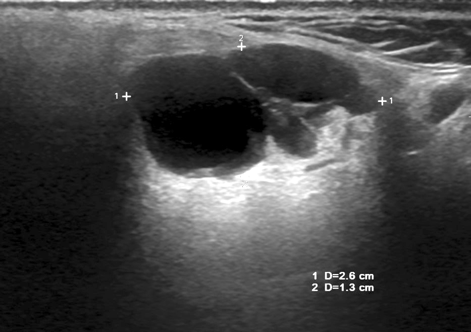
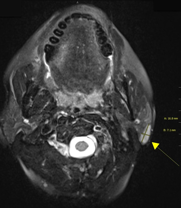
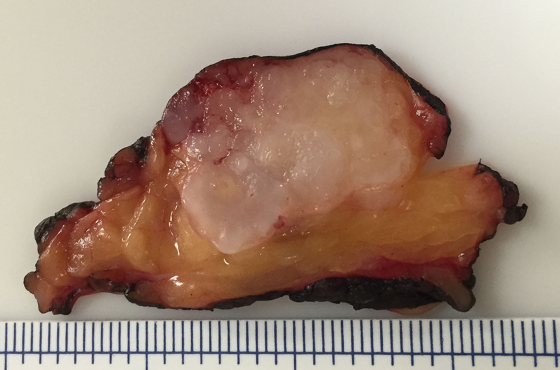
  • pT1: Tumor ≤ 2 cm without extraparenchymal extension
  • pT2: Tumor > 2 cm but ≤ 4 cm without extraparenchymal extension
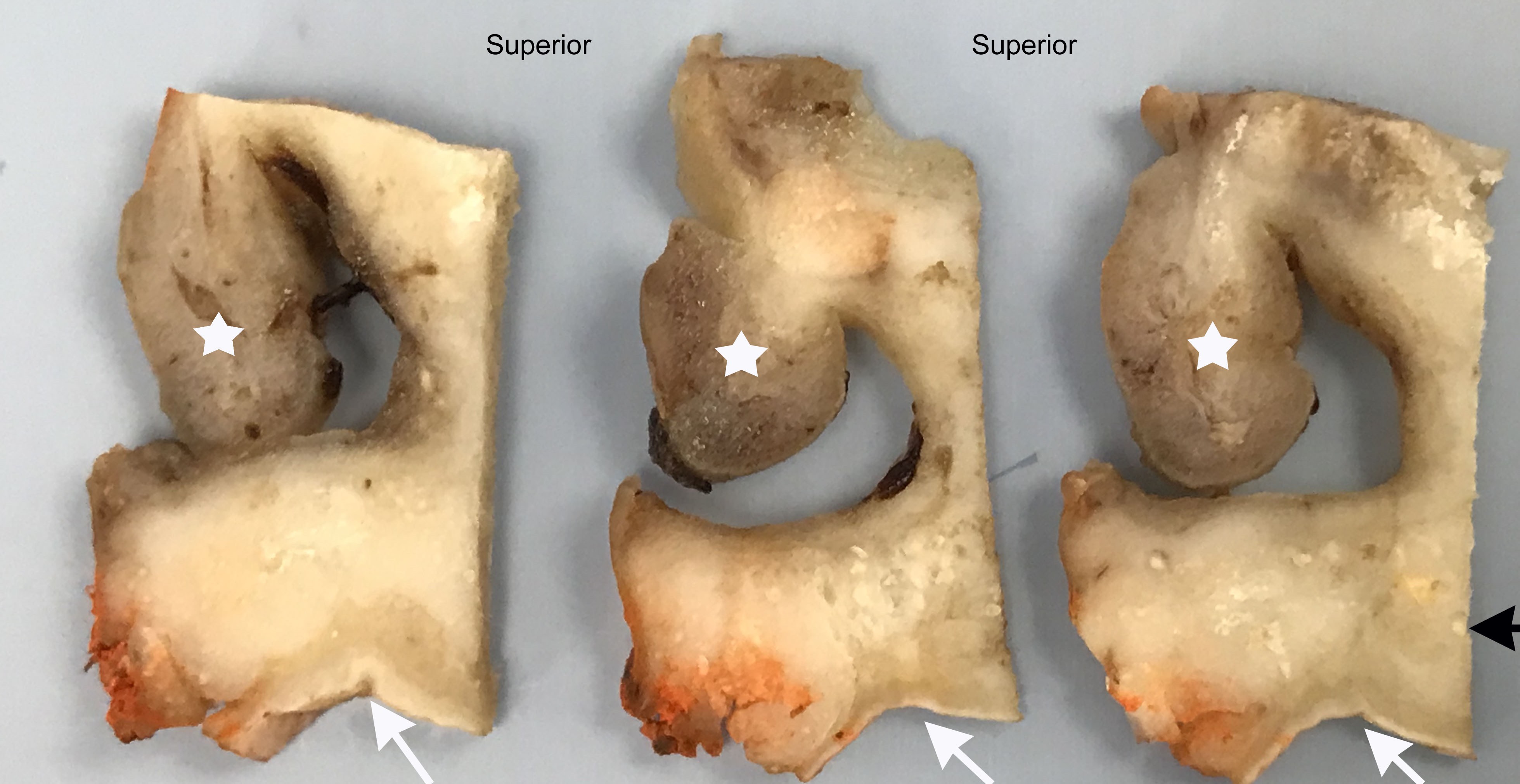
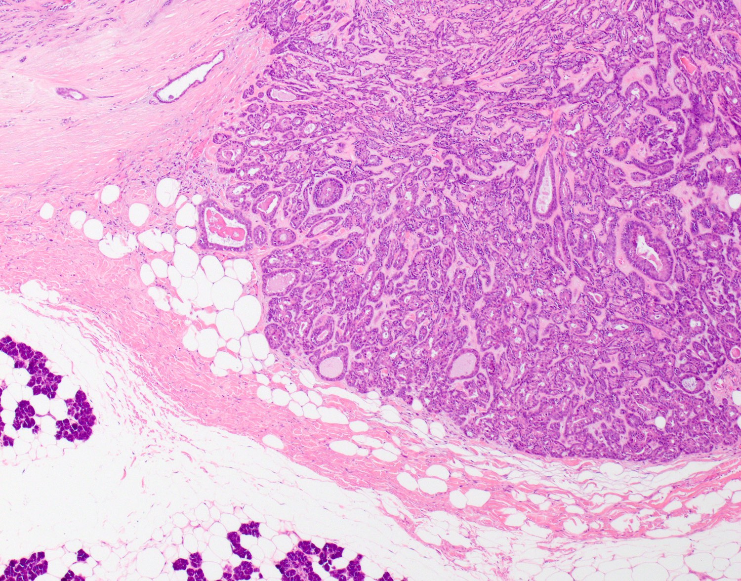
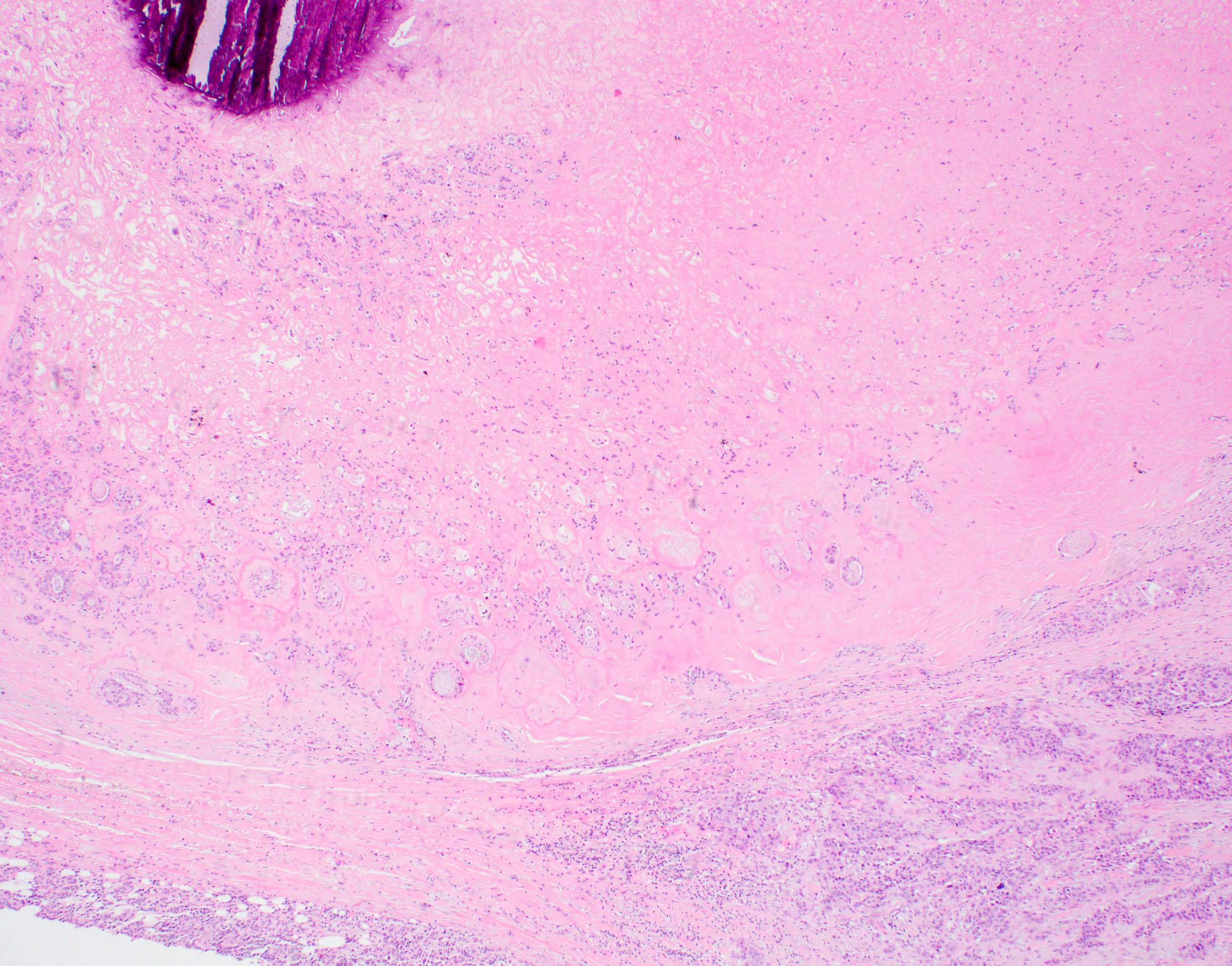
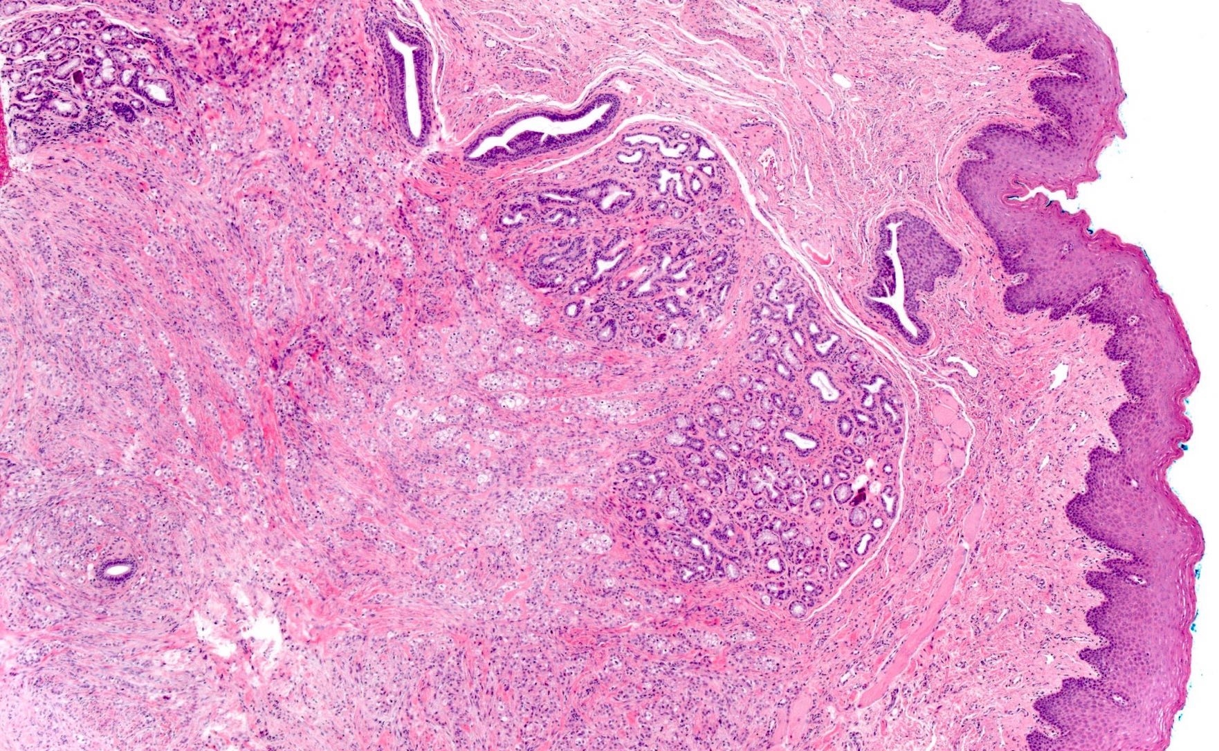
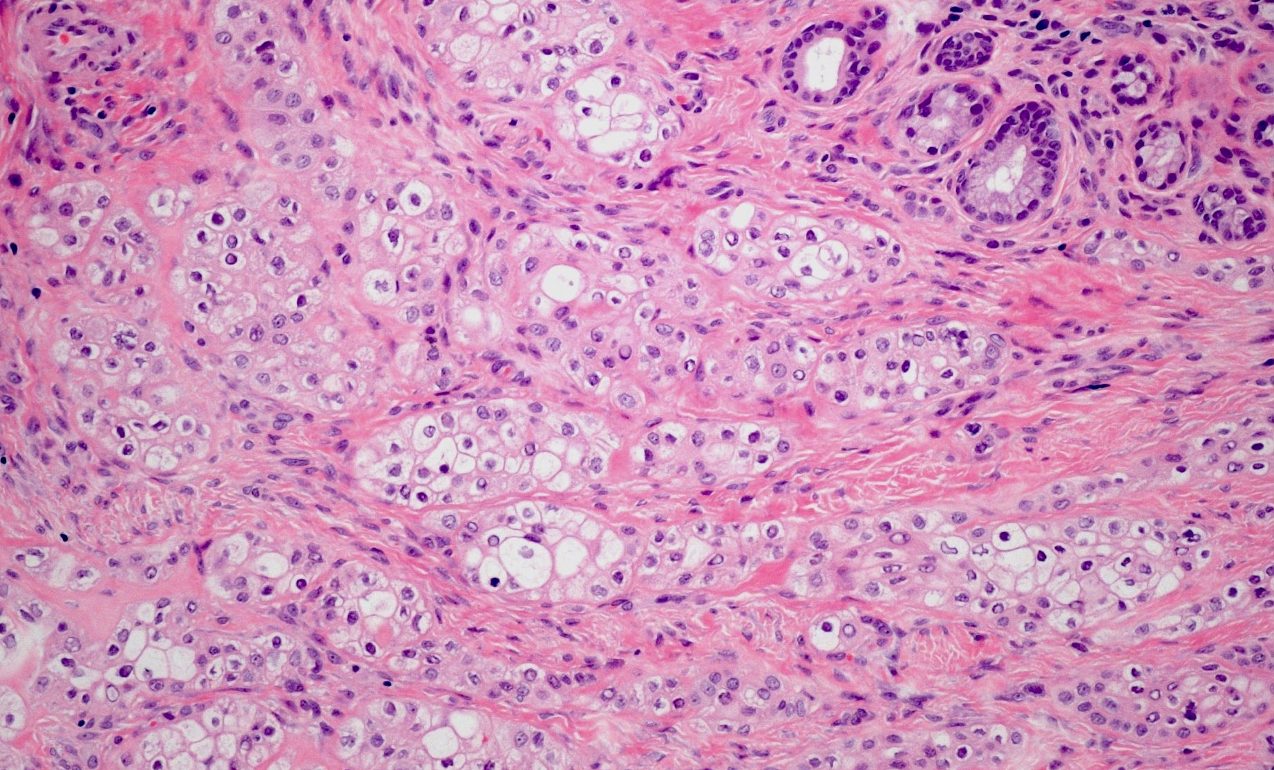
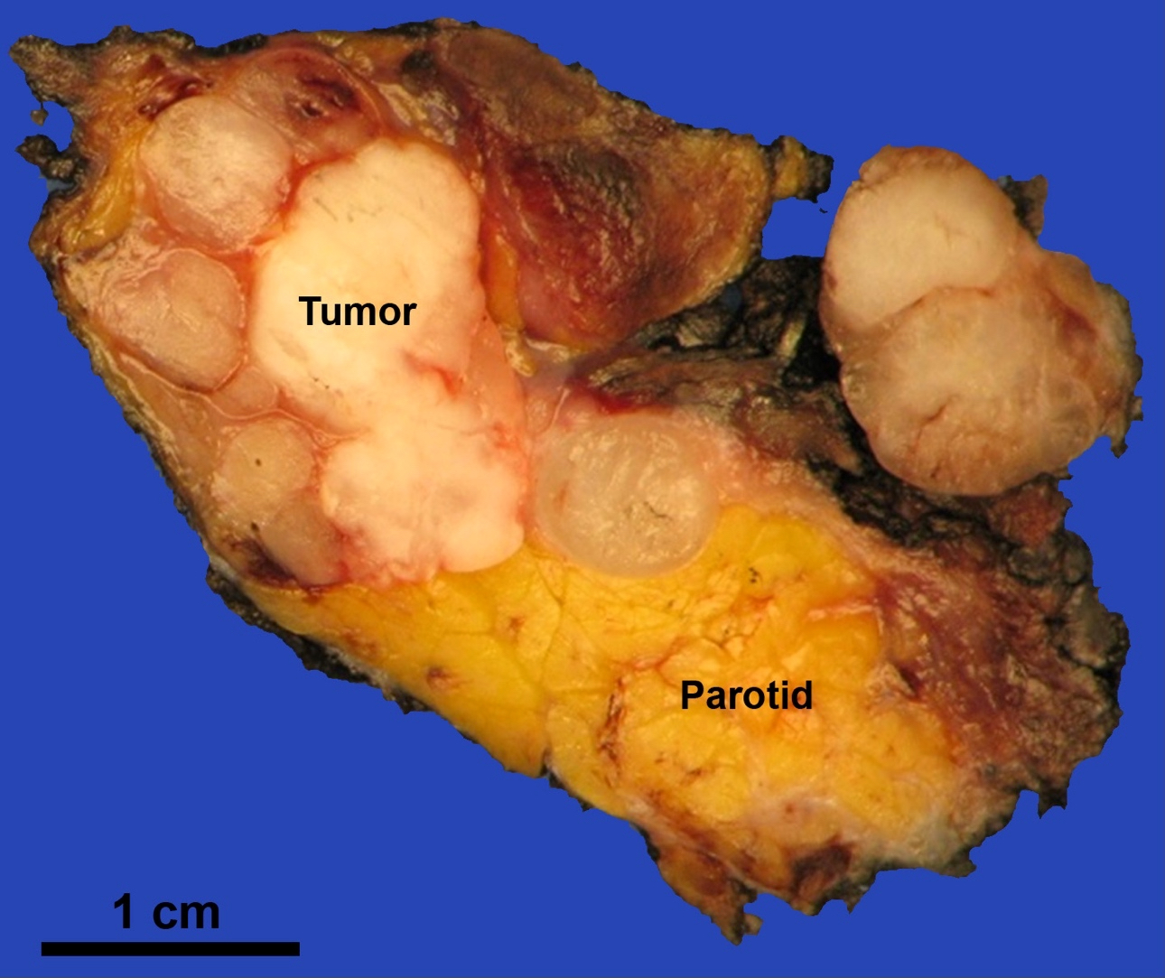
  • pT3: Tumor > 4 cm or tumor with extraparenchymal extension
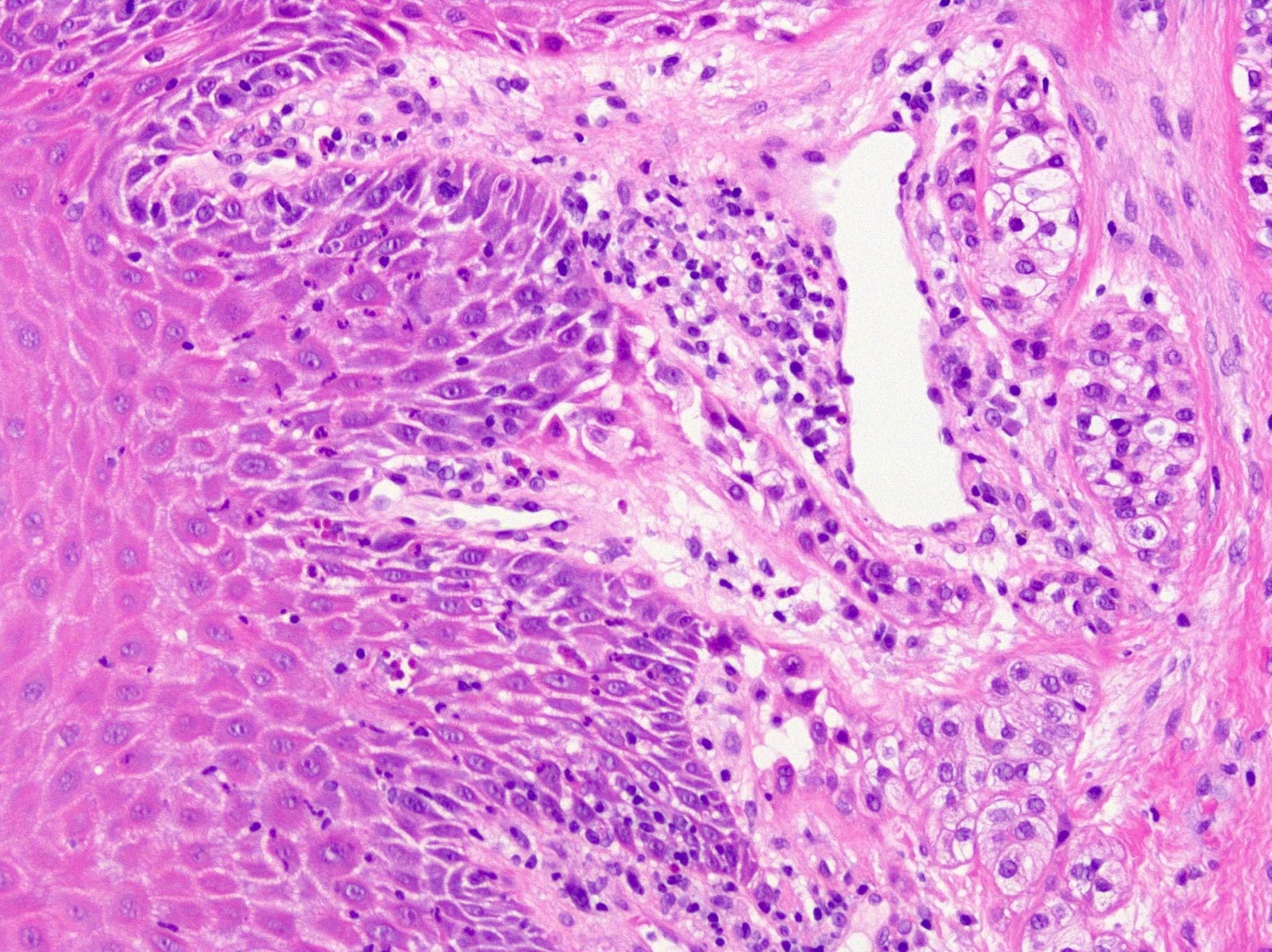
  • pT4a: Tumor of any size invading skin, mandible, ear canal or facial nerve
  • pT4b: Tumor of any size invading skull base or pterygoid plates or encases carotid artery
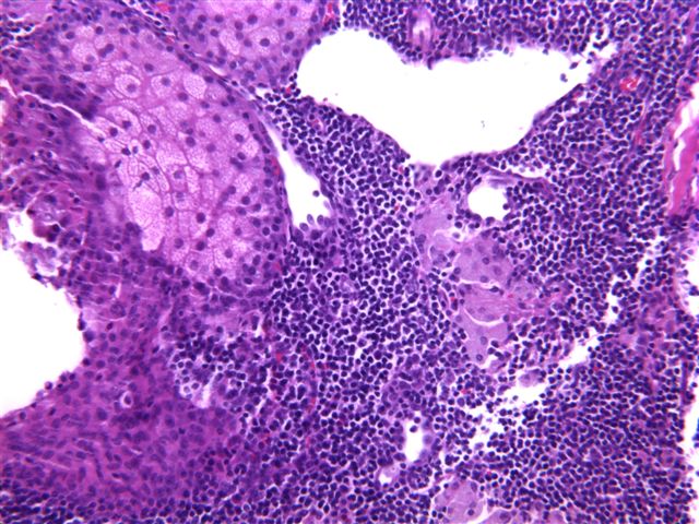
Notes:
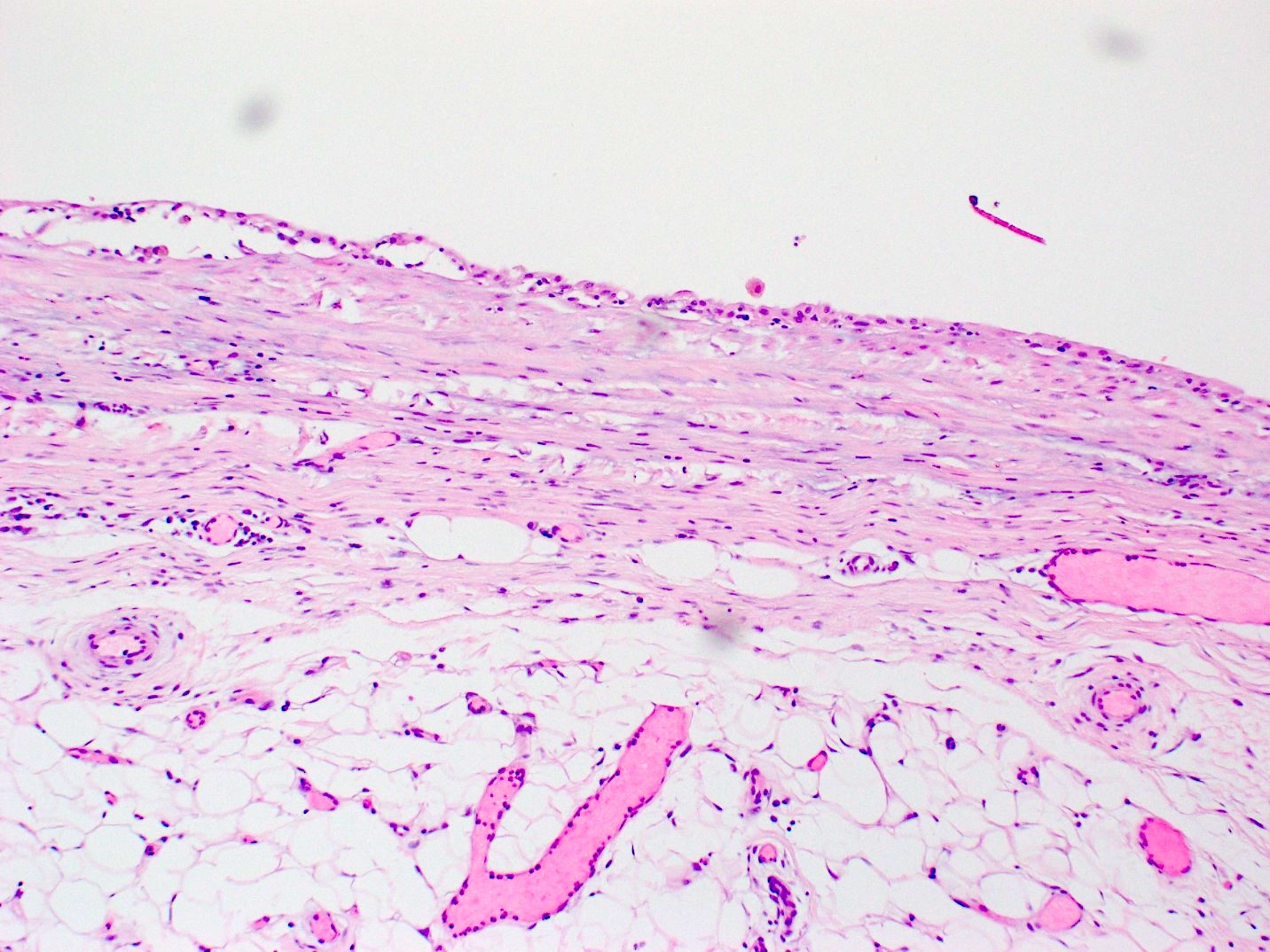
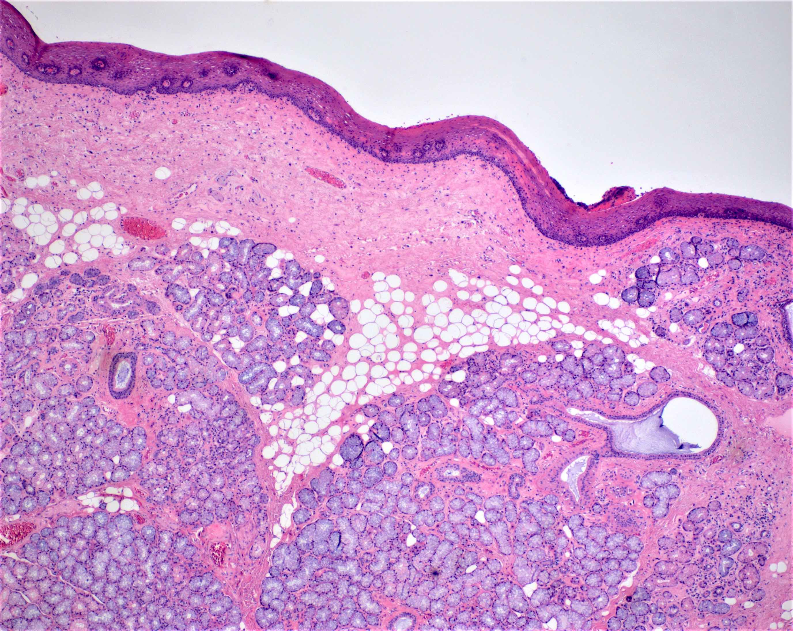
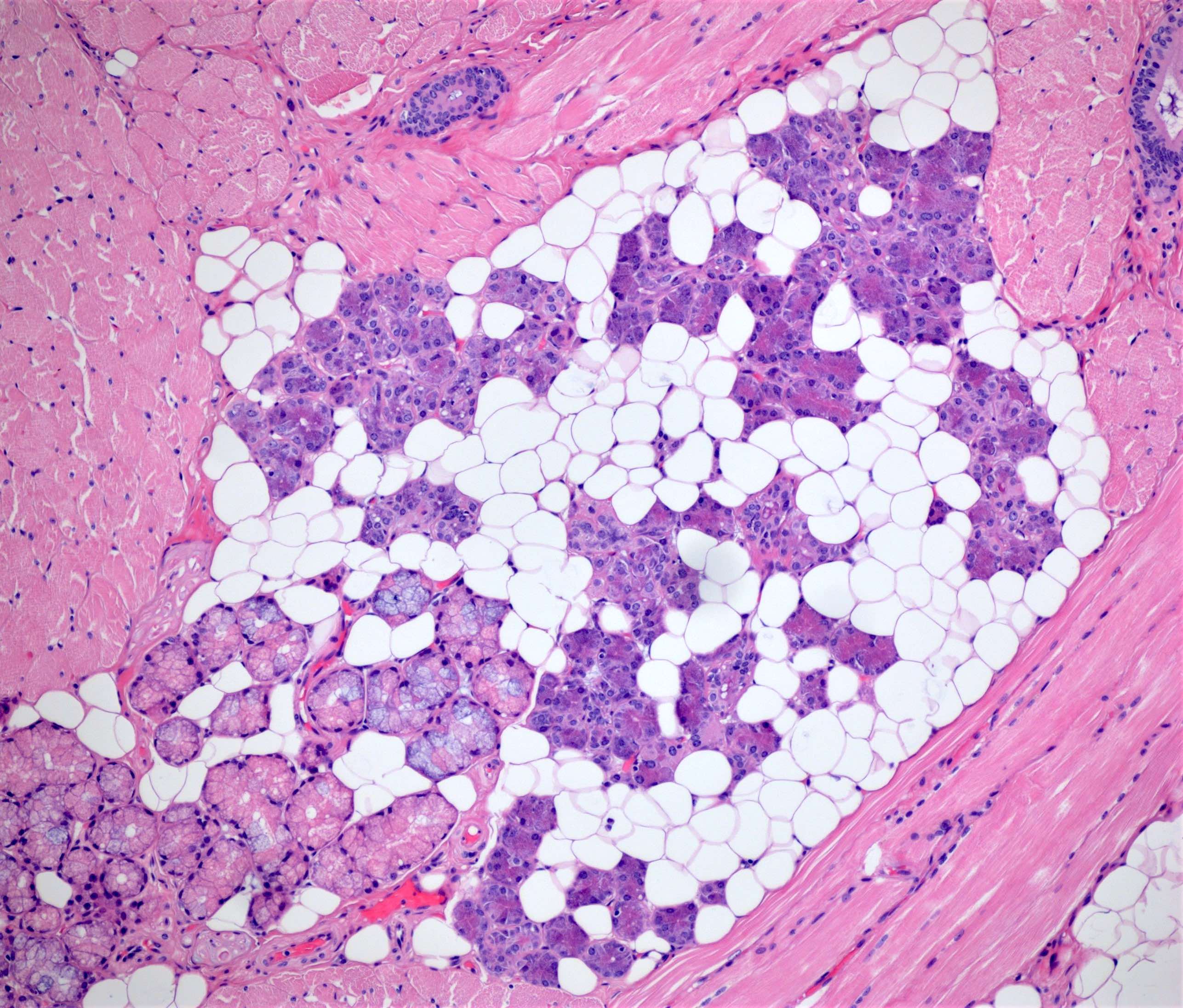
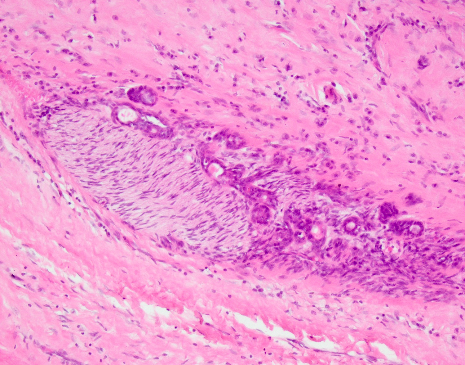
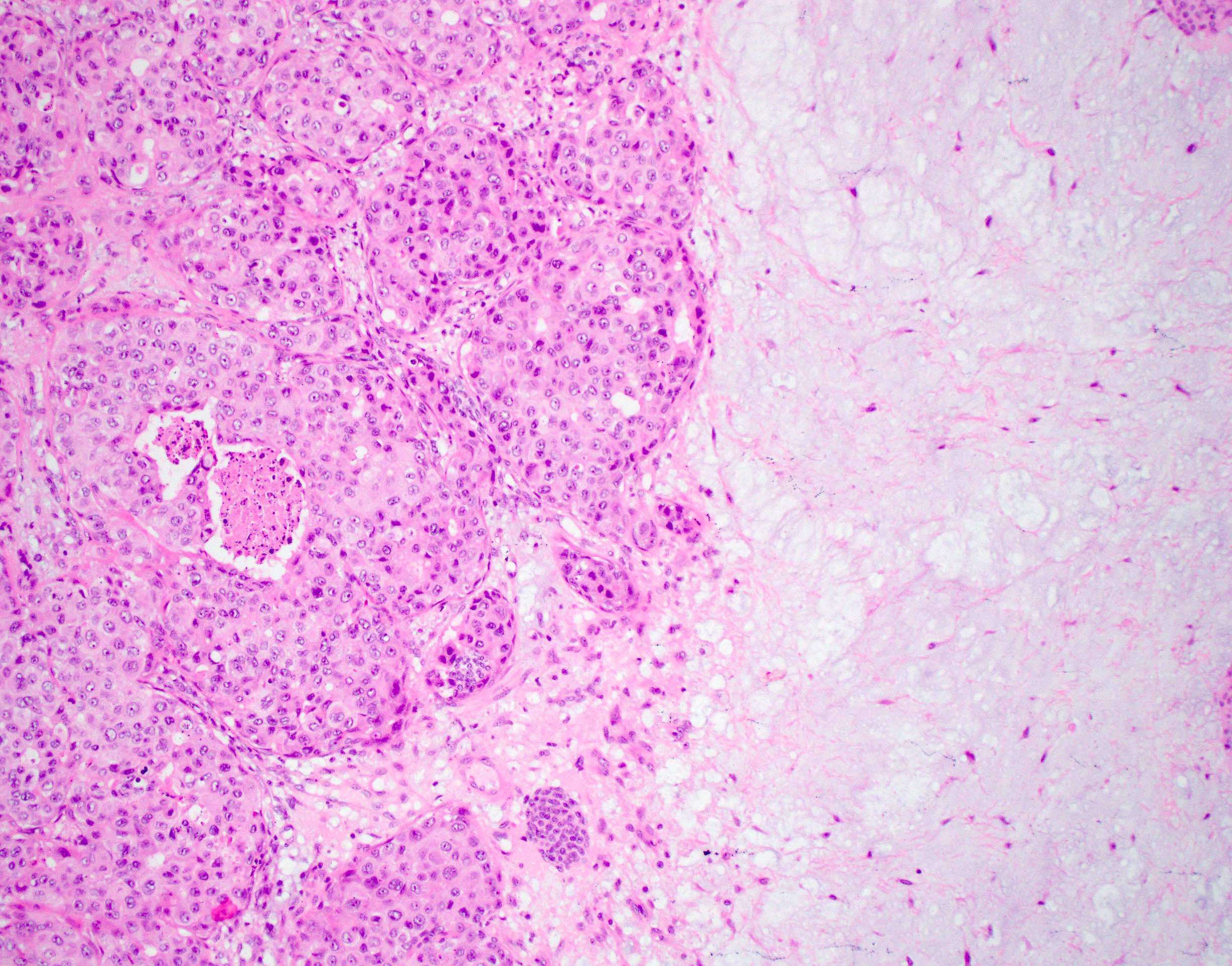
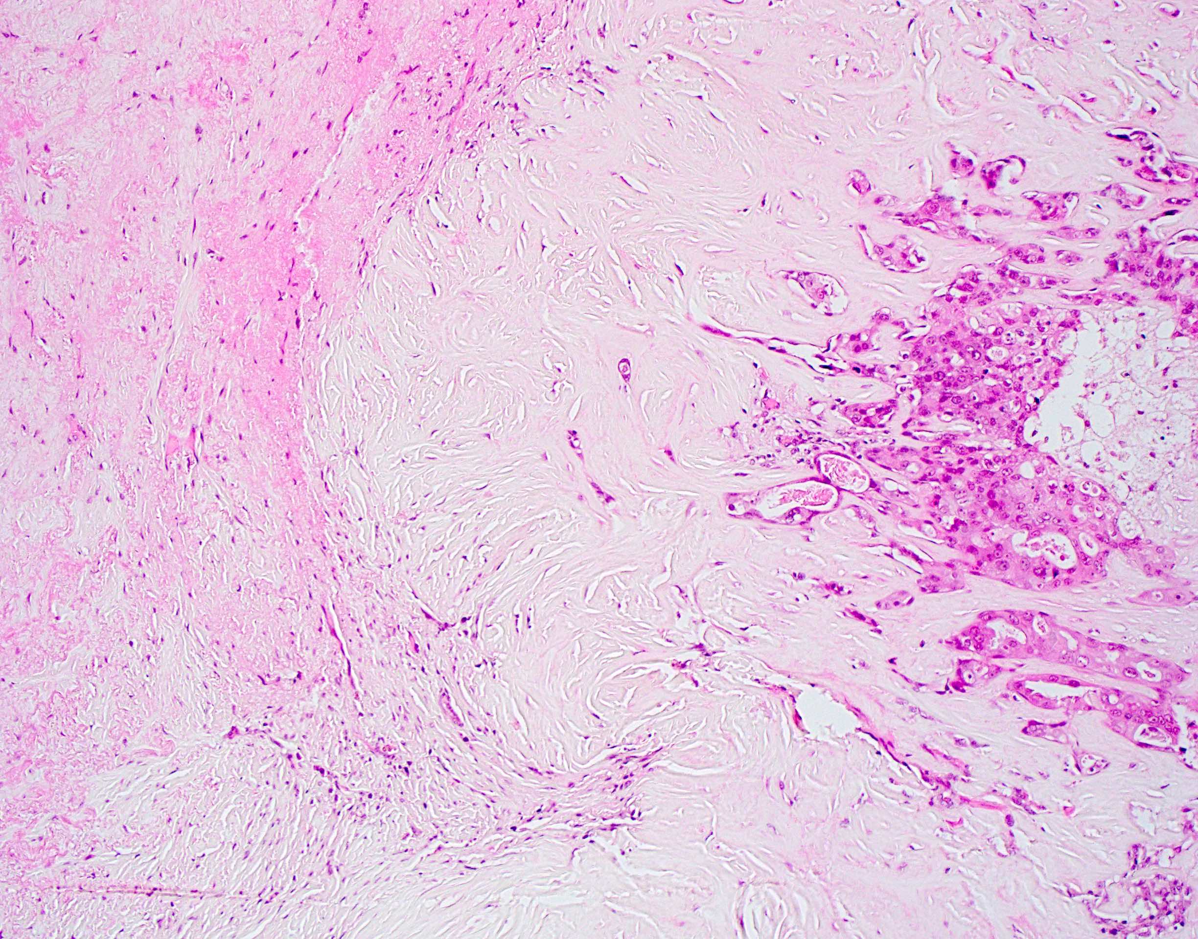
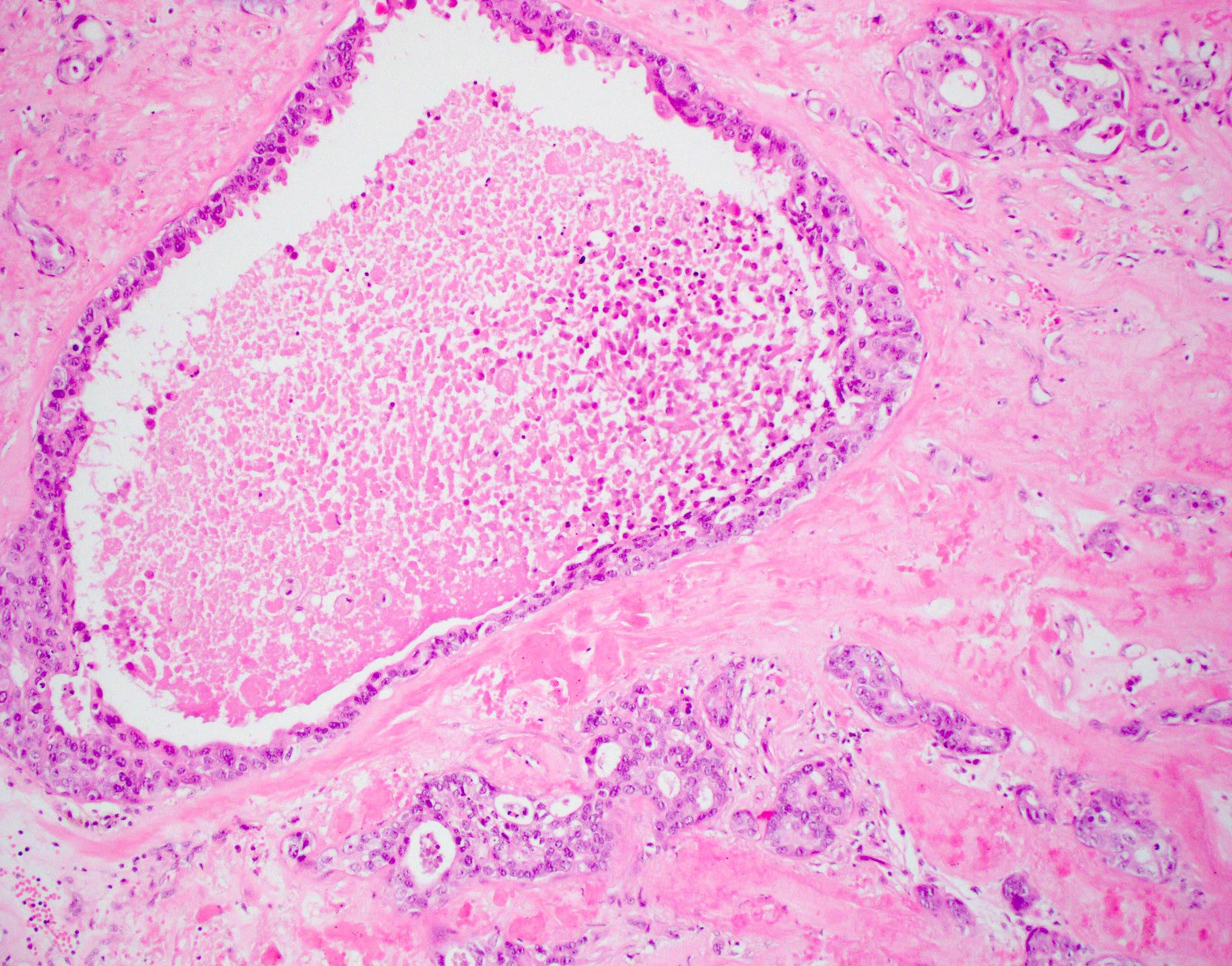
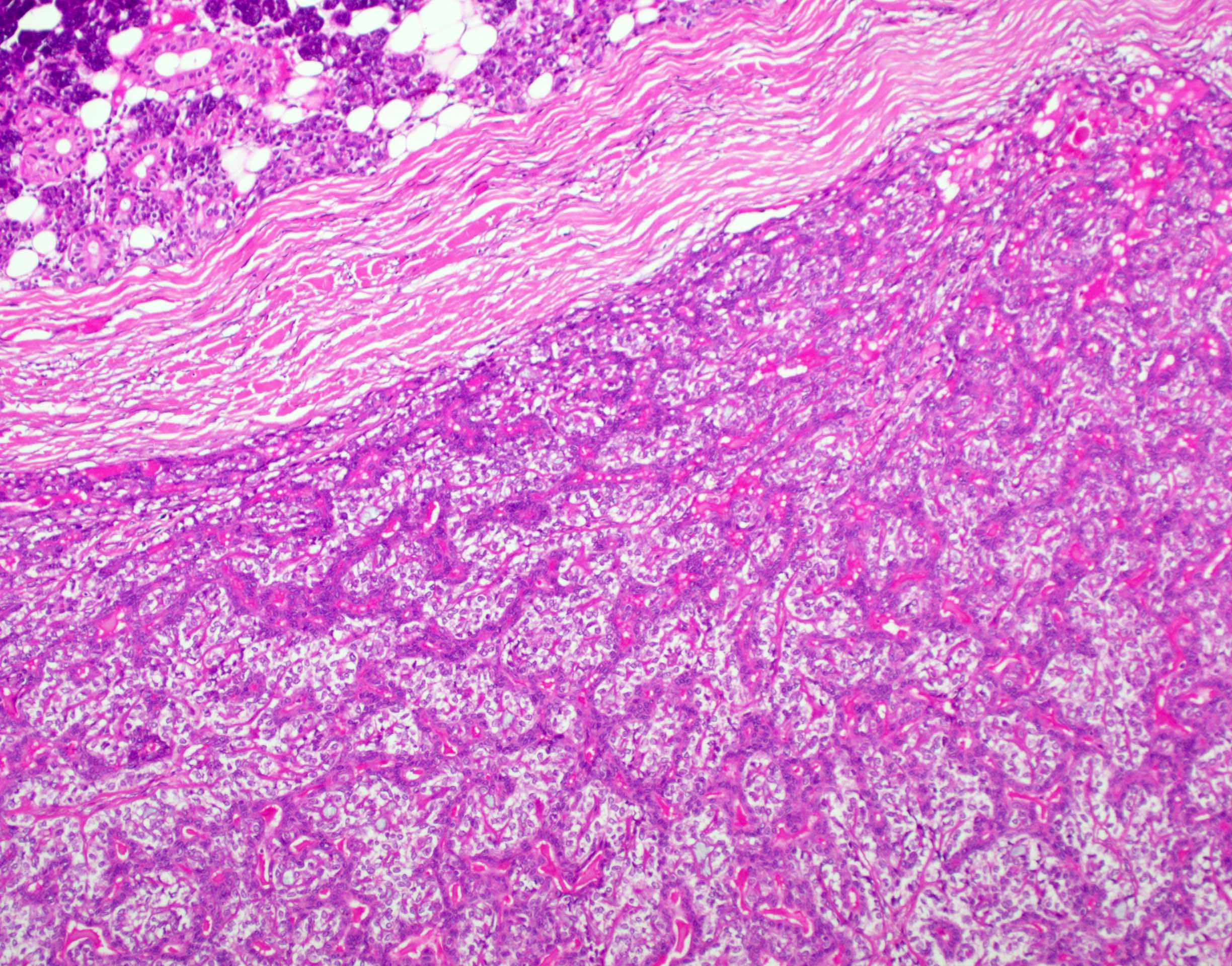
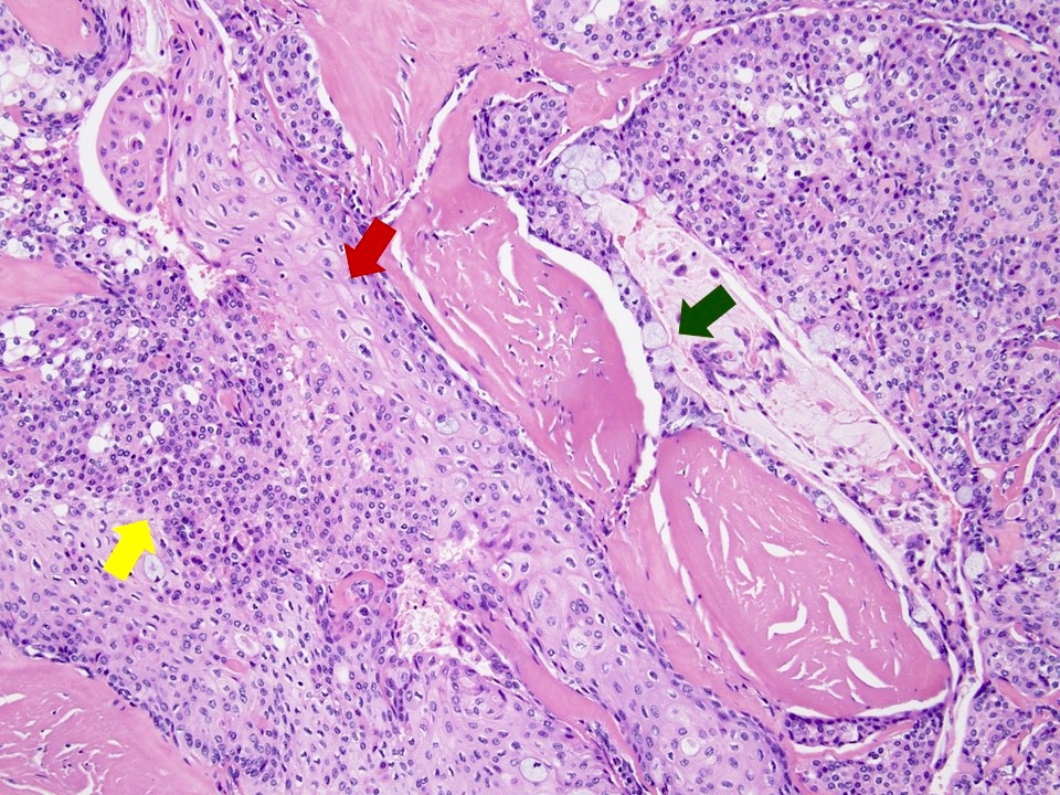
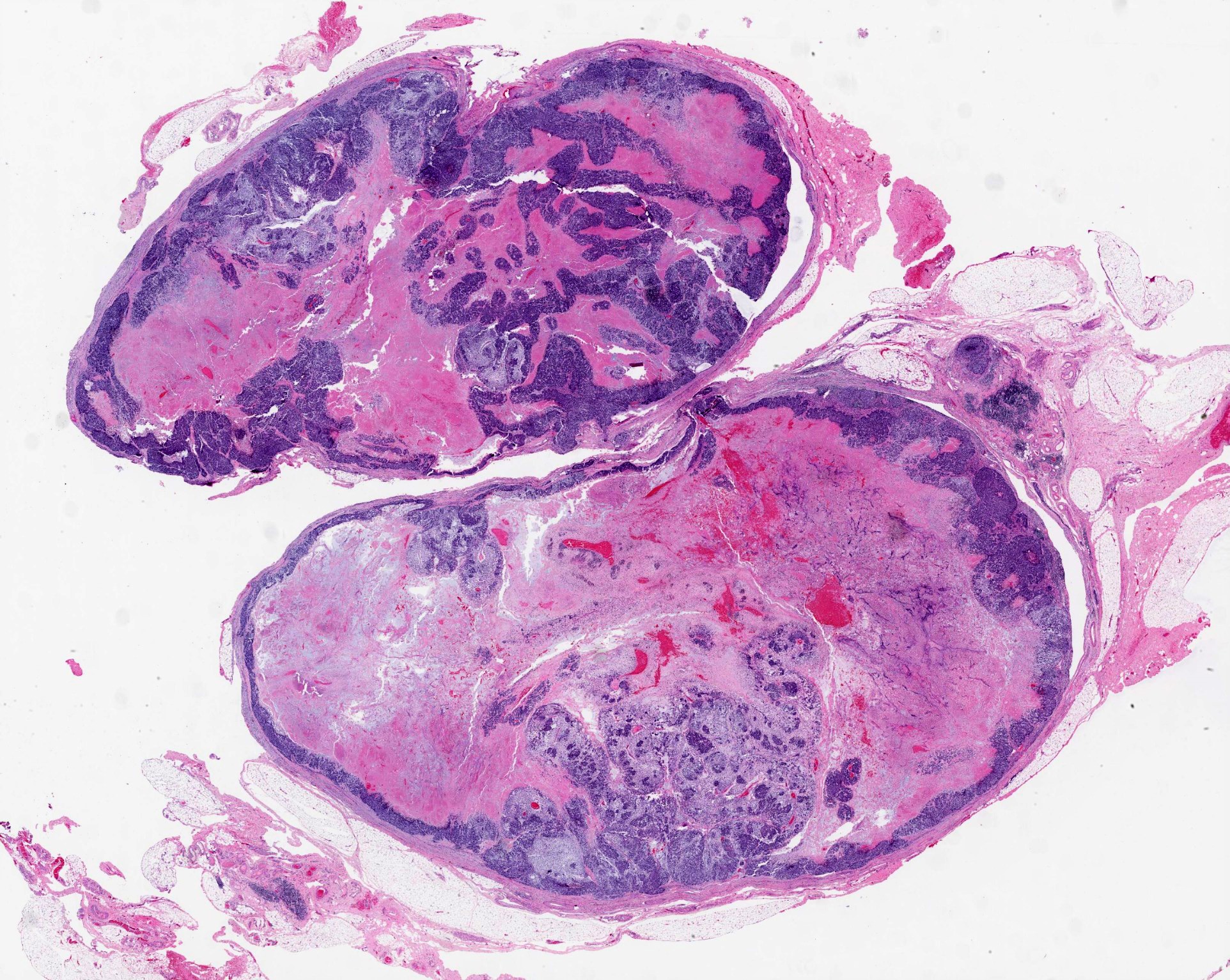
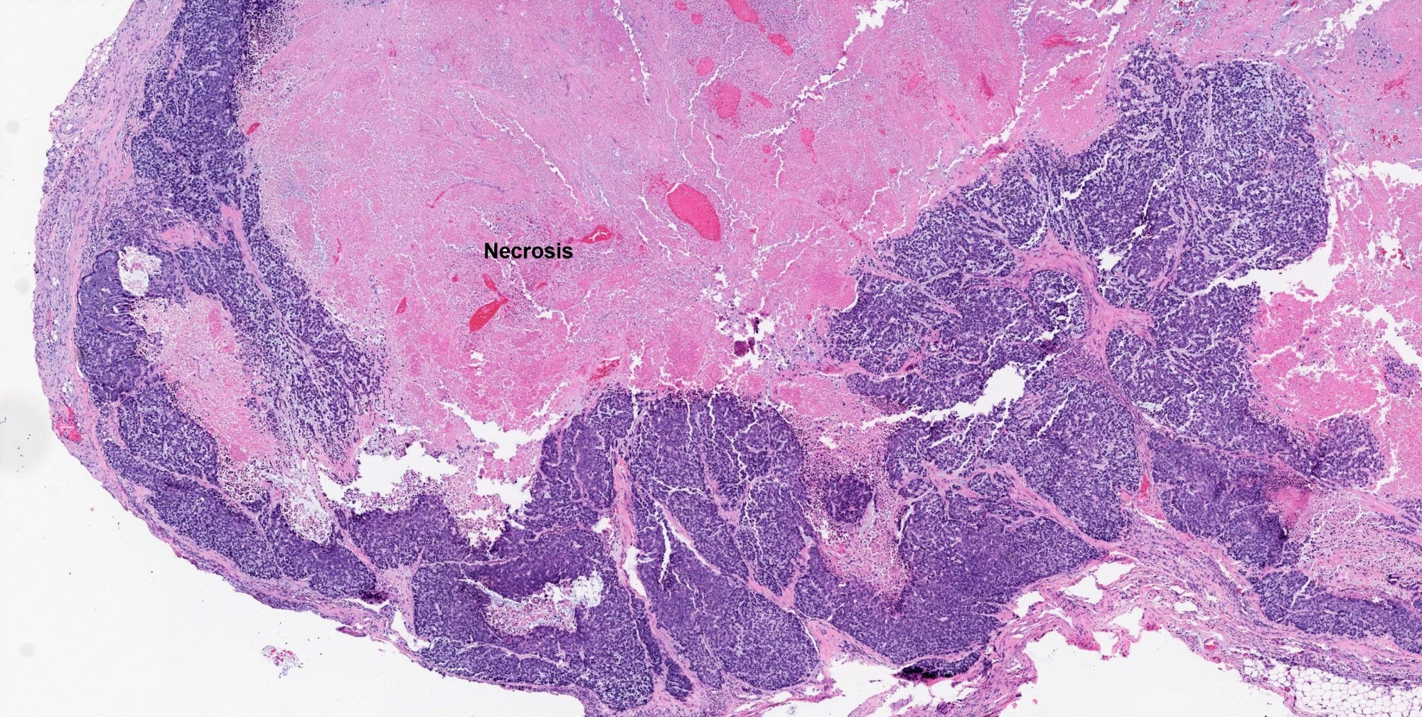
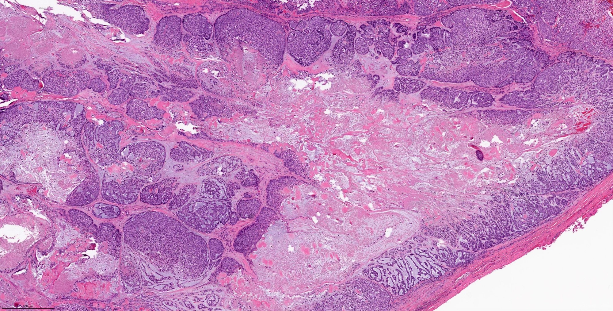
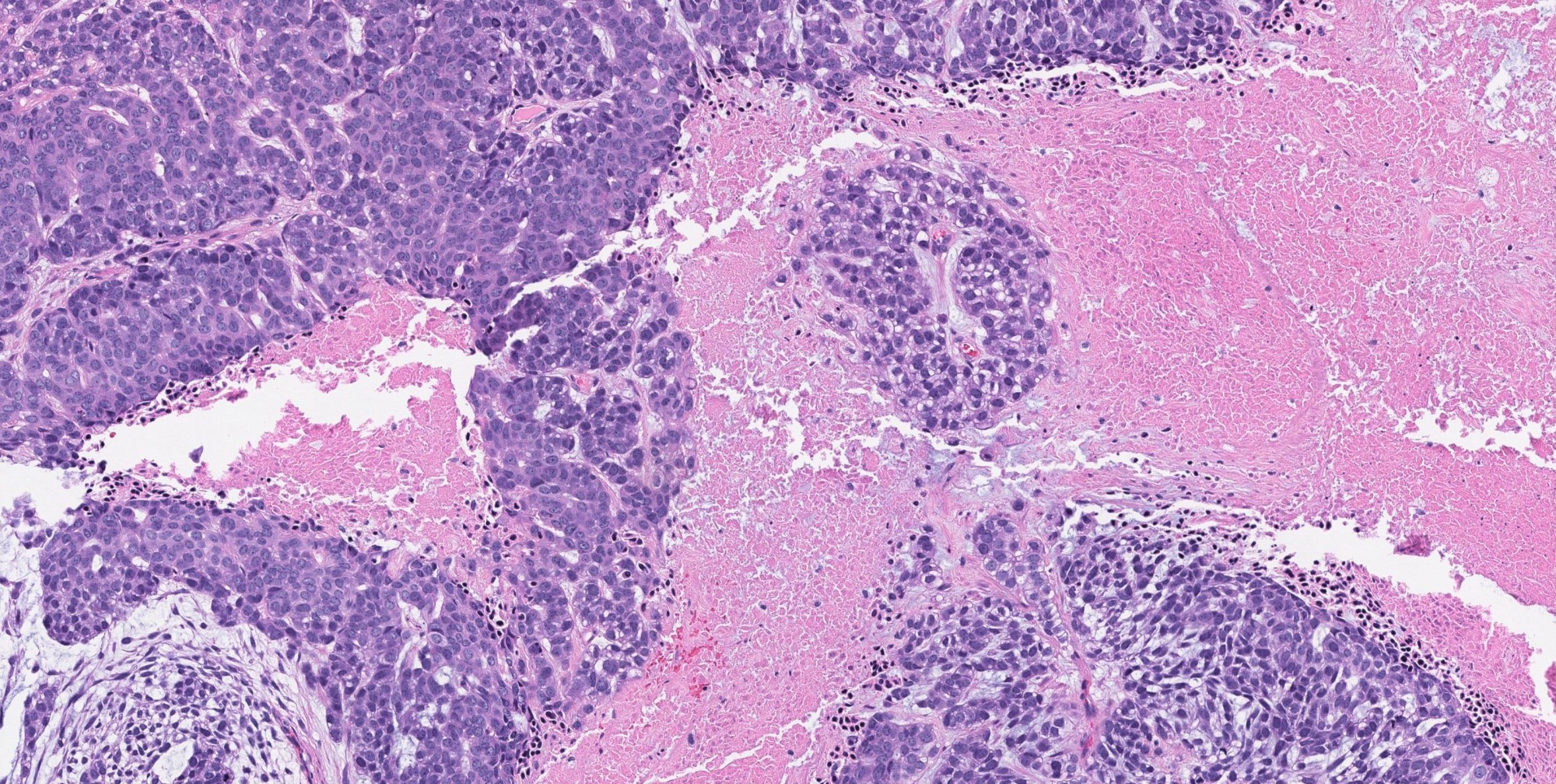
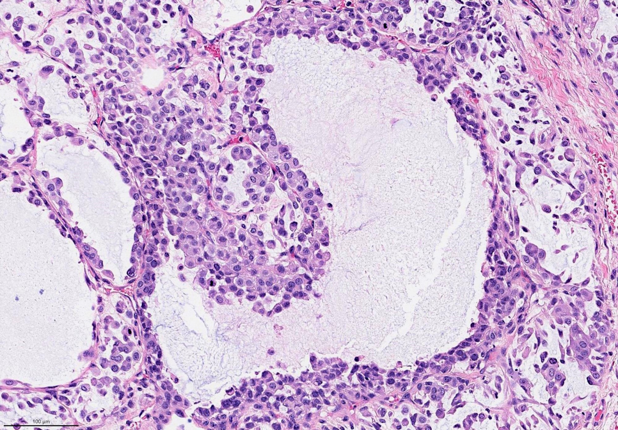
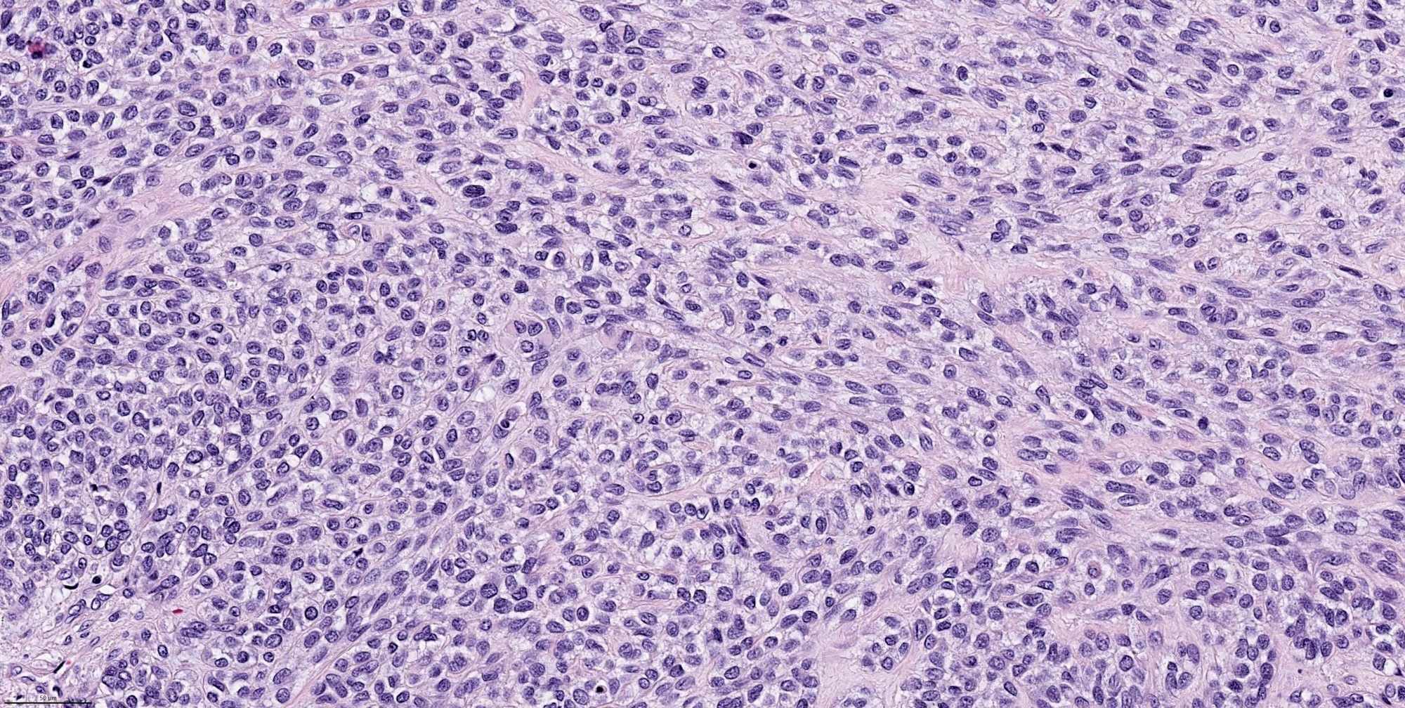
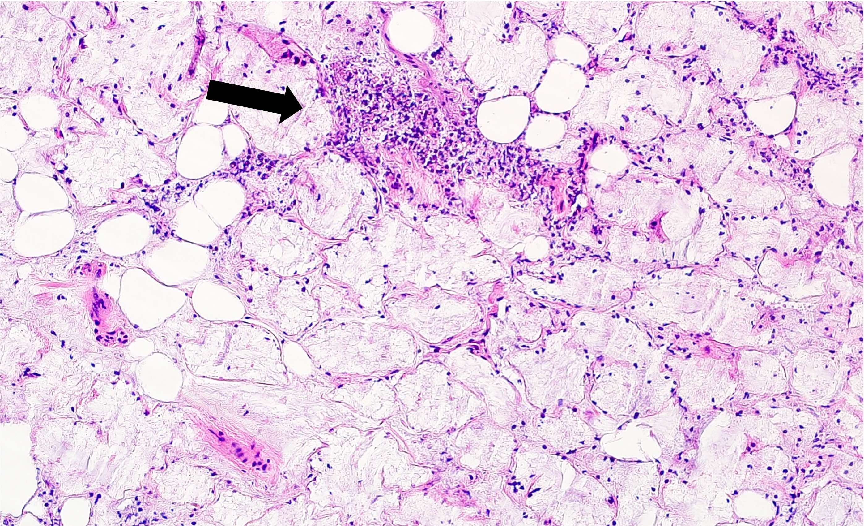
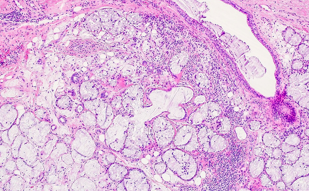
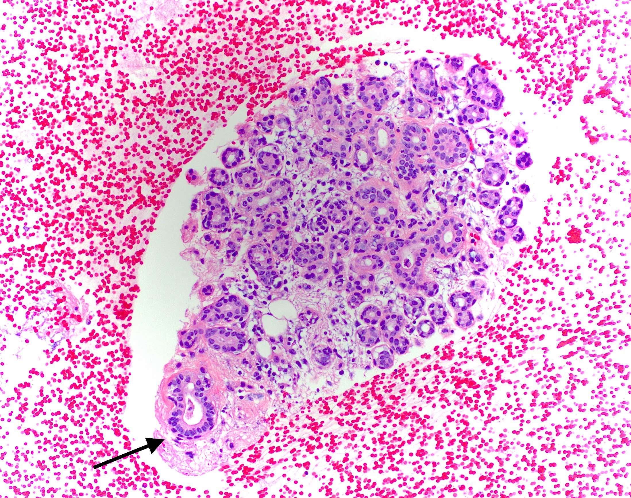
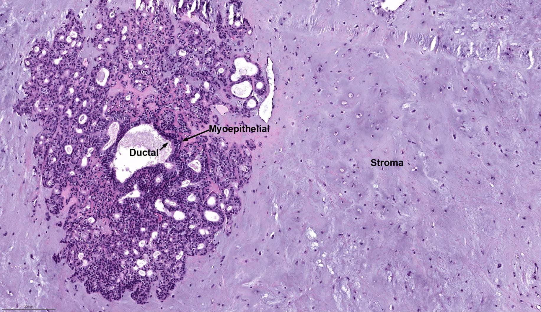
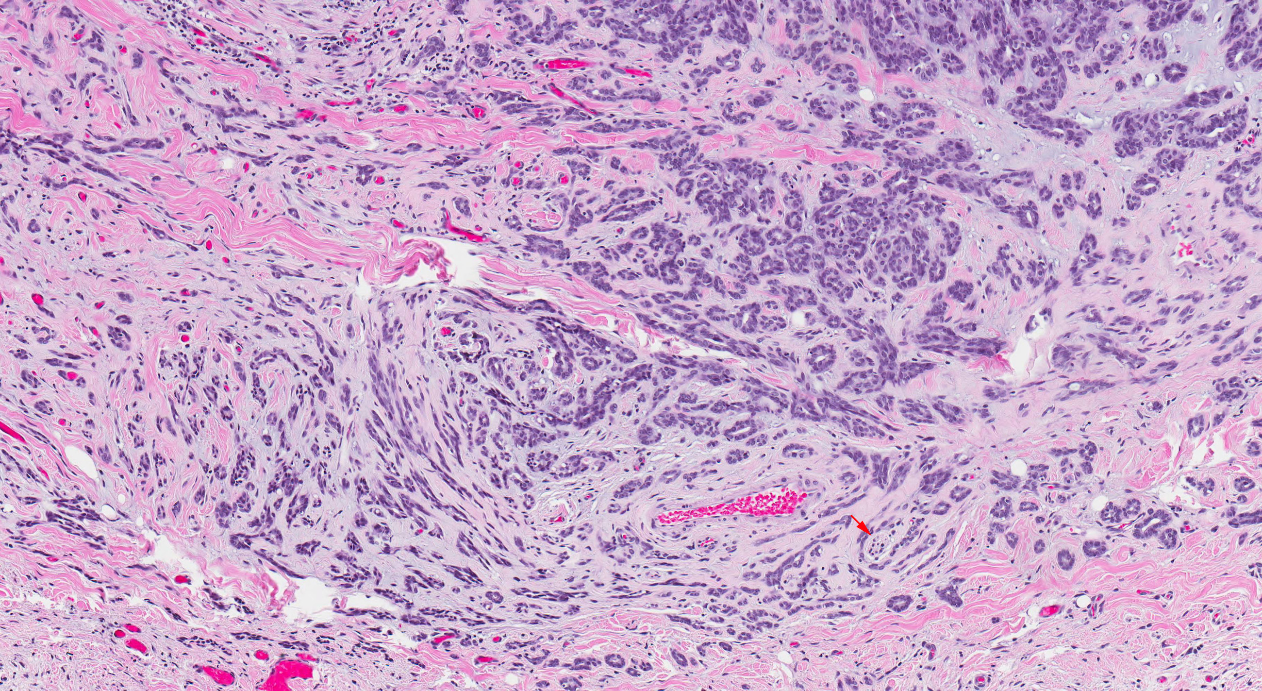
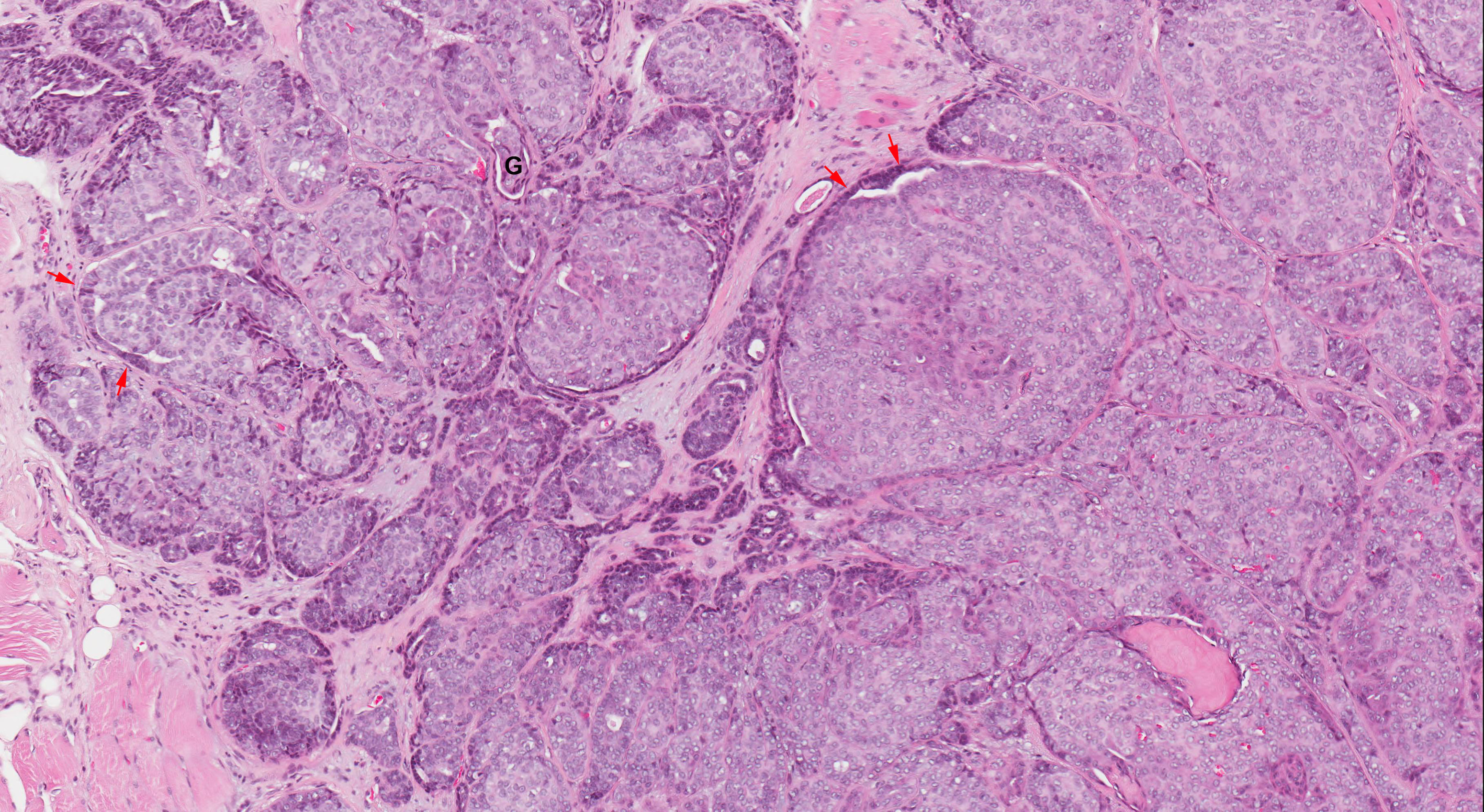
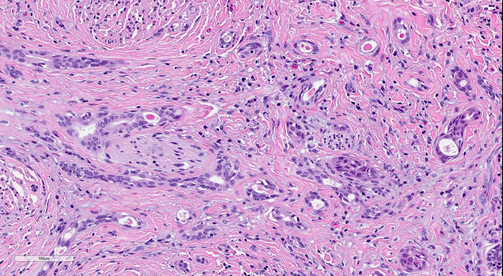
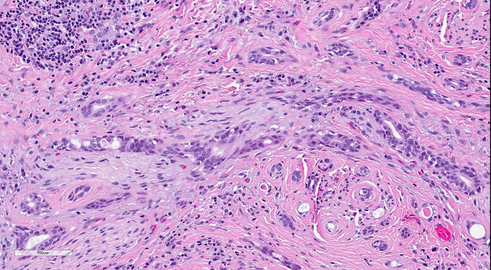
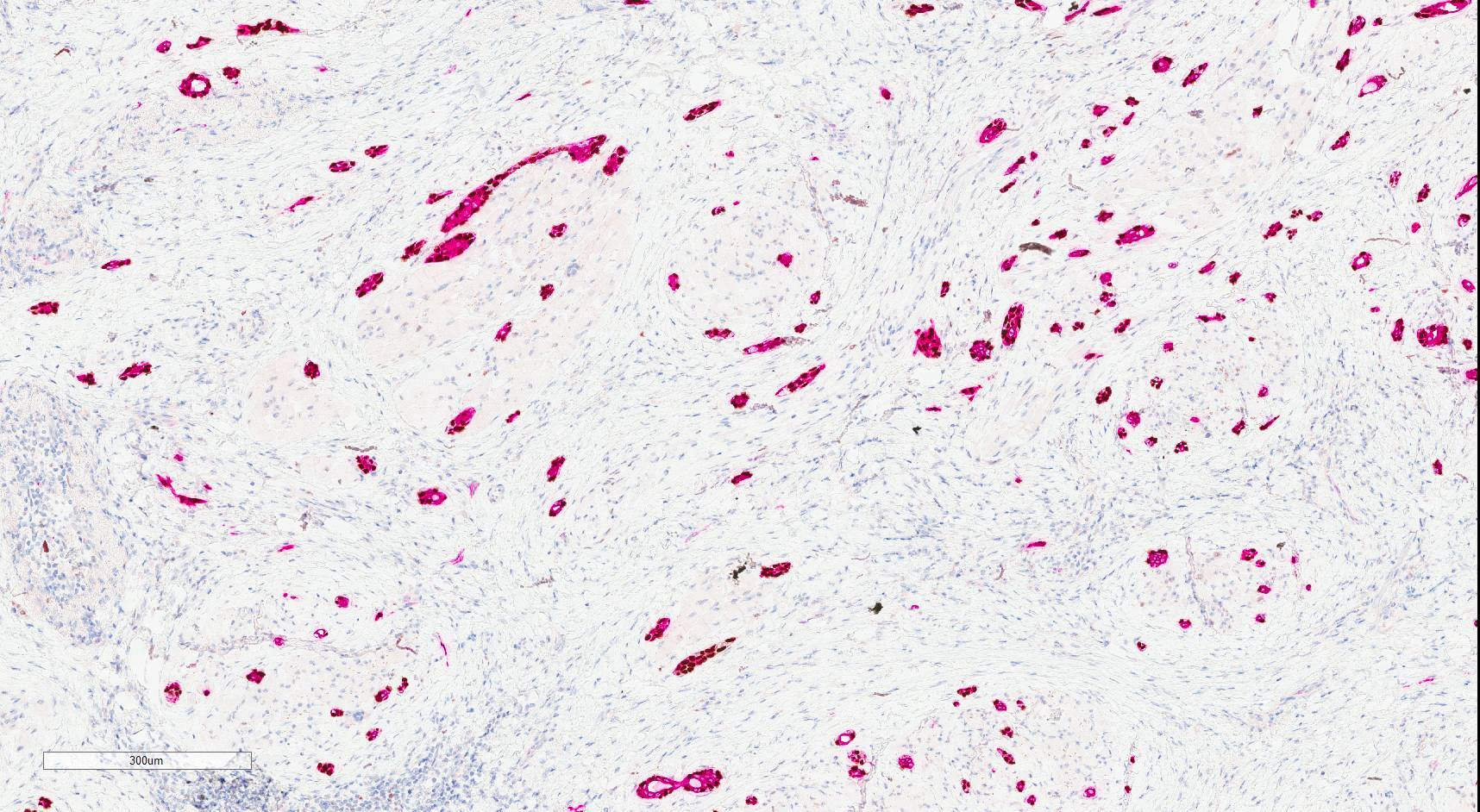
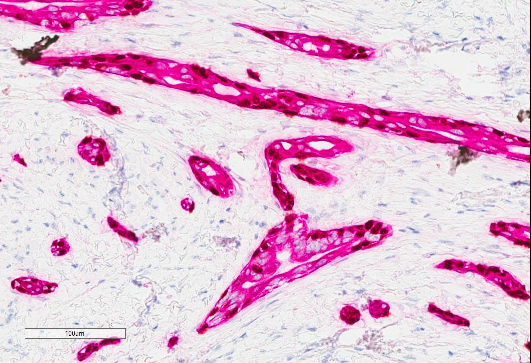
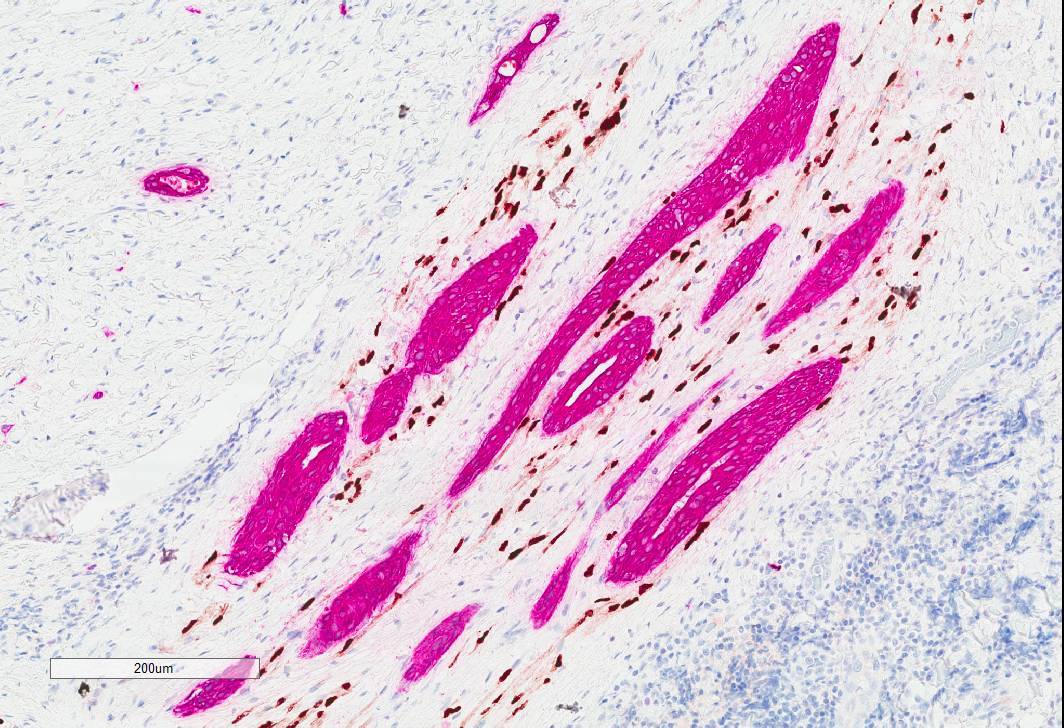
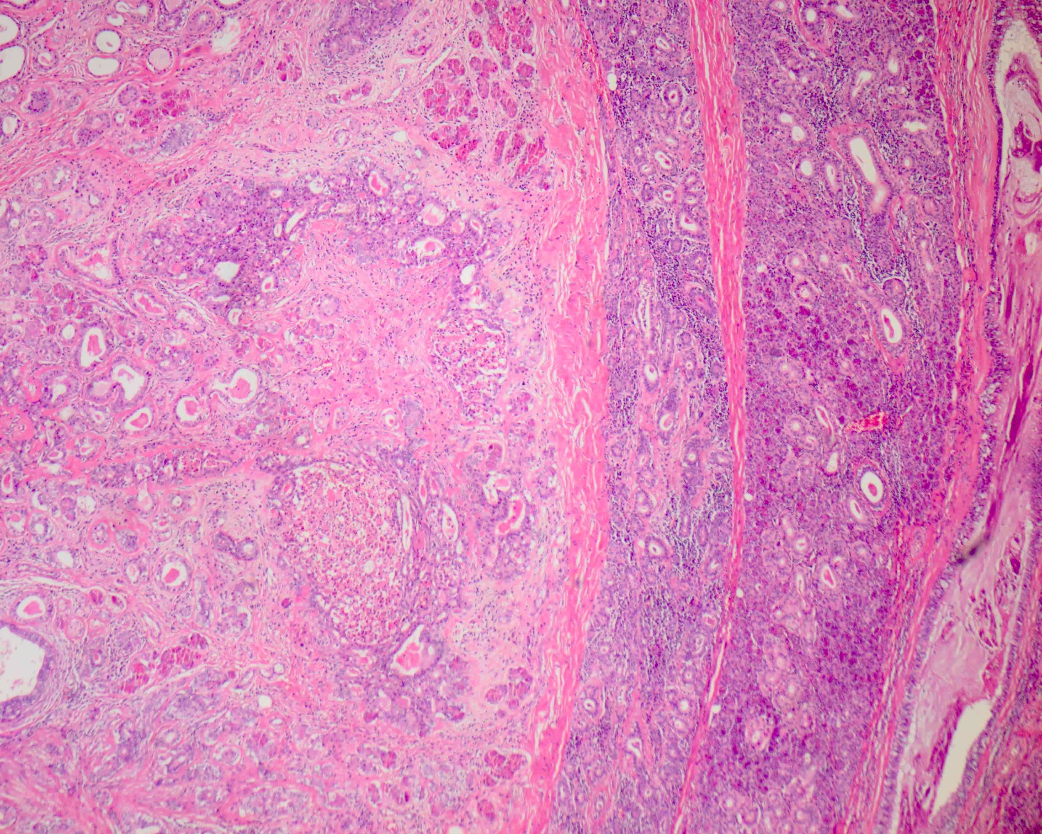
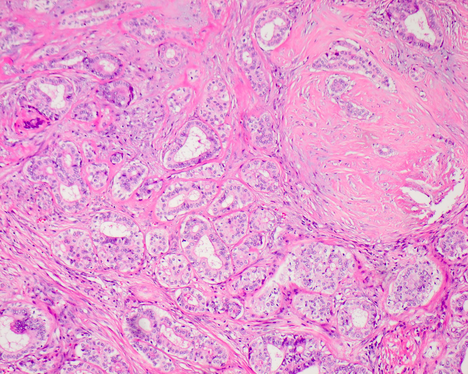
  • Extraparenchymal extension is clinical or macroscopic evidence of invasion of soft tissues
  • Microscopic evidence alone does not constitute extraparenchymal extension for classification purposes
Regional lymph nodes (pN)
  • pNX: Regional lymph nodes cannot be assessed
  • pN0: No regional lymph node metastasis
  • pN1: Metastasis in a single ipsilateral lymph node ≤ 3 cm without extranodal extension
  • pN2a: Single ipsilateral lymph node metastasis ≤ 3 cm with extranodal extension or > 3 cm but ≤ 6 cm without extranodal extension
  • pN2b: Metastasis in multiple ipsilateral lymph nodes ≤ 6 cm without extranodal extension
  • pN2c: Metastasis in bilateral or contralateral lymph node(s) ≤ 6 cm without extranodal extension
  • pN3a: Metastasis in a lymph node > 6 cm without extranodal extension
  • pN3b: Extranodal extension in a single ipsilateral lymph node > 3 cm or single contralateral node or multiple nodes with metastases, any with extranodal extension

Notes:
  • A selective neck dissection will include 10+ lymph nodes and a comprehensive neck dissection (radical or modified radical neck dissection) will include 15+ lymph nodes
  • Negative pathologic examination of a smaller number of nodes still mandates a pN0 designation
  • Midline nodes are considered ipsilateral nodes
  • Measurement of tumor metastasis:
    • Cross sectional diameter of the largest lymph node metastatic deposit (not the lymph node itself) is measured in the gross specimen at the time of macroscopic examination or if necessary, using the histologic slide
  • Extranodal extension (ENE):
    • AJCC 8th edition introduces the use of ENE in pN categorization
    • Must be clearly defined as tumor present within the confines of the lymph node and extending through the lymph node capsule into the surrounding connective tissue, with or without associated stromal reaction
Distant metastasis (pM)
  • pM0: No distant metastasis
  • pM1: Distant metastasis
AJCC prognostic stage grouping
Stage 0: Tis N0 M0
Stage I: T1 N0 M0
Stage II: T2 N0 M0
Stage III: T3 N0 M0
T0 - 3 N1 M0
Stage IVA: T4a N0 - 1 M0
T0 - 4a  N2 M0
Stage IVB: Any T N3 M0
T4b Any N M0
Stage IVC:Any T Any N  M1
Registry data collection variables
  • Extranodal extension clinical (present versus absent)
  • Extranodal extension pathological (present versus absent)
  • Extent of microscopic extranodal extension (distance of extension from the native lymph node capsule to the farthest point of invasion in extranodal tissue)
  • Perineural invasion
  • Lymphovascular invasion
  • p16 / HPV status
  • Performance status
  • Tobacco use and pack years
  • Alcohol use
  • Depression diagnosis
Histopathologic type
  • Mucoepidermoid carcinoma
  • Adenoid cystic carcinoma
  • Acinic cell carcinoma
  • Polymorphous adenocarcinoma
  • (Mammary analogue) secretory carcinoma
  • Salivary duct carcinoma
  • Carcinoma ex pleomorphic adenoma
  • Epithelial myoepithelial carcinoma
  • (Hyalinizing) clear cell carcinoma
  • Adenocarcinoma, not otherwise specified
    • Includes cystadenocarcinoma, intestinal type adenocarcinoma and mucinous adenocarcinoma
  • Basal cell adenocarcinoma
  • Carcinosarcoma
  • Intraductal carcinoma
  • Lymphoepithelial carcinoma
  • Myoepithelial carcinoma
  • Oncocytic carcinoma
  • Poorly differentiated carcinoma
    • Large cell neuroendocrine carcinoma
    • Small cell neuroendocrine carcinoma
    • Undifferentiated carcinoma
  • Sebaceous adenocarcinoma
  • Squamous cell carcinoma

Notes:
  • In this current classification, sialoblastoma is designated as a tumor of uncertain malignant potential
  • Metastasizing pleomorphic adenoma has been collapsed under pleomorphic adenoma
  • Since the biologic behavior of these still overlaps with the other malignant tumors, these can be reported under "other"
Board review style question #1
Regarding the following parotid gland primary salivary gland adenocarcinoma, which of the pT category criteria in the 8th edition AJCC staging guide is satisfied by the examination of this gross resection specimen photograph?


  1. pT1 by depth of invasion
  2. pT2 by tumor size
  3. pT3 by presence of necrosis
  4. pT3 by extraparenchymal extension
Board review style answer #1
D. pT3 by extraparenchymal extension (white arrows point to border of skeletal muscle, yellow arrows point to border of malignancy, black arrow points to tumor invading skeletal muscle)

Comment Here

Reference: Pathologic TNM staging of major salivary glands (AJCC 8th edition)

Striated duct adenoma (pending)
[Pending]

Suspicious for malignancy
Definition / general
Essential features
  • FNA that shows cytologic features suggestive of malignancy and meets some but not all of the criteria for malignancy
  • FNA should be subcategorized if possible (e.g., suspicious for a primary salivary gland malignancy, suspicious for metastasis, suspicious for lymphoma)
Terminology
  • Suspicious for malignancy (category V)
CPT coding
  • 88172 - determination of adequacy of specimen
  • 88173 - FNA interpretation
Sites
Clinical features
  • Frequency among all the Milan system categories: 4%
  • Risk of malignancy (ROM) is 83% in the 2nd edition of Milan System, with a large study demonstrating a ROM of 83.3% (Cancer Cytopathol 2018;126:94)
Laboratory
Radiology description
Cytology description
  • FNA should be subcategorized if possible (e.g., suspicious for a primary salivary gland malignancy, suspicious for a metastasis, suspicious for lymphoma) (Diagn Cytopathol 2017;45:820)
  • Significant proportion will be suboptimal samples of a high grade carcinoma
  • Reasons that a salivary gland FNA leads to an interpretation of suspicious for malignancy include
    • Presence of markedly atypical cells with poor smear preparation, cell preservation, fixation artifact or obscuring blood or inflammation
    • Presence of limited cytologic features of a specific type of malignancy
    • Scant cellularity but presence of markedly atypical or suspicious cytologic features (prominent nucleoli, anisonucleosis, increased N:C ratio, nuclear molding, prominent nuclear pleomorphism, atypical mitoses and coarse chromatin)
    • Scant sample with features suggestive of a neuroendocrine neoplasm
    • Scant sample with features suggestive of a metastatic lesion
  • Many of the aspirates of lymphoma classified as suspicious for malignancy lack sufficient material for ancillary studies; some of the cytomorphologic features suggestive of lymphoma include
    • Prominent population of large atypical lymphocytes with coarse chromatin and prominent nucleoli, seen in large cell lymphomas
    • Monomorphic population of small to intermediate sized lymphocytes, seen in follicular lymphoma, mantle cell lymphoma, small lymphocytic lymphoma
    • Heterogeneous lymphoid population with atypical forms, seen in extranodal marginal zone lymphomas (EMZL), in particular in patients with history of Sjögren syndrome
Cytology images

Contributed by Manish Mahadeorao Bundele, M.B.B.S., M.D. and Rebecca Tian Mei Au, M.B.B.S.
Suspicious for acinic cell carcinoma Suspicious for acinic cell carcinoma

Suspicious for acinic cell carcinoma

Suspicious for adenoid cystic carcinoma Suspicious for adenoid cystic carcinoma

Suspicious for adenoid cystic carcinoma


Suspicious for mucoepidermoid carcinoma Suspicious for mucoepidermoid carcinoma

Suspicious for mucoepidermoid carcinoma

Suspicious for large cell lymphoma Suspicious for large cell lymphoma

Suspicious for large cell lymphoma


Suspicious for metastatic carcinoma Suspicious for metastatic carcinoma

Suspicious for metastatic carcinoma

Suspicious for high grade basal cell adenocarcinoma Suspicious for high grade basal cell adenocarcinoma Suspicious for high grade basal cell adenocarcinoma

Suspicious for high grade basal cell adenocarcinoma

Sample pathology report
  • Parotid, right, fine needle aspiration smears:
    • Satisfactory for evaluation
    • Suspicious for malignancy
    • Rare markedly atypical cell clusters, suspicious for high grade carcinoma

  • Parotid, right, fine needle aspiration smears:
    • Satisfactory for evaluation
    • Suspicious for malignancy
    • Atypical cell clusters in a mucinous background, suspicious for low grade mucoepidermoid carcinoma

  • Parotid, right, fine needle aspiration smears:
    • Satisfactory for evaluation
    • Suspicious for malignancy
    • Monomorphic population of small lymphoid cells, suspicious for non-Hodgkin lymphoma (see comment)
    • Comment: Additional tissue sampling either by repeat FNA or other tissue biopsy is recommended for further evaluation with ancillary studies, including flow cytometry.

  • Parotid, right, fine needle aspiration smears:
    • Evaluation limited by low yield of well preserved cells
    • Suspicious for malignancy
    • Cyst contents with atypical squamous cells with enlarged hyperchromatic nuclei / dyskeratotic cells, suspicious for metastatic keratinizing squamous cell carcinoma
Differential diagnosis
  • Aspirates with scant cellularity but presence of markedly atypical cells:
    • Benign
      • Reactive and reparative atypia
      • Squamous, oncocytic or other metaplastic changes
    • Malignant
      • Salivary duct carcinoma:
        • Highly cellular with 3 dimensional clusters and single discohesive cells in a dirty / necrotic background and with overtly malignant cytologic features with abundant finely granular cytoplasm, nuclear pleomorphism, prominent nucleoli and frequent mitoses
      • Metastasis:
        • Metastatic cystic keratinizing squamous cell carcinoma:
          • Cyst contents with atypical squamous cells and dyskeratotic cells
      • Melanoma:
        • Wide range of cytomorphologic appearances; classically shows discohesive pleomorphic cells with eccentric nuclei, prominent nucleoli and fine to coarsely granular cytoplasmic melanin pigment
  • Lymphoid rich aspirates:
  • Cellular aspirates with oncocytic / oncocytoid cells or vacuolated cells:
  • Cellular aspirates with basaloid cells:
    • Benign
      • Cellular Pleomorphic adenoma:
        • Cellular basaloid neoplasm with absent to scant fibrillary matrix
      • Basal cell adenoma:
        • Cellular basaloid neoplasm with absent to scant hyaline stroma
        • Nonspecific cytomorphologic features; will need histological evaluation to exclude invasive features
      • Myoepithelioma:
        • Cellular basaloid neoplasm with minimal to no matrix
        • Nonspecific cytomorphologic features; will need histological evaluation to exclude invasive features
    • Malignant
      • Adenoid cystic carcinoma:
        • Monotonous, small basaloid cells with dark angulated nuclei and indistinct cytoplasm
        • Arranged in sheets or tubular pattern
        • Homogenous acellular nonfibrillary and intensely metachromatic matrix
      • Carcinoma ex pleomorphic adenoma:
        • Focal component of classic pleomorphic adenoma may be seen
        • Carcinomatous component most often salivary duct carcinoma or high grade adenocarcinoma, NOS
      • Epithelial myoepithelial carcinoma:
        • Variable proportions of ductal and myoepithelial cells
        • Predominant population of clear myoepithelial cells
        • Laminated acellular stromal cores
      • Myoepithelial carcinoma:
        • Cells with myoepithelial differentiation with cell types including plasmacytoid, spindled, clear and epithelioid forms
        • Metachromatic stromal material
        • Will need histological evaluation to demonstrate invasive features
      • Basal cell adenocarcinoma:
        • Cellular basaloid neoplasm with absent to scant hyaline stroma
        • Nonspecific cytomorphologic features; will need histological evaluation to demonstrate invasive features
      • Polymorphous adenocarcinoma:
        • Cytomorphologic features which lack specificity for a definitive diagnosis
        • Ductal phenotype with cellular monotony and architectural heterogeneity
  • Mucin containing aspirates:
    • Benign
      • Mucocele / mucus retention cyst:
        • Pooled mucin surrounded by a simple cyst lined by an intact, attenuated, cuboidal, columnar or squamous epithelium lining
      • Warthin tumor with focal mucinous metaplastic change:
        • Mucinous background with sheets of oncocytes and small lymphocytes
    • Malignant
  • Board review style question #1
    What is the risk of malignancy on excision if a diagnosis of suspicious for malignancy is given on a fine needle aspiration (FNA) of the parotid?

    1. < 20%
    2. ~40%
    3. ~60%
    4. > 80%
    Board review style answer #1
    D. > 80%. The risk of malignancy is 83%, as per the Milan System for Reporting Salivary Gland Cytopathology. Answers A - C are incorrect because the risk on excision is higher than 80% if reported as suspicious for malignancy on FNA.

    Comment Here

    Reference: Suspicious for malignancy
    Board review style question #2

    This is a representative image from a parotid gland fine needle aspiration. On cell block immunostaining, the epithelial cell clusters are positive for GATA3, AR and negative for DOG1, while the spindle cells are positive for p63 and SOX10. What category should this aspirate be placed in according to the Milan System for Reporting Salivary Gland Cytopathology?

    1. Atypia of undetermined significance
    2. Benign neoplasm
    3. Malignant
    4. Suspicious for malignancy
    Board review style answer #2
    D. Suspicious for malignancy. As mentioned above, on cell block immunostaining the atypical epithelial cell clusters are positive for GATA3, AR and negative for DOG1. These features raise the possibility of carcinoma with oncocytoid / apocrine features, such as salivary duct carcinoma. The background bland spindle myoepithelial cells are positive for p63 and SOX10 suggests a pleomorphic adenoma component. Given the limited cluster of cells with carcinoma morphology and admixed with bland myoepithelial cells, the correct answer is suspicious for malignancy. Answer B is incorrect because atypical cells are seen. Answer A is incorrect because the degree of atypia seen in these few atypical cell clusters is of high grade and is generally beyond this category. Answer C is incorrect because qualitative and quantitative features of atypical cells are insufficient for this category.

    Comment Here

    Reference: Suspicious for malignancy

    Warthin tumor
    Definition / general
    • Benign salivary gland tumor that is composed of oncocytic epithelial cells lining papillary and cystic structures in a lymphoid stroma
    • Second most frequent benign tumor of the parotid gland (after pleomorphic adenoma) (Head Neck Pathol 2020;14:412)
    • Infarcted / metaplastic subtype may occur following FNA biopsy (Clin Otolaryngol Allied Sci 1989;14:205)
    Essential features
    • Benign salivary gland tumor
    • Bilayered papillae and cysts lined by oncocytic cells and flattened basal cells
    • Lymphoid stroma
    Terminology
    • Papillary cystadenoma lymphomatosum, adenolymphoma, cystadenolymphoma
    ICD coding
    • ICD-10: D11.9 - benign neoplasm of major salivary gland, unspecified
    Epidemiology
    Sites
    Pathophysiology
    Etiology
    Clinical features
    Radiology description
    Prognostic factors
    Case reports
    Treatment
    Gross description
    Gross images

    Contributed by Sapna Balgobind, M.B.B.Ch.
    Cut surface

    Cut surface

    Microscopic (histologic) description
    • Varying proportions of papillary cystic structures lined by bilayered oncocytic epithelial cells and surrounded by a lymphoid stroma including germinal centers
    • Epithelial component is comprised of inner columnar and outer cuboidal cells
    • Limited foci of squamous, mucous, ciliated and sebaceous cells can be present
    • Infarcted / metaplastic subtype (Am J Surg Pathol 2013;37:1743, Histopathology 1999;35:432):
      • Bilayered epithelium is replaced by squamous metaplastic epithelium with no atypia
      • Mucinous metaplasia may also be present
      • Biopsy site reaction may be present with necrosis, squamous metaplasia and a stromal reaction (including granuloma formation)
    • Stroma poor tumors have bilayered oncocytic lining but minimal lymphoid stroma (Oral Oncol 2002;38:163)
    Microscopic (histologic) images

    Contributed by Sapna Balgobind, M.B.B.Ch. and @AnaPath10 on Twitter
    Parotid gland with circumscribed tumor

    Parotid gland with
    circumscribed
    tumor

    Bilayered oncocytic epithelium with lymphoid background Bilayered oncocytic epithelium with lymphoid background

    Bilayered oncocytic epithelium with lymphoid background

    Mucinous metaplasia

    Mucinous metaplasia


    Squamous metaplasia

    Squamous metaplasia

    Warthin tumor

    Warthin tumor

    p40 staining

    p40 staining

    p63 staining

    p63 staining

    Cytology description
    Cytology images

    Contributed by Sapna Balgobind, M.B.B.Ch.
    Oncocytic cells in groups

    Oncocytic cells in groups

    Oncocytic cells

    Oncocytic cells

    Proteinaceous debris

    Proteinaceous debris

    Oncocytic cells with dense cytoplasm

    Oncocytic cells with dense cytoplasm

    Positive stains
    Negative stains
    Electron microscopy description
    Molecular / cytogenetics description
    • Absent MAML2 fusion (positive in mucoepidermoid carcinoma)
    Sample pathology report
    • Salivary gland, parotidectomy:
      • Warthin tumor (see comment)
      • Comment: Sections show a well circumscribed tumor with a papillary architecture. The papillae are lined by bilayered oncocytic epithelial cells with a surrounding lymphoid stroma, containing germinal centers.
    Differential diagnosis
    Board review style question #1

    A 49 year old man presents with bilateral, slow growing parotid masses. An H&E section of the tumor is pictured above. What is the most likely diagnosis?

    1. Classic Warthin tumor
    2. Lymphoepithelial cyst
    3. Oncocytoma
    4. Warthin-like variant of mucoepidermoid carcinoma
    Board review style answer #1
    A. Classic Warthin tumor. The photograph shows features of a classic Warthin tumor with papillary structures lined by bilayered oncocytic epithelium. The other entities on the list do not match the entity shown on the H&E photograph provided.

    Comment Here

    Reference: Warthin tumor
    Board review style question #2
    Which of the following is a risk factor for developing a Warthin tumor?

    1. Alcohol abuse
    2. Cigarette smoking
    3. Intravenous drug use
    4. MAML2 translocation
    Board review style answer #2
    B. Cigarette smoking. The other risk factors listed have not been proven to be associated with Warthin tumor.

    Comment Here

    Reference: Warthin tumor

    WHO classification (pending)
    [Pending]
    Back to top
    Recent Salivary glands Pathology books

    Bishop: 2021

    Cardesa: 2016

    Carlson: 2015

    Faquin: 2023

    Franchi: 2020

    García : 2019

    Gnepp: 2021

    IARC: 2024

    Jiang: 2023

    Mody: 2022

    Neville: 2023

    Stelow: 2020

    Thompson: 2022

    VandenBussche: 2017

    Wenig: 2017


    Find related Pathology books: cytopathology, head & neck/endocrine
    Image 01 Image 02