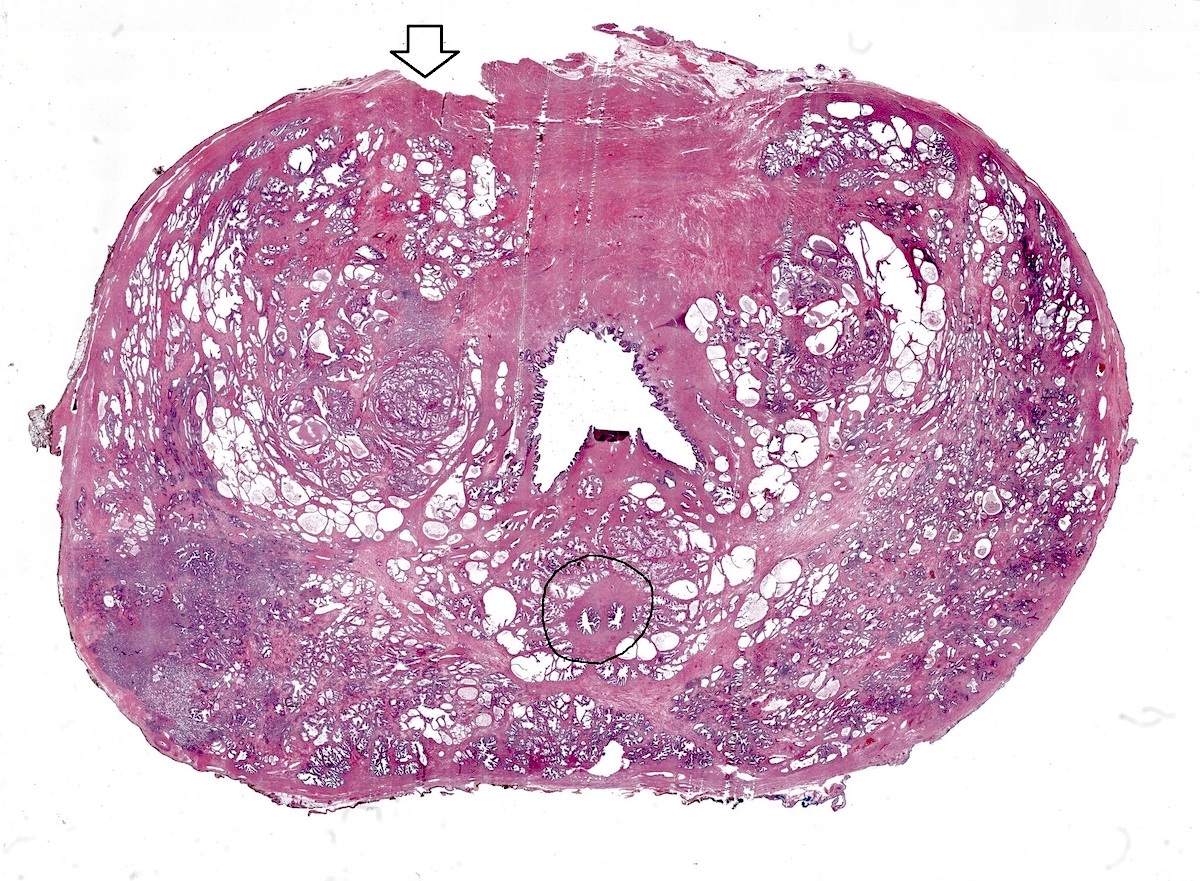
Superpage
Prostate gland & seminal vesicles


Copyright: 2003-2024, PathologyOutlines.com, Inc.

GU related: Jobs, Fellowships, Conferences, Cases, CME, Board Review

Related chapters: Bladder, Penis and scrotum, Testis

Editorial Board oversight: Bonnie Choy, M.D. (last reviewed November 2023)
Page views in 2023: 100
Page views in 2024 to date: 18

Aberrant p63 positivity
Definition / general
  • A small subset of prostatic acinar carcinoma is characterized by strong p63 nuclear immunoexpression; distinctive morphologic and molecular phenotypes have been described in these cases
Essential features
  • p63 protein is normally present in basal cells of benign acini but is absent in usual type acinar or ductal cancers
  • However, starting in 2008, a rare subset of cancers with diffuse p63 positivity has been described
  • Atrophic and basaloid phenotype has been noted on H&E slides
  • Immunostains for cytokeratin 34 beta E12 (negative) and AMACR / P504s (positive) can help resolve problematic foci
  • References: Am J Surg Pathol 2008;32:461, Am J Surg Pathol 2013;37:1401
ICD coding
  • ICD-O: 8140/3 - acinar adenocarcinoma
  • ICD-10: C61 - malignant neoplasm of prostate
  • ICD-11: 2C82.0 & XH4PB1 - adenocarcinoma of prostate & acinar adenocarcinoma of prostate
Epidemiology
Diagnosis
  • Immunostaining is performed with either the triple immunostain (high molecular weight cytokeratin, p63 and p504S) or with p63 alone
Laboratory
Prognostic factors
  • The first description of this entity was comprised of all organ confined cases (Am J Surg Pathol 2008;32:461)
  • A subsequent report described 76% as organ confined, with relatively low Gleason scores, although 38% were 3 + 5 = 8 (Am J Surg Pathol 2013;37:1401)
  • However, one case with metastasis has been reported (Cureus 2022;14:e22753)
    • Thus, most but not all cases seem to have favorable pathology
    • Long term outcome has not been studied yet
  • Parenthetically, p63 staining can assume predominant cytoplasmic reactivity in prostate cancer (unlike its strong nuclear staining in basal cells) but this is a separate matter
Case reports
Microscopic (histologic) description
  • The first series in 2008 by Osunkoya et al. described 21 cases on needle biopsy, including 8 with matched prostatectomy in which > 70% of cancer cells were p63 positive (Am J Surg Pathol 2008;32:461)
    • Distinctive, atrophic-like morphology was noted
  • 21 matched biopsy / prostatectomy cases were characterized (Am J Surg Pathol 2013;37:1401)
    • Besides atrophic-like change, salient features that have been described include a basaloid appearance and high N:C ratio, with multilayered and sometimes spindled nuclei
    • Notably, at prostatectomy, 86% of cases had coexisting usual type p63 negative adenocarcinoma while 14% were pure
Microscopic (histologic) images

Contributed by Kenneth A. Iczkowski, M.D.
Atrophic-like dilated cancer acini

Atrophic-like dilated cancer acini

Basaloid appearance

Basaloid appearance

Atrophic, mucinous cancer

Atrophic, mucinous cancer


Strong diffuse nuclear p63 expression Strong diffuse nuclear p63 expression

Strong diffuse nuclear p63 expression

Few p63 positive acini

Few p63 positive acini

Cytoplasmic p63

Cytoplasmic p63

Positive stains
Negative stains
Molecular / cytogenetics description
  • 37 p63 positive cancer cases were compiled to investigate the molecular phenotype (Mod Pathol 2015;28:446)
    • These tumors were almost all positive for ΔNp63 isoform by immunofluorescence and p63 mRNA by in situ hybridization
    • 100% of these tumors lacked ERG rearrangement at the molecular level by FISH and the protein level by immunostaining; moreover, 100% lacked PTEN loss
      • These 2 features are found in ~50% and 33% of usual acinar carcinomas, respectively
    • Tumors frequently express GSTP1 (14/19; higher than usual)
  • In one case report, 5 tumor foci in a prostatectomy were present, of which one was p63 positive; this focus did not show a TMPRSS::ERG translocation by FISH, while the other foci did (Int J Surg Pathol 2011;19:131)
  • ETS2 tumor suppressor gene is highly expressed in 95% (18/19) of p63 expressing prostatic carcinomas and benign prostate basal cells, with lower to undetectable expression in luminal cells and primary usual type adenocarcinomas (Prostate 2018;78:896)
Sample pathology report
  • Prostate, right mid, biopsy:
    • Prostatic adenocarcinoma, Gleason 3 + 4 (grade group 2) (see comment)
    • Comment: The tumor demonstrates a basaloid appearance with aberrant p63 nuclear reactivity. This is a rare subtype of prostatic adenocarcinoma and cancer is confirmed by lack of immunoreactivity for cytokeratin 34 beta E12.
Differential diagnosis
Board review style question #1

Which of the following are features of the rare type of prostatic carcinoma pictured above?

  1. Basaloid H&E appearance and negative for all luminal and basal markers
  2. Basaloid H&E appearance and positive for basal markers such as cytokeratin 34 beta E12, CK5/6, CK14 and CK15
  3. Basaloid H&E appearance and positive for luminal markers such as CK8, CK18, androgen receptor, NKX3.1, prostein and P504S
  4. Luminal H&E appearance and positive for basal markers such as cytokeratin 34 beta E12, CK5/6, CK14 and CK15
  5. Luminal H&E appearance and positive for luminal markers such as CK8, CK18, androgen receptor, NKX3.1, prostein and P504S
Board review style answer #1
C. Basaloid H&E appearance and positive for luminal markers such as CK8, CK18, androgen receptor, NKX3.1, prostein and P504S. The H&E appearance of these rare tumors is basaloid and atrophic but paradoxically they retain luminal marker expression and are negative for basal cell markers such as cytokeratin 34 beta E12.

Comment Here

Reference: Adenocarcinoma with aberrant p63
Board review style question #2

What is the p63 stain morphology shown above expected to display?

  1. Aberrant diffuse nuclear p63 positivity
  2. Lack of ERG rearrangement
  3. Lack of PTEN loss
  4. Positive reactivity for cytokeratin 34 beta E12
  5. Reduced cancer specific survival and high grade
Board review style answer #2
E. Reduced cancer specific survival and high grade. This cytoplasmic staining is not to be confused with diffuse nuclear p63, which is the topic of this section. The cytoplasmic staining is associated with a worse outcome. B, C and D are incorrect because usual type tumors (without nuclear p63) do not necessarily show lack of ERG rearrangement or lack of PTEN loss and should have loss of basal markers such as cytokeratin 34 beta E12.

Comment Here

Reference: Adenocarcinoma with aberrant p63

Abscess
Definition / general
  • Complication resulting from the acute infectious process of the prostate (prostatitis) characterized by a focal accumulation of purulent material within the prostate (BMC Urol 2016;16:38)
Essential features
  • Infection is caused mainly by gram negative rods
  • Neutrophils and necrotic debris fill prostatic ducts and acini
Terminology
  • Acute inflammatory disease of the prostate
ICD coding
  • ICD-10:
    • N41 - inflammatory diseases of prostate
      • N41.2 - abscess of prostate
  • ICD-11:
    • GA91 - inflammatory and other diseases of prostate
      • GA91.1 - abscess of prostate
Epidemiology
  • Incidence of prostatic abscess is 0.5% of all urologic diseases and 6% of all acute bacterial prostatitis
  • Mortality rate is 1 - 16%
  • Most commonly in fifth to sixth decade but can occur at any age
  • Older males > younger males
  • Prostatic abscess due to sexually transmitted organisms: younger males > older males (Int Braz J Urol 2017;43:835)
Sites
Pathophysiology
  • Most common way of developing is a reflux of infected urinary contents into prostatic ducts (Saudi J Kidney Dis Transpl 2011;22:298)
  • Risk factors: indwelling catheter, underlying voiding dysfunction, poorly controlled diabetes, end stage renal disease, cirrhosis, immunosuppression (Nat Rev Urol 2011;8:207)
  • Hematogenous dissemination from a distant primary (septic foci in respiratory, digestive, urinary tracts, skin and soft tissue) is rare (Arch Esp Urol 2011;64:62)
Etiology
Clinical features
  • Systemic (Urol Int 1992;48:358)
    • Headache, fever, chills and general malaise, low back pain
  • Prostate (local) (Korean J Urol 2012;53:860)
    • Severely tender prostate with areas of fluctuation on digital rectal examination
    • Perineal pain
    • Dysuria, urinary urgency or frequency, hematuria, purulent urethral discharge
    • Obstructive urinary symptoms
Diagnosis
  • History and physical findings
  • Complete blood count with differential, urinalysis
  • Imaging with transrectal ultrasound (the diagnostic method of choice), CT, MRI (rarely used) (Korean J Urol 2012;53:860)
  • Blood culture, urine culture
  • PCR (detection of sexually transmitted organisms)
Laboratory
  • > 10 white blood cells per high power field and pyuria
  • Bacterial culture and antibiotic susceptibility identification
  • DNA sequencing and nucleic acid amplification by PCR
  • Can raise the serum prostate specific antigen (PSA) above normal (Hinyokika Kiyo 1993;39:445)
Radiology description
  • Transrectal ultrasound (TRUS) (Korean J Urol 2012;53:860)
    • 1 or more hypoechoic areas with well defined walls
    • Thick pus primarily in the transition zone and in the central zone of the prostate
  • Computed tomography (CT scan) of the abdomen and pelvis (AJR Am J Roentgenol 1987;148:899)
    • Can better delineate the spread of infection to adjacent organs
  • Magnetic resonance imaging (MRI) (Indian J Radiol Imaging 2011;21:46)
    • Hypointense signal on T1 and hyperintense on T2 image
Radiology images

Images hosted on other servers:

Abdominal
ultrasound showing
prostatic abscess

TRUS showing hypoechoic lesion

Pelvic CT showing an abscess in the prostate

Prognostic factors
  • Depends on the timely diagnosis and treatment
  • Also depends on underlying medical conditions
  • Good prognosis if recognized and treated early
  • Poor prognostic factors: age > 65 years, fever > 100.4°F, benign prostatic hypertrophy, chronic catheterization (BMC Urol 2016;16:38)
Case reports
Treatment
Clinical images

Images hosted on other servers:

Transurethral unroofing of a prostatic abscess

Abscess drainage with transurethral resection

Microscopic (histologic) description
Microscopic (histologic) images

Contributed by Kenneth A. Iczkowski, M.D.
Prostatic microabscess Prostatic microabscess

Prostatic microabscess

Cytology description
Positive stains
Sample pathology report
  • Prostate, biopsy:
    • Prostatic abscess (see comment)
    • Comment: Numerous neutrophilic infiltrates in lumen spaces and around glands consistent with acute inflammation. Some glands are partly damaged by microabscesses. Gram negative rods are noticed on Gram stain.
Differential diagnosis
  • Acute bacterial prostatitis:
    • Clinically and histologically similar (neutrophils are mostly in lumens and epithelium)
    • No fluctuant mass during a rectal examination
    • No hypoechoic areas on ultrasound
  • Chronic bacterial prostatitis:
    • Similar clinical symptoms but lower intensity
    • Predominant lymphocytic infiltration (lymphocytes are mostly in the stroma)
  • Granulomatous prostatitis:
    • Fever and chills are common
    • Irritative voiding symptoms of urgency, frequency
    • May have history of bladder cancer Bacillus Calmette Guerin (BCG) treatment
    • Necrotizing or nonnecrotizing granulomas (cohesive clusters of histiocytes, usually with admixed lymphocytes, eosinophils, neutrophils)
  • Prostate carcinoma:
    • May mimic benign acini with reactive inflammatory atypia
    • Typically lack background inflammation
    • Small glands, sometimes medium to large glands, papillary or cribriform glands or solid growth, single cells or necrosis
    • Nucleomegaly, prominent and multiple nucleoli, lack basal cell layer

Differential diagnosis Histological changes Clinical presentation
Prostatic abscess Neutrophilic infiltrate with glands destruction; abscess formation; neutrophils fill prostatic ducts and acini Severely tender prostate with areas of fluctuation on digital rectal examination
Acute bacterial prostatitis Predominant neutrophilic infiltrates mostly in lumens and epithelium; no abscess formation Severely tender prostate without areas of fluctuation on digital rectal examination
Chronic bacterial prostatitis Predominant lymphocytic infiltrates mostly in the stroma Mildly tender prostate without areas of fluctuation on digital rectal examination
Granulomatous prostatitis Necrotizing or nonnecrotizing granulomas (cohesive clusters of histiocytes, usually with admixed lymphocytes, eosinophils, neutrophils with or without necrosis in the center) Mildly tender prostate on digital rectal examination; irritative voiding symptoms of urgency, frequency
Prostate carcinoma Typically lack background inflammation; small glands with papillary or cribriform or solid growth or single cells Firm/nodular, nontender prostate on digital rectal examination; blood in the urine
Board review style question #1

A 64 year old man comes to the clinic with the complaint of severe dysuria for the past week. Just a week before the beginning of these symptoms, the patient was treated with a short course of antibiotics for a urinary tract infection. The patient also complains of burning micturition for a week. He has a history of benign prostatic hypertrophy, causing urinary retention, sometimes needing catheterization in the past. On admission, he has a high grade fever. Rectal examination reveals an enlarged prostate that is tender to palpation. Transrectal ultrasound suggests a 2.3 cm collection in the prostate. What histological finding is most common for this entity?

  1. Eosinophilic infiltration
  2. Lymphocytic infiltration
  3. Microabscesses with neutrophils
  4. Necrotizing granulomas
  5. Nonnecrotizing granulomas
Board review style answer #1
C. Microabscesses with neutrophils. Acute inflammation of the prostate with abscess formation due to bacterial infection can show neutrophilic infiltrates in lumen spaces and in epithelium, consistent with gland destruction and microabscesses.

Comment Here

Reference: Abscess

Adenocarcinoma
Definition / general
  • Most common malignancy of the prostate gland
  • Originates from prostatic secretory epithelium
Essential features
  • Clinical and radiological features neither sensitive nor specific
  • Often diagnosed by nontargeted needle biopsies investigating raised serum prostate specific antigen (PSA)
  • Absence of basal cell layer is a pathognomonic histological feature
  • Pathognomonic diagnostic features: circumferential perineural invasion, glomerulations and collagenous micronodules (mucinous fibroplasia)
  • Other histological features: infiltrative architecture, nucleolar prominence, amphophilic cytoplasm and some intraluminal contents (crystalloids, blue mucin, pink amorphous material)
Terminology
  • Prostate cancer
  • Prostate adenocarcinoma
  • Subtypes of prostatic adenocarcinoma: acinar adenocarcinoma, ductal adenocarcinoma, atrophic adenocarcinoma, pseudohyperplastic adenocarcinoma, microcystic adenocarcinoma, foamy gland adenocarcinoma, mucinous adenocarcinoma, signet ring variant of adenocarcinoma, pleomorphic giant cell adenocarcinoma, sarcomatoid adenocarcinoma
ICD coding
  • ICD-O: C61.9 - prostate, NOS
  • ICD-10: C61 - malignant neoplasm of the prostate
Epidemiology
Sites
  • Most tumors are multifocal (Eur Urol 2019;75:498)
  • 75 - 80% are posterior / posterolateral peripheral zone
  • Approximately 13 - 20% are in transition (periurethral) zone (Prostate 1997;30:130, Urology 1994;43:11)
  • Most clinically significant cancers arise in the peripheral zone that is sampled by needle biopsies
  • Transition zone prostate cancer is associated with favorable pathologic features and better recurrence free survival (Prostate 2015;75:183)
  • Less frequently involved anterior prostate most likely due to inadequate sampling using standard biopsy approach (Histopathology 2012;60:142)
Pathophysiology
Etiology
Clinical features
  • Generally asymptomatic unless locally advanced or metastatic
  • Often discovered following investigation of nonspecific lower urinary tract symptoms
  • Digital rectal examination (DRE): prostate may feel normal or may be enlarged / asymmetrical / hard / have a palpable nodule present
Diagnosis
  • Generally diagnosed by systematic transrectal ultrasound guided prostate biopsies
  • Transperineal needle biopsies increasingly used as associated with lower risk of infection
  • Prebiopsy MRI followed by systematic biopsies supplemented with targeted biopsies from any radiological abnormality leads to better identification of clinically significant prostate cancer than systematic prostate biopsy alone (Lancet 2017;389:815)
  • Incidental prostate cancer sometimes diagnosed in transurethral resections
  • Immunohistochemistry with basal cell markers (HMWCK, p63) and AMACR used to establish the diagnosis in equivocal cases
Laboratory
  • Raised serum PSA
  • Different PSA cutoffs have been used to prompt prostate needle biopsy
  • Age specific cutoffs, PSA velocity (rate of change in PSA over time) and PSA density (PSA per unit prostate volume - ng/mL/cc) may increase sensitivity and specificity of PSA testing (NCCN Guidelines: Prostate Cancer Early Detection [Accessed 10 December 2021])
  • U.S. Preventative Services Task Force (USPSTF) recommends against PSA based screening for prostate cancer in men 70 years and older
  • American Urological Association (AUA) does not recommend PSA screening in men under age 40 years or in men aged 40 - 54 years at average risk
    • For men age 55 - 59 years, shared decision making is desirable
    • For men aged 70 years and over or men with < 10 - 15 year life expectancy, PSA screening is not recommended (J Urol 2013;190:419)
  • Potential urine biomarker for prostate cancer is PCA3 (Adv Anat Pathol 2020;27:11)
Radiology description
  • Ultrasound scan (USS) generally used to guide prostate biopsies; prostate cancer may appear hypoechoic but USS neither sensitive nor specific
  • Multiparametric MRI commonly used for local tumor staging; may also be used to identify abnormalities for targeting at biopsy
  • MRI abnormalities generally reported using either PI-RADS (Prostate Imaging - Reporting and Data System) or Likert score
  • CT scan used to identify metastatic disease in lymph nodes
  • Bone scan used to detect bony metastases
  • PET scan used to detect micrometastatic disease in selected patients, such as men with raised PSA levels after treatment
Prognostic factors
  • Biopsy: tumor extent (mm or percentage core involvement), grade (Gleason score and grade group), perineural invasion, extraprostatic extension
  • Radical prostatectomy: tumor size, Gleason score and grade group, stage, margin status
  • Cribriform morphology and intraductal carcinoma associated with invasive prostate cancer are adverse prognostic indicators (Transl Androl Urol 2018;7:145)
  • Small cell carcinoma component is associated with aggressive behavior and treated differently
  • Some expert groups recommend incorporating intraductal component into the Gleason score while others recommend reporting it separately in a comment (Am J Surg Pathol 2020;44:e87, Arch Pathol Lab Med 2021;145:461, Histopathology 2021;78:231)
Case reports
Treatment
  • Preoperative risk stratification based on serum PSA, clinical stage, biopsy parameters (tumor extent, grade, cribriform morphology, intraductal carcinoma, perineural invasion)
  • Primary treatment options based on preoperative risk stratification:
    • Active surveillance
    • Focal therapy (cryotherapy, high intensity ultrasound)
    • Radical prostatectomy
    • Brachytherapy
    • External beam radiotherapy
    • Hormone therapy (e.g., luteinizing hormone releasing hormone [LHRH] analogues, antiandrogens)
    • Orchidectomy (rare in contemporary practice)
    • Chemotherapy (for metastatic disease)
  • Postprostatectomy options:
    • Generally, PSA monitoring and early salvage therapy if rising serum PSA
    • Less commonly adjuvant therapy for high stage disease or margin positivity
Gross description
  • Often grossly inapparent
  • May form a cream mass
Gross images

Contributed by Debra Zynger, M.D. and Kenneth A. Iczkowski, M.D.
Missing Image

Prostatectomy specimen

Missing Image Missing Image

Extraprostatic extension (pT3a)

Missing Image Missing Image

Seminal vesicle invasion (pT3b)

Anterior horn of
peripheral zone

Microscopic (histologic) description
  • Gleason grading is based on the architecture of the tumor
  • Gleason grades represent a morphological spectrum from well formed glands (pattern 3) to increasingly complicated glandular proliferations (pattern 4) to almost no glandular differentiation (pattern 5) (Diagnostic Histopathology 2019;25:371)
  • Glandular crowding and infiltrative growth pattern
  • Nuclear enlargement, nucleolar prominence
  • Round generally monomorphic nuclei
  • Amphophilic cytoplasm
  • Mitoses
  • Apoptotic bodies
  • Stromal desmoplasia
  • Intraluminal contents: crystalloids, pink amorphous secretions, blue mucin
  • Glomerulations, collagenous micronodules (mucinous fibroplasia)
  • Absence of basal cell layer (generally requires immunohistochemical confirmation)
  • Reference: Cold Spring Harb Perspect Med 2017;7:a030411
Microscopic (histologic) images

Contributed by Murali Varma, M.B.B.S.
Infiltrative growth pattern

Infiltrative growth pattern

Glomerulations

Glomerulations

Prominent nucleoli

Prominent nucleoli

Perineural invasion

Perineural invasion

Atrophic variant of prostate cancer Atrophic variant of prostate cancer

Atrophic variant of prostate cancer


Atrophic variant of prostate cancer Atrophic variant of prostate cancer

Atrophic variant of prostate cancer

Pseudohyperplastic variant of prostate cancer Pseudohyperplastic variant of prostate cancer

Pseudohyperplastic variant of prostate cancer

Foamy gland variant of prostate cancer

Foamy gland variant of prostate cancer

Crystals in gland lumens

Crystals in gland lumens


Pink amorphous material within gland lumens Pink amorphous material within gland lumens

Pink amorphous material within gland lumens

Amphophilic cytoplasm

Amphophilic cytoplasm

Mitosis

Mitosis

Collagenous micronodules

Collagenous micronodules

Virtual slides

Images hosted on other servers:

Prostate adenocarcinoma Gleason 4+3=7

Prostate adenocarcinoma Gleason 4+4=8

Prostate ductal adenocarcinoma

Cytology description
  • Urine cytology for detecting prostate cancer has a very low sensitivity (Prostate Cancer Prostatic Dis 2019;22:362)
  • Urine cytology is not used clinically in the diagnosis of prostate cancer
  • FNA of metastatic prostate cancer to a lymph node may show microacinar complexes / cell clusters / single cells with fragile cytoplasm and prominent nucleoli (Diagn Cytopathol 2007;35:565)
Positive stains
Negative stains
Molecular / cytogenetics description
  • Prostate cancer is a heritable disease
  • Family history of a first degree relative with prostate cancer increases the risk of developing prostate cancer by 2 fold (Nat Rev Urol 2014;11:18)
  • 30 - 40% of familial risk is due to genetic factors (Adv Anat Pathol 2020;27:11)
  • Genetic factors include highly penetrable rare variants and more common low to moderate risk variants (Adv Anat Pathol 2020;27:11)
  • Highly penetrant variants occur in BRCA2 and HOXB13
  • Over 280 SNPs have been identified as prostate cancer risk factors (Adv Anat Pathol 2020;27:11)
  • For most SNPs, the molecular mechanism of cancer association is generally unknown, as they occur in noncoding regions of the genome (Adv Anat Pathol 2020;27:11)
  • Somatic mutations occur in genes such as ERG, ETV1/4, FLI1, SPOP, FOXA1, IDH1, PTEN, TP53, MYC, CDH1 (Cell 2015;163:1011, Adv Anat Pathol 2020;27:11)
  • Most common somatic genomic rearrangement is fusion of the androgen regulated gene TMPRSS2 with a member of the ETS transcription family (Adv Anat Pathol 2020;27:11)
  • Somatic mutation profiles of prostate cancer are associated with clinical and pathological outcomes
    • There are 7 major subtypes, which are defined by either specific gene fusions of ETS transcription family members (ERG, ETV1, ETV4 and FLI1) or mutations (SPOP, FOXA1, IDH1) (Oncotarget 2018;9:14723)
  • Different subtypes have different molecular profiles, for example (Oncotarget 2018;9:14723):
    • ETS subset (59% of cases) are enriched in PTEN mutations
    • SPOP mutant subset (11%) of cases have distinct somatic copy number alteration profiles, including deletions of CHD1, 6q and 2q
Sample pathology report
  • Prostate core biopsies:
    • Acinar adenocarcinoma (see comment)
    • Comment:
      • Number of cores involved:
        • Right 1/6 cores
        • Location(s): right apex
        • Left 0/6 cores
        • Total number of cores involved: 1/12
      • Greatest length of cancer in a core: 5 mm (40%)
      • No evidence of perineural invasion or extraprostatic extension
      • Gleason score: 3+4=7 (10% pattern 4, no cribriform morphology)
      • Grade group 2
  • Radical prostatectomy:
    • Histological tumor type: acinar adenocarcinoma
    • Gleason score:
      • Primary Gleason grade: 3
      • Secondary Gleason grade: 3
      • Tertiary Gleason grade (< 5%): not applicable
      • Gleason score: 3+3=6
      • Grade group: 1
    • Location of dominant tumor: right apex
    • Extraprostatic extension: not identified
    • Bladder neck: not involved
    • Seminal vessels: not involved
    • Margin status: not involved
    • Lymphovascular invasion: not identified
    • Regional lymph node status:
      • Number of nodes examined: 9
      • Number of positive lymph nodes: 0
    • Primary tumor: pT2 pN0
Differential diagnosis
Board review style question #1

A 74 year old man with urinary hesitancy was found to have a PSA of 8 ng/mL. He had a transrectal ultrasound (TRUS) and prostate biopsy. A representative image is above. Which of the following is the typical immunoprofile of such a tumor?

  1. CK7- CK20- NKX3.1-
  2. CK7- CK20- NKX3.1+
  3. CK7- CK20+ NKX3.1-
  4. CK7+ CK20- NKX3.1-
  5. CK7+ CK20+ NKX3.1+
Board review style answer #1
B. CK7- CK20- NKX3.1+

Comment Here

Reference: Prostate adenocarcinoma
Board review style question #2

A prostate biopsy shows this tumor. What is its grade?

  1. Gleason score 2+3, grade group 1
  2. Gleason score 3+2, grade group 1
  3. Gleason score 3+3, grade group 1
  4. Gleason score 3+3, grade group 2
  5. Gleason score 3+4, grade group 2
Board review style answer #2
C. Gleason 3+3, grade group 1

Comment Here

Reference: Prostate adenocarcinoma

Adenocarcinoma
Definition / general
  • Very rare, must confirm microscopically
  • Primary tumors should be localized primarily to seminal vesicle; must rule out invasion from prostate (do PSA / PAP), rectum or other sites
  • Usually a papillary adenocarcinoma resembling architecture of normal seminal vesicle
  • Resembles prostatic duct adenocarcinoma Gleason patterns 3 or 4 or mucinous (colloid) carcinoma
  • Usually unresectable and patients die within 2 years
  • Adenocarcinoma: very rare, must rule out prostate primary; often mucin producing, ulcerates through skin of scrotum
Case reports
Gross images

Images hosted on other servers:

Low grade epithelial stromal tumor

Microscopic (histologic) images

Images hosted on other servers:

Metastatic hepatocellular carcinoma

Low grade epithelial stromal tumor

Positive stains
Negative stains
Differential diagnosis

Adenoid cystic (basal cell) carcinoma
Definition / general
  • Also known as adenoid basal, adenoid cystic-like tumor, adenoid basal proliferation of uncertain significance
  • Resembles to some extent the salivary gland tumor
  • Usually indolent; cases with aggressive behavior often have large solid nests with central necrosis, high Ki67%, less staining with basal cell markers (Am J Surg Pathol 2007;31:697)
Case reports
Microscopic (histologic) description
  • Expansive (not circumscribed) growth pattern
  • Multinodular, basaloid cells in clusters, some with punched out lumens
  • Surrounding fibromyxoid stroma, squamous differentiation, merges with basal cell hyperplasia
  • May be associated with minor component of usual type prostatic adenocarcinoma
  • Considered malignant if extensive infiltration between normal prostate gland, extension out of the prostate, perineural invasion, necrosis or metastases
  • Gleason grading of these tumors is not recommended
Microscopic (histologic) images

Contributed by Daniel Athanazio, M.D., Ph.D. and Maiara Souza, M.D.

Solid growth - basaloid morphology

Infiltrative growth

Basaloid morphology

Combination of basaloid and luminal cells


BCL2

p63

PSA

Synaptophysin

Positive stains
Negative stains

Adenosis / atypical adenomatous hyperplasia
Definition / general
Essential features
  • Benign lobular proliferation of small crowded glands
  • Similar to prostatic adenocarcinoma; in adenosis, cells may show small or medium size nucleoli, crystalloids, intraluminal secretions, minimal infiltration and positive immunostaining for AMACR (racemase)
  • In contrast to prostatic adenocarcinoma, in adenosis, glands have more pale cytoplasm, merge with adjacent benign glands, commonly have corpora amylacea (Am J Surg Pathol 1994;18:863, Surg Pathol Clin 2008;1:1)
  • Basal cells can be identified on H&E slides or with immunohistochemistry for p63, cytokeratin 5/6 or HMWCK (Pathology 2013;45:251)
  • AMACR (racemase) can be focally or diffusely expressed in up to 18% of cases of adenosis (Am J Surg Pathol 2002;26:921)
  • Lack of ERG expression in adenosis supports the notion that it is not a precursor lesion of adenocarcinoma (Hum Pathol 2013;44:1895)
Terminology
ICD coding
  • ICD-10: N40.2 - nodular prostate without lower urinary tract symptoms
Epidemiology
  • 2 - 20% of transurethral resections of the prostate, 1% of prostate needle biopsies
Sites
Pathophysiology
Clinical features
  • Morphologic variant of benign crowded glands, a mimicker of low grade prostatic adenocarcinoma
  • Does not have clinical implications
Diagnosis
  • Detected on needle biopsy or transurethral resection
Radiology description
  • Multiparametric magnetic resonance imaging (mpMRI) may be difficult to interpret because it shows overlapping features with low grade prostatic adenocarcinoma (Eur Radiol 2017;27:2095)
Prognostic factors
  • Not a precursor lesion of prostatic adenocarcinoma (Hum Pathol 2013;44:1895)
  • Comparative genomic hybridization and multiplex PCR did not find common alterations between adenosis and accompanying cancer foci and concluded that adenosis should not be considered as an obligate premalignant lesion (Int J Oncol 2005;26:267)
  • Diffuse adenosis of the peripheral zone should be considered a risk factor for prostate cancer (Am J Surg Pathol 2008;32:1360)
Case reports
Treatment
  • Treatment not needed
Microscopic (histologic) description
Microscopic (histologic) images

Contributed by Cristina Magi-Galluzzi, M.D., Ph.D.
Well circumscribed nodule

Well circumscribed nodule

Mixed small and large acini

Mixed small and large acini

Basal cells

Basal cells

Crowded glands

Crowded glands

Bland nuclear features

Bland nuclear features

Fragmented basal cells

Fragmented basal cells

Positive stains
  • Basal cell markers (p63, CK5/6 and HMWCK) are positive in a patchy fashion (discontinuous staining; mixture of glands with and without basal cell layer) (Pathology 2013;45:251)
Negative stains
Molecular / cytogenetics description
Sample pathology report
  • It is not recommended to report adenosis
  • Prostate, needle biopsy or transurethral resection:
    • Benign prostatic tissue
Differential diagnosis
  • Low grade (Gleason score 3 + 3 = 6, grade group 1) prostatic adenocarcinoma:
    • Cytologic atypia, including prominent nucleoli and lack of basal cells
  • Partial atrophy:
    • Glands are partially atrophic with undulating luminal surface with papillary infolding; cytologic features are benign
Board review style question #1

Which of the following statements is true about the lesion depicted above?

  1. Atypical small acinar proliferation, favor benign
  2. Atypical small acinar proliferation, suspicious for malignancy
  3. Benign small acinar proliferation
  4. Prostatic carcinoma, Gleason score 3 + 3 = 6
Board review style answer #1
C. Benign small acinar proliferation. This is an example of adenosis (atypical adenomatous hyperplasia) with small acini lacking cytologic atypia and retaining basal cells.

Comment Here

Reference: Adenosis / atypical adenomatous hyperplasia
Board review style question #2
Which of the following statements is true about adenosis?

  1. A fragmented and discontinuous basal cell layer is present
  2. AMACR is always negative
  3. It is a precursor lesion of prostatic adenocarcinoma
  4. More commonly in the peripheral zone
Board review style answer #2
A. A fragmented and discontinuous basal cell layer is present. Adenosis more commonly involves the transition zone; AMACR can be focally or diffusely expressed in up to 18% of cases of adenosis. Adenosis is not a precursor lesion of prostatic adenocarcinoma.

Comment Here

Reference: Adenosis / atypical adenomatous hyperplasia

Adenosquamous carcinoma
Definition / general
  • Adenosquamous carcinoma of the prostate is characterized by presence of both glandular / acinar and squamous components
Essential features
  • Adenosquamous prostatic carcinoma is a very rare aggressive tumor
  • Critical to exclude other sources of the squamous component through clinical history, examinations, endoscopy and immunostains
  • 67% of cases are associated with prior androgen deprivation therapy or radiotherapy
  • Diagnosis requires proper morphology and immunostaining with prostate specific markers (prostate specific antigen [PSA], prostate specific membrane antigen [PSMA], prostatic specific acid phosphatase [PSAP] and prostatic acid phosphatase [PAP]), which should be positive in the glandular and negative in the squamous component
ICD coding
  • ICD-O: C61.9 - prostate, NOS
  • ICD-10: C61 - malignant neoplasm of the prostate
Epidemiology
Sites
  • Prostate, often with extraprostatic extension and seminal vesicle involvement
Pathophysiology
  • Theories
    • Metaplastic transformation of adenocarcinoma frequently after treatment; this is supported by a case of adenosquamous carcinoma in which the squamous component rose from 5% before adjuvant radiotherapy to predominantly squamous after radiotherapy (Eur J Cancer 2018;95:109)
    • Collision tumor: intermingling of squamous and glandular components with no abrupt transition argues against this, as does the observation that squamous component may be PSAP reactive (Hum Pathol 1984;15:87, Int J Urol 2005;12:319, Scientific World Journal 2006;6:2491)
    • Arises from pluripotent stem cells capable of multidirectional differentiation (Urol Case Rep 2019;29:101084)
Etiology
  • Often association with status postandrogen ablation or prostatic radiotherapy
Clinical features
Diagnosis
  • Histologic findings are established on transurethral resection (consistent with frequent obstructive uropathy) or biopsy
  • Imaging cannot distinguish it from generic prostate cancer
Laboratory
Radiology description
Radiology images

Images hosted on other servers:
Tumor abutting anorectal canal

Tumor abutting anorectal canal

Prognostic factors
  • 30% 5 year survival (Rare Tumors 2010;2:e47)
  • For patients undergoing prostatectomy it is 63%, whereas for those not undergoing prostatectomy 1 year survival is 39%
Case reports
Treatment
Microscopic (histologic) description
  • In addition to the usual glandular prostate cancer component, there is a component of cells with eosinophilic cytoplasm
  • Keratin pearls and intercellular bridges are required to diagnose squamous cell carcinoma
  • Additional sarcomatoid transformation has been rarely reported (Histopathology 2020;77:890, Scientific World Journal 2006;6:2491)
Microscopic (histologic) images

Contributed by Susan Prendeville, M.D. and Gladell Paner, M.D.
Biopsy, prominent squamous areas

Biopsy, prominent squamous areas

Same, with obvious keratin

With obvious keratin

Mixed components, postradiation setting

Mixed components, postradiation setting

Transition, squamous to glandular

Transition, squamous to glandular

Negative stains
Molecular / cytogenetics description
Sample pathology report
  • Prostate, left apex, biopsies:
    • Prostatic adenosquamous carcinoma, Gleason 4 + 4 (score = 8) (see comment)
    • Comment: 30% of the tumor is a squamous component with cells showing intracellular and extracellular keratin and intercellular bridges. This finding correlates with the documented antiandrogen dosing the patient underwent after diagnosis of a Gleason 3+3 carcinoma 4 years ago. Adenosquamous carcinoma is an aggressive tumor.
Differential diagnosis
  • Secondary involvement by squamous neoplasms arising in bladder, urethra or anorectal region:
    • Clinical endoscopic findings negative in these sites
    • Normal serum PSA
    • Limited use for immunostains
  • Pure squamous carcinoma:
    • Absence of a contiguous or separate focus of glandular cancer in the specimen
  • Admixed prostate cancer with urothelial neoplasms arising in bladder or urethra:
    • Keratin pearls and intercellular bridges are required to diagnose squamous cell carcinoma
    • If these findings are absent and if GATA3 or uroplakin II staining are present, the lesion is urothelial and not prostatic
  • Basal cell hyperplasia with squamous features (Hum Pathol 2005;36:531):
    • Lack of atypia of squamous cells
    • Absent invasive glandular component
    • Cytologic features of basal cells including nuclear vacuoles
Board review style question #1

Which of the following findings would exclude a definite diagnosis of prostatic adenosquamous carcinoma in a biopsy?

  1. Close intermingling of squamous and glandular tumor components
  2. Lack of a history of hormone deprivation or radiotherapy for prostate cancer
  3. Prior history of muscle invasive urothelial carcinoma with squamous features
  4. PSA staining usually negative in the squamous component
  5. Staining of the squamous tumor with cytokeratin 34 beta E12
Board review style answer #1
C. Prior history of muscle invasive urothelial carcinoma with squamous features. Prior history of a squamous cancer in the vicinity of the prostate is critical. A urothelial carcinoma, including that with squamous features, can easily invade the prostate. Answer D is incorrect because indeed the squamous component does lose reactivity for PSA in most adenosquamous tumors. Answer E is incorrect because the squamous component of adenosquamous carcinoma should stain with cytokeratin 34 beta E12. Answer B is incorrect because while many cases of adenosquamous carcinoma do have an association with hormonal or radiotherapy, quite a few reported cases do not. Answer A is incorrect because indeed there often is close intermingling of squamous and glandular components of the tumor.

Comment Here

Reference: Adenosquamous carcinoma

Amyloid
Definition / general
  • Primary amyloidosis of the prostate is a rare disease
  • Involves seminal vesicles in about 10% of radical prostatectomies, usually represents a localized form
  • Amyloidosis develops subepithelially spreading to include the wall of seminal vesicles and ejaculatory ducts; appears to be related to advanced age (Ann Diagn Pathol 2008;12:235)
  • Note: corpora amylacea may stain positive with Congo red
Essential features
  • Pale amorphous hyaline, eosinophilic substance that accumulates and can pressure the adjacent epithelium
  • Often displays processing cracks
  • More common in seminal vesicles and vas deferens
  • Subepithelial and vascular deposits
Epidemiology
  • Occurs in 2-10% of radical prostatectomies (Turk Patoloji Derg 2012;28:44)
  • Incidence increases with age, reaching 21% in men age 75 years and older (Histopathology 1993;22:173, Am J Pathol 1983;110:64)
  • Vascular amyloid deposits are present in 2% - 10% of prostates with nodular hyperplasia or adenocarcinoma
  • Higher incidence of amyloid deposits in patients with myeloma, primary amyloidosis of kidney or chronic diseases
  • Amyloidosis of the seminal vesicles involves 10% of radical prostatectomy specimens
Sites
  • More common in seminal vesicles and vas deferens
  • Deposits are more commonly subepithelial and vascular
Etiology
  • Although immunohistochemistry often detects lactoferrin (Ann Pathol 2004;24:236), amyloid apparently derives from semenogelin I, the major secretory product of the seminal vesicles (J Lab Clin Med 2005;145:187)
  • Semenogelin I and II are mainly responsible for immediate gel formation of freshly ejaculated semen, and are degraded by the proteolytic action of prostate specific antigen/PSA (J Androl 1996;17:17)
Pathophysiology
  • Abnormal folding of proteins that deposit as fibrils in the extracellular tissue and may accumulate preventing normal function
  • Amyloidosis includes multiple biochemically distinct proteins but with similar morphologic appearance
  • Different forms of amyloidosis include:
    • Primary systemic amyloidosis (no evidence of preceding or coexisting disease, paraproteinemia or plasma cell neoplasia)
    • Amyloidosis associated with multiple myeloma
    • Secondary to coexisting previous chronic inflammatory or infectious conditions, hemodialysis
    • Localized form
Clinical features
  • Most commonly asymptomatic
  • Can simulate prostate or bladder cancer invasion of seminal vesicles on MRI
Diagnosis
  • Histology: amorphous pale eosinophilic material often with cracks from processing
  • Histochemical stain with Congo red shows green birefringence on polarized microscopy
Radiology description
  • Can simulate prostate or bladder cancer invasion of seminal vesicles on MRI
Case reports
Treatment
  • Based on the underlying condition
Gross description
  • Usually not seen grossly
  • When involvement is massive, the organ can be enlarged and firm and cut section could show a waxy appearance
Microscopic (histologic) description
  • Pale amorphous hyaline, eosinophilic substance that accumulates and can pressure the adjacent epithelium
  • Often displays processing cracks
  • Subepithelial location
  • Can compress the adjacent epithelium
Microscopic (histologic) images

Contributed by Andres Matoso, M.D., @katcollmd on Twitter and Case #85
Missing Image

Various images

Amyloid Amyloid

Amyloid

Amyloid Amyloid

Amyloid


66 year old man with radical prostatectomy for adenocarcinoma

Trichrome stain

Positive stains
  • Trichrome (stains amyloid dusky gray), Congo Red, immunohistochemistry for specific amyloid forms
Electron microscopy description
  • Electron microscopy shows nonbranching amyloid fibrils that measure 7.5 to 10 nm (Mod Pathol 1989;2:671)
Differential diagnosis
  • It is important to exclude an underlying etiology including plasma cell neoplasia or an inflammatory condition

Anatomy & histology-Cowper glands
Definition / general
  • Cowper glands are also called bulbourethral glands because they are located surrounding the bulbar portion of the urethra, outside of the prostate
Epidemiology
  • Most commonly, they can be found in TURP specimens, but occasionally they may be seen in needle biopsies or in biopsies from urethral stricture
Sites
  • Bulbar urethra
Pathophysiology
  • They are the homologous of Bartholin glands in women
  • They secrete clear fluid prior to ejaculation that helps to lubricate the urethra and a small proportion of the fluid of the ejaculate
Microscopic (histologic) description
  • They are composed of mucinous acini arranged in a lobular pattern with intercalated ducts
  • When present in needle biopsies, they may resemble foamy gland prostate adenocarcinoma
  • They are surrounded by skeletal muscle, further mimicking cancer
  • They can be positive for prostate markers PSA, PSAP and NKX3.1
  • The correct diagnosis lies in recognizing the dual population of acini and ducts surrounded by skeletal muscle as opposed to benign prostatic glands
  • Immunohistochemistry for high molecular weight cytokeratin is positive in basal cells and can help in the differential diagnosis with prostate cancer
Microscopic (histologic) images

Contributed by Andres Matoso, M.D. and @ThatGlassTho on Twitter
Missing Image

Low power of Cowper glands

Missing Image

Glands and skeletal muscle

Missing Image

Mucicarmin+

Missing Image

PAS+

Missing Image

NKX3.1+


Anatomy & histology-Cowper glands Anatomy & histology-Cowper glands

Anatomy & histology-Cowper glands

Positive stains
  • By immunohistochemistry, Cowper glands can be positive for prostate markers PSA, PSAP and NKX3.1
  • Special stains mucicarmin and PAS are also positive
Differential diagnosis
Additional references

Anatomy & histology-paraganglion
Definition / general
  • Group of neuroendocrine cells derived from neural crest
Epidemiology
  • Usually located in posterolateral soft tissue outside of prostate
  • Seen in radical prostatectomy specimens and rarely in prostate needle biopsies, or in smooth muscle of bladder neck in transurethral resections of prostate or bladder
Microscopic (histologic) description
  • Group of cells with clear, vacuolated or amphophilic cytoplasm with a delicate blood vessel meshwork
  • Nuclei may show prominent nucleoli or degenerative endocrine type atypia
Microscopic (histologic) images

Contributed by Andres Matoso, M.D.
Missing Image

Chromogranin A+

Differential diagnosis
  • The main differential diagnosis is with Gleason pattern 5 prostate cancer
    • The degenerative type atypia in paraganglions is helpful when present; also absence of surrounding prostate glands
    • Immunohistochemistry for prostate markers and cytokeratin is helpful

Anatomy & histology-prostate
Definition / general
  • Knowledge of the normal prostate morphology can help with orientation and avoid confusion of these findings with cancer
Essential features
  • Benign hyperplasia expands the transition zone and obstructs the bladder outlet; cancer occurs mostly in the peripheral zone where it does not obstruct the bladder outlet
  • Secretory cells and basal cells are the main 2 layers of benign acini (glands) and the loss of basal cells is a key diagnostic feature of prostate cancer; other cell types in the epithelium include neuroendocrine cells and lymphoid cells
  • Prostate specific antigen is the most useful marker of secretory cells and 34 beta E12 / high molecular weight keratin is the most useful marker of basal cells
Anatomy
  • Apex contains some skeletal fibers from urogenital diaphragm
  • Seminal vesicles extend from posterior prostate to posterior surface of bladder
  • Ampulla of vas (ductus) deferens and terminal seminal vesicle duct form ejaculatory duct, join prostatic utricle to open into prostatic urethra
  • Denonvillier's fascia (also called rectovesical septum or rectoprostatic fascia): thin layer of connective tissue that separates prostate and seminal vesicles from rectum
  • Prostatic urethra begins on superior surface, descends almost vertically, with continuous prostatic utricle extending to posterior prostatic wall, exits anteriorly
    • Divided into halves by sharp 35° angle midway, at site of verumontanum (bulge along posterior proximal urethra, site of emptying of ejaculatory, central and transition zone ducts)
  • Peripheral zone ducts empty into distal urethra
  • Prostatic nervous plexus supplies prostate, seminal vesicles, corpus spongiosum, corpora cavernosum and urethra
Terminology
  • Regions (Eur Urol 1991;20:261):
    • Transition zone:
      • 5% of prostatic volume
      • 2 pear shaped lobes surrounding proximal urethra
      • Expands after age 50, causing nodular prostatic hyperplasia; may expand to 95% of gland (Urology 2017;105:136)
      • Site of 10% of prostate cancer
      • Contains moderately compact fascicles of smooth muscle
    • Central zone:
      • 25% of prostatic volume
      • Surrounds transition zone to angle of urethra to bladder base
      • Site of 5% of prostate cancer
      • Unlike peripheral and transition zones, ducts are large and irregular
      • Glands are complex with tall columnar, pseudostratified lining, papillary infoldings and epithelial bridging (can mimic high grade prostatic intraepithelial neoplasia or cribriform cancer)
      • Striking basal cell layer with eosinophilic cytoplasm
      • Stroma is densest in central zone, least dense in peripheral zone, in between for transition zone (Hum Pathol 2002;33:518)
    • Peripheral zone:
      • 70% of prostatic volume, from apex posterior to base, surrounds transition and central zones
      • Site of > 80% of prostate cancer
      • Has loose fibromuscular stroma with widely spaced smooth muscle bundles, moderate gland complexity
Physiology
  • Function: conduit for urine, adds nutritional secretions to sperm to form semen during ejaculation; role in orgasm (Clin Anat 2018;31:81)
Diagrams / tables

Images hosted on other servers:
Missing Image

Fundus

Missing Image

Seminal vesicles and ducts

Missing Image

McNeal zones

Missing Image

Sagittal view

Missing Image

Axial views

Clinical features
  • Embryologic model: 5 lobes, 2 lateral plus posterior, middle, anterior lobes
  • Other model (not used): 2 lateral lobes, small median lobe (contains posterior lobe, forms floor of urethra)
  • Current model (McNeal): transition, central, peripheral, periurethral zones (Prostate 1981;2:35)
    • Outer (cortical) zones are termed peripheral and central
    • Central is towards base
    • Inner (periurethral) zone is termed transition
Laboratory
  • Prostate specific antigen
Gross description
  • In young men: 20 g, funnel shaped, 4 x 3 x 2 cm
  • Within true pelvis, between bladder neck (base of prostate) proximally and urogenital diaphragm / levator ani muscle (apex of prostate) distally
  • Seminal vesicles are posterior and superior
  • Reference: Am J Clin Pathol 2001;115:39
Gross images

Contributed by Kenneth A. Iczkowski, M.D. and AFIP
Missing Image

Cross section of gland

Normal gland

Normal gland

Microscopic (histologic) description
  • Secretory cells:
    • Located along glandular lumen
    • Columnar (except if atrophic)
    • Nucleoplasm is dark purple
  • Basal cells:
    • Separate secretory cells from basement membrane
    • Consist of low cuboidal epithelium and columnar mucus secreting cells
    • Nucleoplasm is lighter blue and may have prominent nuclear grooves, vacuoles
    • Are reserve cells (stem cells) and can undergo myoepithelial metaplasia but are not myoepithelial cells
    • Undergo hyperplasia causing multilayering and possibly prominent nucleoli in reactive / inflamed prostates
    • Their presence distinguishes benign conditions (basal cells present) from well differentiated adenocarcinoma (basal cells absent), although prostatic intraepithelial neoplasia and intraductal carcinoma retain at least focal basal cells; also, invasive cancer can retain rare basal cells (Am J Surg Pathol 1999;23:147)
  • Neuroendocrine cells:
    • Sparse and irregularly distributed population, function is not understood; not perceptible without stains (Precis Clin Med 2021;4:25)
  • Immune cells:
    • Modest component of lymphocytes is normal in prostates of all ages, mainly T cells, which are more numerous in the stroma than the epithelium; neutrophils are abnormal and may correlate with clinical prostatitis and raised serum PSA (Prostate 2003;55:187)
Microscopic (histologic) images

Contributed by Kenneth A. Iczkowski, M.D.
Missing Image

Whole mount histology

Missing Image

Close up, anterior aspect

Missing Image

Basal versus secretory cells

Missing Image

Basal cell hyperplasia

Missing Image

Differential diagnosis of a multilayered epithelium

Missing Image

Hyperplastic central zone

Virtual slides

Images hosted on other servers:
Missing Image

Benign prostatic glands, stroma

Board review style question #1
Which of these immunostains marks luminal cells but not basal cells in normal prostate?

  1. Androgen receptors
  2. CK5/6
  3. Cytokeratin 903 (34 beta E12 / high molecular weight keratin)
  4. p63
  5. PSA
Board review style answer #1
E. PSA will be positive in luminal cells but is not part of the basal phenotype; proliferations and tumors derived from basal cells are also PSA negative. Customary markers used for basal cells include nuclear p63 and cytoplasmic basal cell cytokeratin (34 beta E12) and CK5/6. They also mark with androgen receptors.

Comment Here

Reference: Prostate gland & seminal vesicles - Anatomy & histology
Board review style question #2

Thinking about the above image of benign reactive basal cells, what histologic feature is characteristic of a secretory cell but not a basal cell?

  1. Acquiring prominent nucleoli
  2. Nuclear vacuoles
  3. Nuclei may have prominent nuclear grooves
  4. Nucleoplasm is darker purple
  5. Nucleoplasm is lighter blue
Board review style answer #2
D. Nucleoplasm is darker purple. The secretory cells are less metabolically active than basal cells, giving them a more condensed nucleoplasm. Nuclear vacuoles and grooves, however, are more characteristic of basal cells. Answer A is wrong because basal cells can get somewhat prominent nucleoli if they are reactive; in this case, in response to acute inflammation. However, prominent nucleoli in secretory cells signal PIN or cancer.

Comment Here

Reference: Prostate gland & seminal vesicles - Anatomy & histology

Anatomy & histology-seminal vesicles / ejaculatory duct
Definition / general
  • Seminal vesicles: a pair of highly coiled tubular glands positioned below the posterior wall of the urinary bladder; produces fluid and nutrient constituents of semen (Pawlina: Histology - A Text and Atlas, 8th Edition, 2018)
  • Nonneoplastic diseases of the seminal vesicles, including but not limited to congenital anomalies of seminal vesicles, seminal vesiculitis and seminal vesicle calculi
Essential features
  • Seminal vesicles are paired, highly coiled, tubular structures composed of ducts and glands lined by pseudostratified cuboidal to columnar epithelial cells and basal cells
  • Epithelial cells stain positive for MUC6, PAX2, PAX8, GATA3, CK7, AE1 / AE3, CAM 5.2, CK5/6, CK19, p63 and CD10
  • CK20, cytokeratin 34 beta E12 and NKX3.1 immunomarkers are negative (Am J Surg Pathol 2003;27:519, Hum Pathol 2010;41:1145, Ann Diagn Pathol 2020;49:151644)
  • Nonneoplastic diseases of the seminal vesicles include congenital anomalies (agenesis, hypoplasia, malformations), seminal vesicle cysts, seminal vesiculitis, calcification and calculi
Terminology
  • Vesicular glands (seminal glands)
  • Agenesis of seminal vesicle
  • Congenital anomalies of the seminal vesicles
  • Seminal vesicle cysts
  • Seminal vesiculitis
  • Seminal vesicle calculi
ICD coding
  • ICD-9
    • 608.0 - vesiculitis (seminal)
    • 608.89 - other specified disorders of the male genital organs
  • ICD-10: N50.89 - other specified disorders of the male genital organs
Epidemiology
Sites
  • Seminal vesicles are a pair of convoluted tubular glands located in the space between the urinary bladder and the rectum
  • Glands are positioned superior to the rectum, inferior to the fundus (posteroinferior part) of the bladder and posterior to the prostate
  • They are separated from the rectum by Denonvilliers fascia
  • Reference: J Anat 1985;143:45
Pathophysiology
  • Congenital anomalies of the seminal vesicles
    • During the tenth week of fetal development, the distal mesonephric duct proliferates and forms the epididymis, ductus deferens and a bullous outpouching, of which the seminal vesicles develop under the influence of testosterone (J Anat 1985;143:45)
    • Complete or partially complete failure to develop results in agenesis, hypoplasia or cysts (Urology 1997;49:313)
  • Acute seminal vesiculitis: retrograde infection with or without indwelling urinary catheter, commonly associated with prostatitis and epididymitis (Br J Urol 1991;67:632)
  • Seminal vesicle calcification and calculi: may be caused by reflux of urine (Curr Urol 2019;12:113)
Etiology
  • Congenital anomalies of seminal vesicle: a developmental anomaly of the mesonephric duct (Urology 1997;49:313)
  • Seminal vesicle cysts: congenital or acquired
  • Seminal vesiculitis: inflammation of the seminal vesicles
  • Seminal vesicle calculi: inflammatory (after seminal vesiculitis) or noninflammatory (Curr Urol 2019;12:113)
Diagrams / tables

Images hosted on other servers:
Seminal vesicles and prostate

Seminal vesicles and prostate

Clinical features
  • Congenital anomalies
    • Agenesis and hypoplasia (Urology 2021;149:e44)
      • Infertility: agenesis often associated with decreased semen volume, hypospermia or azoospermia, abnormal sperm motility and absence of coagulative activity (Can Urol Assoc J 2014;8:E266)
    • Bilateral agenesis: mostly seen in patients with cystic fibrosis (J Clin Pathol 1969;22:725)
    • Unilateral duplication of seminal vesicle (Urology 1999;54:162)
    • Commonly associated with maldevelopment of other mesonephric growth
  • Seminal vesicle cysts (Arch Esp Urol 2004;57:165)
  • Seminal vesiculitis
    • Irritative voiding symptoms, fever, scrotal and testicular pain, perineal and rectal pain and purulent ejaculation may occur (Actas Urol Esp 2005;29:523)
    • Acute vesiculitis: caused by retrograde infection
    • Chronic vesiculitis: associated with chronic prostatitis
    • Schistosomiasis: usually with S. haematobium infection of the bladder
    • Viruses (cytomegalovirus), fungi, parasites (Int J Surg Pathol 2016;24:720)
    • Echinococcal (hydatid) cyst (Int J Urol 2006;13:308)
  • Calcification and calculi
    • Hematospermia and painful ejaculation (Scand J Urol 2017;51:237)
    • Often associated with postinfection (particularly tuberculosis)
    • Dystrophic calcifications: associated with diabetes mellitus or uremia (J Urol 1971;105:542)
Diagnosis
  • Symptoms and signs: hematospermia, lumbosacral and perineal pain, groin pain, painful ejaculation, dysuria, hematuria, oligospermia, azoospermia (Int J Reprod Biomed 2016;14:293)
  • Digital rectal examination (DRE): detection of enlarged seminal vesicle cysts
  • Transrectal ultrasound (TRUS): normal seminal vesicles show elongated mass superior to prostate (Int J Reprod Biomed 2016;14:293)
  • Computed tomography: contrast enhanced CT shows fluid filled structure with a thin septa
  • Magnetic resonance imaging (MRI): normal seminal vesicles show elongated, fluid filled structure with thin septa (Semin Roentgenol 1993;28:83)
  • Transurethral seminal vesiculoscopy
Laboratory
  • Seminal fluid analysis (Fertil Steril 2015;103:e18)
    • Low semen volume: < 1.5 mL
    • Low sperm count: < 10 million/mL
    • Sperm vitality: high number of immotile and nonviable sperm
    • Sperm morphology: abnormal sperm morphology suggestive of abnormal spermatogenesis
    • Other cells: assessed with peroxidase activity and leukocyte markers
Radiology description
  • MRI shows normal seminal vesicles with clustered, grape-like appearance (J Clin Imaging Sci 2014;4:61)
  • Axial contrast enhanced CT scan and MRI show absence of the seminal vesicle in agenesis of the organ (AJR Am J Roentgenol 2007;189:130)
  • Cysts within the seminal vesicle may be seen on a CT scan or MRI
  • MRI shows mild asymmetric dilation of the seminal vesicle with focal areas of wall thickening in acute seminal vesiculitis (J Clin Imaging Sci 2014;4:61)
Radiology images

Images hosted on other servers:
Normal seminal vesicles

Normal seminal vesicles

Agenesis

Agenesis

Cyst

Cyst

Seminal vesiculitis

Seminal vesiculitis

Case reports
Treatment
  • Antibiotics therapy for bacterial seminal vesiculitis
  • Complete surgical excision of seminal vesiculitis complicated by abscess, fistula or stricture
  • Surgical excision of symptomatic seminal vesicle cysts (Int J Surg Case Rep 2020;73:61)
  • Transurethral seminal vesiculoscopy and lithotripsy for small calculi
  • Transperitoneal laparoscopic procedure for large calculi (Scand J Urol 2017;51:237)
Clinical images

Images hosted on other servers:
Seminal vesicles

Cyst

Stones

Stones

Gross description
  • Normal seminal vesicle (Mills: Histology for Pathologists, 3rd Edition, 2006)
    • 2 highly convoluted, lobulated and tubular structures posterolateral to base of the urinary bladder and parallel with the ampulla of vas deferens
    • 3.5 - 7.5 cm in length; 1.2 - 2.4 cm in thickness
    • Duplication of main duct, each measuring 10 - 15 cm, may present (10%)
    • Main duct has 8 first order branching ducts and several secondary ducts
    • Short excretory duct joins the ampulla of the vas deferens to form ejaculatory duct
    • Muscular layers, including outer longitudinal and inner circular, are thinner than that of the vas deferens
  • Normal ejaculatory duct (Mills: Histology for Pathologists, 3rd Edition, 2006)
    • Short paired ducts, each measuring 1.5 cm
    • Convergence of the excretory duct and the ampulla of vas deferens
    • Extends into the prostatic central zone and enters the posterior distal prostatic urethra at the verumontanum
  • Agenesis of seminal vesicles (J Urol 1998;160:2126)
    • Absence of seminal vesicles (unilateral or bilateral)
  • Seminal vesicle cysts (Urology 2004;63:584)
    • Usually unilateral and unilocular, lateral to midline, up to 3 times larger than normal seminal vesicle, smaller than Müllerian duct cyst
    • Gigantic cyst may occur
  • Seminal vesicle calcification and calculi (Br J Urol 1991;68:322)
    • Brown stones, measuring up to 1 cm
    • Consists of phosphate and carbonate salts
Gross images

Images hosted on other servers:
Normal seminal vesicle Normal seminal vesicle

Normal seminal vesicle

Acquired cystic dilatation

Acquired cystic dilatation

Cyst Cyst

Cyst

Calculus

Calculus

Microscopic (histologic) description
  • Normal seminal vesicle (Mills: Histology for Pathologists, 3rd Edition, 2006)
    • Seminal vesicle wall consists of a thin, external, longitudinal muscle and a thicker, internal, circular muscle
    • Complex alveolus-like papillary mucosal folds lined by stratified columnar epithelium; basal cells are present
    • Columnar epithelial cells with short microvilli and cytoplasmic golden brown lipofuscin pigment that are also found in ampulla of vas deferens and ejaculatory duct
    • 2 types of lipofuscin pigment
      • Type 1: uniform size (1 - 2 μm); coarse, highly refractile, golden brown granules
      • Type 2: variable size (0.25 - 4 μm); nonrefractile and yellow-brown, gray-brown or blue-pink
    • Atypical monstrous epithelial cells characterized by enlarged hyperchromatic and irregular nuclei; may represent response to hormones (i.e., Arias-Stella reaction) and are increased in aged seminal vesicles (Int J Clin Exp Pathol 2011;4:727)
    • Lumen contains eosinophilic crystalloid secretions and dense, plate-like secretions mimicking that of prostate adenocarcinoma; spermatozoa may be present (Arch Pathol Lab Med 2001;125:141)
    • Eosinophilic hyaline bodies (small, 15 - 20 μm) represent degenerating smooth muscle cells that are present in the muscular wall of seminal vesicles, vas deferens and in fibromuscular tissue of prostate
  • Normal ejaculatory duct (Mills: Histology for Pathologists, 3rd Edition, 2006)
    • Cells lining the epithelium resemble the seminal vesicle and ampulla of the vas deferens
    • Ductal cells may contain lipofuscin pigment and are negative for prostate specific antigen (PSA)
  • Seminal vesicle cysts (Yonsei Med J 2009;50:560)
    • Unilocular cyst is lined by cuboidal or flattened epithelium and consists of fibrous wall
  • Seminal vesicle calcification and calculi: calcified material and calculi in the lumens
Microscopic (histologic) images

Contributed by Faryal Shoaib, M.D., Y. Albert Yeh, M.D., Ph.D. and Andres Matoso, M.D.
Multilobulated tubular structures

Multilobulated tubular structures

Branching side ducts

Branching side ducts

Lining columnar cells

Lining columnar cells

Enlarged epithelial cells

Enlarged epithelial cells

Large atypical cells

Large atypical cells


Alveolar-like mucosal folds

Alveolar-like mucosal folds

Eosinophilic crystalloids

Eosinophilic crystalloids

Stromal hyaline body Stromal hyaline body

Stromal hyaline body

Stromal hyaline body

Stromal hyaline body


Stromal hyaline body

Stromal hyaline body

Monstrous epithelial cells

Monstrous epithelial cells

Prostatic glands

Prostatic glands

Nuclear pleomorphism

Nuclear pleomorphism

Yellow pigment

Yellow pigment

Sample pathology report
  • Prostate, right base, needle biopsy:
    • Benign prostatic tissue and seminal vesicle, negative for carcinoma (see comment)
    • Comment: The prostate biopsy shows prostatic glands and fibromuscular stroma. There is a group of small glands lined by epithelial cells with bland nuclei. A few atypical cells with slightly enlarged nuclei are noted. Immunohistochemical stains MUC6, PAX2, PAX8, CK7 and CK5/6 are positive in this group of small glands. NKX3.1 and PSA are negative. These features are consistent with seminal vesicle tissue. There is no evidence of carcinoma.
Differential diagnosis
  • Normal seminal vesicle:
  • Seminal vesicle cyst:
    • Large, lateral, may contain spermatozoa (Urology 2004;63:584)
      • Diverticulum of ejaculatory duct:
        • Variable in size, usually midline, may contain spermatozoa
      • Prostatic cyst:
        • Variable in size, usually lateral, lack of spermatozoa
      • Müllerian duct cyst:
        • Large, usually midline, lacks spermatozoa
  • Ectopic prostatic tissue (Urology 1996;48:490):
    • Ectopic prostatic or urothelial tissue usually present in the wall of a seminal vesicle cyst
    • Epithelial cells negative for MUC6, PAX2 and PAX8 immunohistochemical stains
Board review style question #1

A 74 year old man was found to have an elevated PSA of 8.0 in a routine health examination. A prostate needle biopsy was performed and revealed a prostatic adenocarcinoma, Gleason score 4 + 3, with perineural invasion. Radical prostatectomy was performed. A photomicrograph of one of the tissue sections is shown above. What is the diagnosis?

  1. Adenocarcinoma of the seminal vesicle
  2. Cystadenoma of the seminal vesicle
  3. High grade prostatic intraepithelial neoplasia
  4. Normal seminal vesicle
  5. Prostatic acinar adenocarcinoma, Gleason score 4 + 3
Board review style answer #1
D. Normal seminal vesicle. The image shows glands lined by pseudostratified, cuboidal secretory cells and basal cells. The ductal and glandular lumens contain many eosinophilic crystalloids with some eosinophilic, plate-like crystalloids. These features are characteristic of normal seminal vesicles. Answer B is incorrect because the alveolar-like mucosal folds are lined by normal cuboidal to columnar epithelial cells. Answers C and E are incorrect because this is not prostatic tissue, although there are some crystalloids in the ductal and glandular lumens. Answer A is incorrect because there are no malignant glands in the seminal vesicle.

Comment Here

Reference: Seminal vesicles / ejaculatory duct
Board review style question #2

A 64 year old man was found to have an elevated PSA of 10.2 in a routine health examination. A prostate needle biopsy was performed and revealed a prostatic adenocarcinoma, Gleason score 4 + 4, with perineural invasion. Radical prostatectomy was performed. A photomicrograph of one of the tissue sections is shown above. The epithelial cells are positive for MUC6 and PAX2 immunohistochemical stains. What is the diagnosis?

  1. Adenocarcinoma of the seminal vesicle
  2. High grade prostatic intraepithelial neoplasia
  3. Normal seminal vesicle
  4. Prostatic acinar adenocarcinoma, Gleason score 4 + 4, tertiary 5
  5. Seminal vesicle with dysplasia
Board review style answer #2
C. Normal seminal vesicle. Seminal vesicles stain positive for MUC6 and PAX2 immunomarkers, while prostatic glands are negative for the 2 biomarkers. Answers B and D are incorrect because this is not prostatic tissue. Answers A and E are incorrect because there are no malignant or dysplastic cells in the seminal vesicle. The atypical monstrous cell with a nuclear invagination represents a degenerative cell usually seen in aging seminal vesicles.

Comment Here

Reference: Seminal vesicles / ejaculatory duct

Atrophic adenocarcinoma
Definition / general
  • Proliferation of malignant acini that architecturally resembles atrophy or postatrophic hyperplasia but retains the diagnostic cytologic features of cancer
  • Usually NOT associated with hormone therapy
Microscopic (histologic) description
  • Acini are round, often dilated and distorted and lined by flattened attenuated epithelium with scant cytoplasm and infiltrative growth pattern
  • Nuclear enlargement and prominent nucleoli present
  • Atrophic features represent mean 25% of total tumor (range 10-90%) in needle biopsies and prostatectomy specimens
  • Usually Gleason score 6-7 (Am J Surg Pathol 1997;21:289, Am J Clin Pathol 1998;109:695)
  • Also luminal eosinophilic proteinaceous secretions, blue mucin, crystalloids, apocrine blebs, collagenous micronodules, high-grade PIN within two high-power fields (Am J Surg Pathol 1997;21:931)
Differential diagnosis
  • Benign atrophy: no associated carcinoma, small and indistinct nucleoli, basal layer present but may be fragmented

Atrophy
Definition / general
  • Atrophy and its less common subtype postatrophic hyperplasia often comprise small acini and are the most frequent histologic mimics of prostatic adenocarcinoma
Essential features
  • Very common microscopic finding: up to 75% of biopsies (Ann Diagn Pathol 2016;24:55)
  • Most frequent mimic of cancer (Am J Surg Pathol 2005;29:874)
  • When associated with fibrosis, the atrophic glands may appear infiltrative
  • Atrophy includes several histologic variants: simple, cystic, partial and postatrophic hyperplasia
  • Radiographic abnormalities have been described and serum prostate specific antigen (PSA) may be increased
  • Immunostains may help in the distinction from cancer but caution is needed in their interpretation
Terminology
  • Benign prostate tissue with atrophy
  • When associated with inflammation and increased Ki67 index, it has been termed proliferative inflammatory atrophy (PIA)
ICD coding
  • ICD-9: 602.2 - atrophy of prostate
  • ICD-10: N42.89 - other specified disorders of prostate
Epidemiology
  • Atrophy is a common incidental finding in prostate biopsy and prostatectomy and increases with age
Pathophysiology
Etiology
  • Increases with age
Diagnosis
  • Diagnosis is by prostate biopsy or transurethral resection
Laboratory
  • Atrophy correlates with serum total or free PSA elevation; for this reason, it may be desirable to report atrophy in a negative biopsy set (Abdom Imaging 2009;34:271)
Radiology description
Radiology images

Images hosted on other servers:

mpMRI findings, cystic atrophy

Prognostic factors
Treatment
  • Does not require treatment
Microscopic (histologic) description
  • 4 subtypes have been categorized (Am J Surg Pathol 2006;30:1281):
    • Simple atrophy:
      • Characterized by its basophilic appearance at low magnification
      • Glands may be crowded or back to back with little intervening stroma
      • When associated with sclerosed stroma, the glands may assume a more angulated shape and infiltrative appearance
    • Simple atrophy with cyst formation:
      • Ballooning large acinar spaces
    • Postatrophic hyperplasia (PAH):
      • Postatrophic hyperplasia also appears basophilic at low power but often surrounds a dilated duct and is arranged in lobules of crowded acini
      • Normally has more mitotic figures than adjacent nonatrophic glands
    • Partial atrophy:
      • In contrast to simple atrophy and postatrophic hyperplasia, acini have a pale appearance and nuclei are more spaced apart
Microscopic (histologic) images

Contributed by Kenneth A. Iczkowski, M.D.
Missing Image

Small acinar atrophy

Missing Image

Dilated acinar atrophy

Missing Image Missing Image

Red doesn’t equal cancer


Missing Image

Sclerosing adenosis

Missing Image Missing Image

Cancer mimicking atrophy

Positive stains
Negative stains
Sample pathology report
  • Prostate, biopsies:
    • Benign prostatic tissue with focal / multifocal / diffuse atrophy
Differential diagnosis
  • Must distinguish from the atrophic variant of prostatic adenocarcinoma, which has these features:
    • Less cytoplasm, dilated lumens, macronucleoli in the majority of cells and absence of basal cells
    • Infiltrative growth between benign glands without atrophy
    • Usually coexists with usual type adenocarcinoma
Board review style question #1
Which of the following is true of prostatic atrophy?

  1. It is a strong predictive finding for subsequent cancer detection
  2. It is characterized exclusively by a small acinar pattern
  3. It cannot be associated with increased serum prostate specific antigen (PSA)
  4. It has no characteristic radiologic findings
  5. It is mimicked by a variant of cancer
Board review style answer #1
E. Atrophy is mimicked by a variant of cancer, called the pseudoatrophic variant. Atrophy is not a predictor of subsequent cancer detection (answer A) and may even lessen the chances of eventual cancer. Atrophy is not exclusive to small acini (answer B) and large acinar (cystic) types exist. Atrophy sometimes can be associated with increased serum prostate specific antigen (PSA) (answer C). Atrophy may have certain characteristic radiologic findings (answer D).

Comment here

Reference: Prostatic atrophy
Board review style question #2

What does the red staining AMACR signal indicate?

  1. Lack of cytologic atypia rules out cancer, even when red AMACR signal is present
  2. It indicates that some of these acini are suspicious for cancer
  3. It indicates cancer
  4. It indicates high grade prostatic intraepithelial neoplasia
Board review style answer #2
A. This red signal is present in benign, nonatypical atrophic acini. Remember that AMACR / P504S can give false positive results and atrophy is one source of such results. The dual stain should be interpreted with caution, based on corresponding hematoxylin eosin findings. High grade PIN should be AMACR positive but would have atypia.

Comment here

Reference: Prostatic atrophy

Atypical intraductal proliferation
Definition / general
  • Atypical intraductal proliferation (AIP) of the prostate is a gland space containing a usually cribriform arrangement of cells that have mild to moderate atypia, bounded by at least a partial basal cell layer
  • It is a diagnosis of uncertainty on the continuum between high grade prostatic intraepithelial neoplasia and intraductal carcinoma, which is most applicable if tissue sampling is limited, as with biopsies
Essential features
  • AIP is a continuum of proliferations, mostly (90%) cribriform (but 10% may be noncribriform) in which a distinction is not possible between high grade prostatic intraepithelial neoplasia (HGPIN) and intraductal carcinoma of prostate (IDC) (Histopathology 2019;75:346)
  • AIP architectural complexity or cytologic atypia exceed that of HGPIN but fall short of the criteria for IDC
  • At least a partial basal cell layer is retained by H&E or immunostain
  • Location of AIP / atypical cribriform lesion (ACL) within 3 mm from infiltrating cancer is considered to favor IDC, while > 3 mm from infiltrating cancer favors HGPIN
  • Exclusion criteria: comedonecrosis or extreme nuclear atypia; total loss of a basal cell layer
  • Most AIP occurrence on needle biopsy is accompanied by invasive carcinoma; whereas if there is only isolated AIP, repeat biopsy may show invasive carcinoma (Histopathology 2019;75:346)
Terminology
ICD coding
  • ICD-9: 602.3 - dysplasia of prostate
Epidemiology
  • Mean age: 65; mean serum PSA: 8 (Histopathology 2019;75:346)
  • Incidence in radical prostatectomy cases: incidence was 17.9% of cases for all lesions, among which 12.8% were isolated lesions; that is, not associated with infiltrating cancer (Am J Surg Pathol 2010;34:470)
  • Incidence in needle biopsies: incidence was estimated at 1% (8/796) (Histopathology 2019;75:346)
    • AIPs unaccompanied by invasive carcinoma comprised only 10% of the above, for a 0.1% incidence
Sites
  • Prostate
Diagnosis
  • Histologic finding of prostate biopsy or prostatectomy; rarely sampled in transurethral resection
  • No radiologic correlates are known
Laboratory
  • Elevated serum PSA
Prognostic factors
  • Histologic findings in prostatectomy specimens
    • AIP was associated with higher Gleason score and larger tumor volume, than in prostates without it (Am J Surg Pathol 2010;34:470)
    • Compared to prostatectomy with high grade PIN, AIP was associated by multivariate analysis with higher Gleason score, larger tumor volume and more advanced stage than in cases with only high grade PIN (Hum Pathol 2014;45:1572)
  • Clinical findings in prostatectomy specimens
    • AIP / ACL showed a risk for biochemical recurrence that was intermediate between cases with high grade PIN and intraductal carcinoma (Hum Pathol 2014;45:1572)
    • High grade PIN only, without AIP, by multivariate analysis indicated a less frequent biochemical recurrence
Treatment
  • On biopsies if there is concomitant invasive carcinoma, definitive treatment or active surveillance is needed
  • On biopsies if the AIP finding is isolated, repeat biopsy is advisable; this may show cancer (4 of 6 cases) or benign prostate (2 of 6) (Histopathology 2019;75:346)
Microscopic (histologic) description
  • Concept of AIP / ACL was introduced in a pair of articles in 2010 (Am J Surg Pathol 2010;34:470, Am J Surg Pathol 2010;34:478)
  • One definition of AIP was a loose cribriform intraductal proliferation with greater architectural complexity when compared to HGPIN but lacking significant nuclear pleomorphism or necrosis (Hum Pathol 2014;45:1572)
  • In another study, cases were divided into isolated ACL and ACL associated with invasive cancer (Am J Surg Pathol 2010;34:470)
    • Isolated ACL almost never (1/23 cases) had a branching contour, none had comedonecrosis and none had 6x nuclear enlargement, standing in contrast to 84%, 33% and 28%, respectively of cases associated with invasive cancer
    • Cases with associated cancer were considered intraductal carcinoma
Microscopic (histologic) images

Contributed by Kenneth A. Iczkowski, M.D.
Basal cells in AIP

Basal cells in AIP

Papillary AIP, with invasive acini

Papillary AIP, with invasive acini

Cocktail immunostain of AIP

Cocktail immunostain of AIP

Isolated AIP on biopsy Isolated AIP

Isolated AIP on biopsy


Cribriform AIP

Cribriform AIP

AIP, favor intraductal

AIP, favor intraductal

Grade 5 foamy glands

Grade 5 foamy glands

Not an AIP

Not an AIP

Not an AIP, immunostain

Not an AIP, immunostain

Positive stains
Molecular / cytogenetics description
  • Break apart FISH probe was used to investigate AIP
    • In prostatectomy lesions that were cribriform HGPIN, 0/16 (0%) had ERG rearrangement
    • ACL lesions with associated invasive cancer were presumed to be intraductal carcinoma and 75% (36/48) had ERG rearrangement
    • Among these, 65% were through deletion and 35% through insertion (Am J Surg Pathol 2010;34:478)
    • Those ACL with marked atypia (comedonecrosis or 6x nuclear enlargement) had a similar incidence of ERG rearrangement to those that did not (78% versus 72%)
  • PTEN loss status was similar to adjacent invasive cancer in 88% of biopsy cases designated AIP and in 91% of cases designated IDC, suggesting 2 different morphologic spectra of IDC (Histopathology 2017;71:693)
Sample pathology report
  • Prostate, right apex, biopsy:
    • Isolated atypical intraductal proliferation, cannot distinguish between prostatic intraepithelial neoplasia and intraductal carcinoma (see comment)
    • Comment: This lesion may be associated with invasive cancer. Repeat biopsy may be advisable to rule out unsampled prostate cancer.
Differential diagnosis
  • Clear cell cribriform hyperplasia:
    • Located in central zone / base of prostatic biopsies
    • Typically clear cytoplasm
    • No cytologic atypia; nucleoli not visible in central cells but may be visible in basal cells
    • Basal cell layer is retained
    • No comedonecrosis
  • High grade prostatic intraepithelial neoplasia (HGPIN):
    • The cribriform pattern is 1 of 4 HGPIN patterns
    • Cytologic atypia is not extreme
    • Usually stratified vesicular nuclei
    • At least a patchy basal cell layer is retained
    • No comedonecrosis
  • Intraductal carcinoma of prostate:
    • Defined as a confluent sheet of contiguous malignant epithelial cells with multiple glandular lumina that are easily visible at low power (objective magnification 10x); there should be no intervening stroma or mucin separating individual or fused glandular structures (Am J Surg Pathol 2020;44:e87)
    • More extreme cytologic atypia and nuclear enlargement (up to 6x)
    • Branching of duct contours is common
    • Possibly comedonecrosis
  • Ductal carcinoma of prostate:
    • Lacks a basal cell layer
    • Pseudostratified columnar epithelium with cytoplasmic tufting
    • May have pagetoid spread
  • Invasive Gleason 4 (or 5) cribriform prostatic adenocarcinoma:
    • Lacks a basal cell layer, unlike AIP or intraductal carcinoma
    • May have comedonecrosis; if so, the grade is elevated to Gleason 5
Board review style question #1

This lesion is found in a prostate biopsy. Which of the following features favor an atypical intraductal proliferation diagnosis?

  1. 6x nuclear enlargement
  2. Comedonecrosis
  3. Loose cribriform arrangement of cells
  4. Total absence of basal cells by immunostain
Board review style answer #1
C. Loose cribriform arrangement of cells. Most atypical intraductal proliferations (or atypical cribriform lesion [ACL]), like this one, do contain a loose cribriform pattern. We do not see 6x nuclear enlargement in this example and if present, it would favor intraductal carcinoma (IDC) (answer A). We do not see comedonecrosis here and again this would favor IDC (answer B). Atypical intraductal proliferation (AIP) should retain at least some basal cells and there probably are some by H&E on the left edge of the proliferation. Absence of basal cells would favor IDC (answer D).

Comment Here

Reference: Atypical intraductal proliferation

Atypical small acinar proliferation suspicious (ASAP)
Definition / general
  • Atypical small acinar proliferation suspicious (ASAP) was devised to represent our inability to render an incontrovertible diagnosis of cancer within a focus of concern that generally has fewer than 2 dozen acini, less than 1 mm (Am J Surg Pathol 1997;21:1489, Urology 1998;51:749)
  • A fallacy among nonpathologists is that it is an early neoplastic lesion or precancerous along with high grade prostatic intraepithelial neoplasia (HGPIN) or a precursor lesion to cancer; rather, it is already cancer or already benign - we cannot tell (Arch Ital Urol Androl 2019;91:153, Curr Urol 2017;10:199, Prostate 2019;79:195)
Essential features
  • 2 - 5% of prostatic needle biopsy sets in numerous series that have been studied contain a final diagnosis wherein the worst finding in any specimen part is atypical small acinar proliferation suspicious for carcinoma
  • Diagnosis represents pathologists’ inability to reach a definitive benign or cancer diagnosis, usually after performing immunostains; immunostains may be omitted if the grade of the suspicious focus is less than the highest grade of definite cancer (which is at least 3 + 4 =7) in the biopsy set; however, the immunostain is worth performing if one site has any grade of cancer and the stain might confirm the same grade cancer in other sites (Am J Surg Pathol 2014;38:e6)
  • 34 - 60% predictive value for detection of cancer on repeat biopsy
  • When cancer is detected on repeat biopsy, about 80% are of low grade (Gleason ≤ 6); most meet the criteria for insignificant cancer but repeat biopsy within 3 - 6 months is probably indicated in most instances, particularly if serum PSA is persistently elevated
Terminology
  • In the line diagnosis, "Atypical small acinar proliferation" should be followed by words such as "suspicious for but not diagnostic of cancer"
  • Atypical small acinar proliferation suspicious synonyms include focal glandular atypia (FGA) and atypical glands suspicious for cancer
ICD coding
  • ICD-10: N42.32 - atypical small acinar proliferation of prostate
  • ICD-9: 602.3 - dysplasia of prostate
Epidemiology
Clinical features
  • Generally, there was a clinical suspicion of cancer to justify the biopsy (elevated PSA or abnormal digital rectal exam)
Diagnosis
  • In order to diagnose definitive cancer in a suspicious focus, consider:
    • Would you remain absolutely confident of this biopsy diagnosis if it were followed by a negative radical prostatectomy?
    • Are you certain there would be diagnostic agreement for cancer if your work was audited by another pathologist?
    • Can the findings in this one core alone support an overall confident diagnosis of cancer for the case?
    • If these questions cannot be answered in the affirmative, atypical small acinar proliferation suspicious for carcinoma is the most appropriate diagnosis
  • Immunohistochemical work up:
    • Immunostains may be omitted if the grade of cancer for which atypical small acinar proliferation is suspicious is less than the highest grade of definite cancer (which is at least 3 + 4 = 7) in the biopsy set
    • Immunostains are worth performing if one site has any grade of cancer and the stain might confirm the same grade cancer in other sites (Am J Surg Pathol 2014;38:e6)
    • Staining for AMACR, HMWK/cytokeratin 34βE12/CK5 and p63 separately or simultaneously using two different colored chromogens can, under ideal conditions, resolve up to 76% of atypical small acinar proliferations per consensus of 3 urologic pathologists studied (Histopathology 2004;45:218)
    • However, in the remaining cases, immunostains cannot resolve a diagnostic dilemma but will support or reinforce a diagnosis of atypical small acinar proliferation suspicious
Laboratory
Prognostic factors
  • A recent study showed that an initial biopsy with atypical small acinar proliferation, high grade prostatic intraepithelial neoplasia or Gleason ≤ 6 insignificant cancer (criteria: ≤ 2 cores, ≤ 50% of a tissue site with cancer and unilateral cancer only) all had a similar rate (14%) of Gleason ≥ 7 cancer upon repeat biopsy (Hum Pathol 2018;79:116)
    • This rate was 9% or 6% in other studies (see table below) (Clin Genitourin Cancer 2017;15:e995, Can J Urol 2017;24:8714)
    • Thus, cancer following an atypical small acinar proliferation diagnosis is skewed toward mostly low grade cancer on repeat biopsy
    • However, these findings do not negate the recommendation for repeat biopsy
Treatment
Microscopic (histologic) description
  • Certain histologic features that most often preclude a definitive diagnosis of malignancy:
    • Small size of the focus (70% of cases)
    • Disappearance on step levels (61%)
    • Lack of significant cytologic atypia such as nucleolomegaly (55%)
    • Associated acute / chronic inflammation (9%)
  • Lack of sufficient findings for a diagnosis of cancer raises the possibility of one of many benign mimics including:
Microscopic (histologic) images

Contributed by Kenneth Iczkowski, M.D.

Quantitative ASAP

Three atypical acini and triple stain

Four atypical acini and triple stain


Qualitative ASAP

Upper left atypical acini and triple stain

Large atypical acini and triple stain

Eight small acini and triple stain


ASAP + HGPIN

ASAP + HGPIN and triple stain

ASAP + HGPIN and triple stain


Contributed by Debra Zynger, M.D.
Straight luminal border

Straight luminal border

Triple stain

Triple stain

Crowded, straight luminal border, mucin and nuceloli Crowded, straight luminal border, mucin and nuceloli Crowded, straight luminal border, mucin and nuceloli

Crowded, straight luminal border, mucin and nuceloli

Positive stains
  • AMACR (alpha-methylacyl CoA racemase, P504S) often positive but reactivity may be weaker than expected for cancer, causing hesitation in diagnosing cancer
Negative stains
  • Prostatic basal cell stains (HMWK/cytokeratin 34βE12/CK5 cytoplasmic and p63 nuclear) often negative or retain a stray basal cell, again causing hesitation in diagnosing cancer
Molecular / cytogenetics description
  • Some molecular markers have been helpful in predicting repeat biopsy outcome; TMPRSS2-ETS family fusions were higher (63%) in atypical small acinar proliferation patients with subsequent cancer diagnosis than in those without (25%), MMP2 upregulation showed a similar trend; these figures were 56% and 27% in another study by the same authors (Prostate 2019;79:195, Gene 2018;645:69)
Sample pathology report
  • Prostate, right mid lateral, biopsy:
    • Atypical small acinar proliferation, suspicious for but not diagnostic of cancer
    • Immunohistochemical results: positive: AMACR; patchy: HMWK, p63
Board review style question #1


Atypical small acinar proliferation, suspicious for malignancy, shown in the image

  1. Carries a predictive value for cancer on repeat biopsy of 15 - 25%
  2. Is a precursor to invasive carcinoma
  3. Usually should be diagnosed only after basal cell and AMACR immunostaining if the worst finding in a prostate needle biopsy case
  4. When followed by repeat biopsy that shows cancer, the cancer grade distribution is the same as for all newly detected cancers
Board review style answer #1
C. Usually should be diagnosed only after basal cell and AMACR immunostaining, unless there is definitive cancer in other sites and the additional diagnosis of carcinoma would not impact patient management. Choice A is wrong because the predictive value is closer to > 40%. Choice B is wrong because ASAP is not a precursor lesion but rather an indeterminate diagnosis. Choice D is wrong because cancer following an ASAP diagnosis is skewed toward mostly low grade cancer on repeat biopsy.

Comment Here

Reference: Atypical small acinar proliferation (ASAP), suspicious for but not diagnostic of malignancy

Basal cell hyperplasia
Definition / general
  • Basal cell hyperplasia (BCH) is a benign histologic finding characterized by the proliferation of the basal cells within the prostatic acini (Urology 2019;129:160)
  • Benign mimicker of adenocarcinoma and basal cell carcinoma of the prostate gland
  • Divided into usual BCH (most common), florid BCH, BCH with prominent nucleoli / atypical hyperplasia, atrophy associated and adenoid cyst / cystic-like hyperplasia (Review Arch Pathol Lab Med 2012;136:721, Urology 2019;129:160)
  • Most commonly seen in the transition zone occupying 10 - 60% volume and often associated with nodular, glandular and stromal hyperplasia (Prostate 2017;77:1344)
  • Peripheral zone BCH is associated with acinar atrophy and chronic inflammation
Essential features
  • Solid nests, small crowded acini or pseudocribriform glands; multilayering of round to polygonal uniform cells with scant eosinophilic to clear cytoplasm and conspicuous nucleoli; can show coarse calcifications or eosinophilic cytoplasmic globules (J Clin Pathol 2005;58:290)
  • HMWCK / 34 beta E12, p63, GATA3 and CK8 / 18 are positive markers
  • AMACR and prostatic markers are typically negative
  • No malignant features, including perineural invasion or infiltration into the periprostatic adipose tissue or seminal vesicle(s), are seen
Terminology
  • Fetalization or embryonal hyperplasia of the prostate gland as the foci of basal cell hyperplasia has its resemblance with fetal prostatic acini
  • Embryonal hyperplasia and atypical basal cell hyperplasia are terms that are no longer used
  • Florid basal cell hyperplasia refers to extensive proliferation of basal cells involving > 100 small crowded acini (per section) forming a nodule (Hum Pathol 2005;36:480)
Epidemiology
  • Incidence ranges from 8 - 10% of needle biopsies to almost 80% in whole prostate mounts (Review Arch Pathol Lab Med 2012;136:721, Mod Pathol 2003;16:598, Urology 2019;129:160)
  • More common in transurethral resection (TURP) specimens (due to sampling from the transition zone); can also be seen in needle biopsies and radical prostatectomies
  • Prevalence of BCH in biopsies varies according to the sampled area (transitional versus peripheral zone) and gland characteristics such as associated benign prostatic hyperplasia (Urology 2019;129:160)
  • No association with ethnicity, BMI, PSA or prostate volume
  • 2% of needle core biopsies may show BCH with prominent nucleoli (> 10% of nuclei)
Sites
  • More common in the transition zone of the prostate gland; can be present in the peripheral zone
  • Although more commonly seen in the TURP specimens, can also be seen in needle biopsies and radical prostatectomies
Pathophysiology
  • Hypothesized to be either a primary response to the luminal epithelial apoptosis or a secondary response to the inflammation
  • Role of BCL2 antiapoptotic protein and hormones (androgen and estrogen) is attributed (Mod Pathol 2003;16:598)
  • Increase in proliferation index with a diminished apoptotic index
  • Gene expression study suggests metaplastic BPH nature with conversion of the single layer of basal epithelial to a stratified squamous epithelium (Prostate 2017;77:1344)
  • Diffuse BCH along with squamous and urothelial metaplasia are observed in benign prostate glands following androgen deprivation therapy
Etiology
Clinical features
Diagnosis
Laboratory
  • Serum PSA usually within normal limits
  • Decrease in PSA may be seen in a post androgen deprivation setting due to ongoing antiandrogen therapy / androgen ablation
Radiology description
  • Sometimes calcification may be visible, though very rare
Prognostic factors
  • Not associated with adverse prognosis or high risk of prostatic cancer
Case reports
Microscopic (histologic) description
  • At low magnification, basal cell hyperplasia has a basophilic appearance of nodular proliferation and back to back uniform glands with stratified crowded nuclei
  • Solid nests of benign appearing round to elongated to polygonal acinar epithelial cells, with similar cells having clear to eosinophilic scant cytoplasm
  • Piling up of the nuclei within the lumen, ranging from a double cell layer in a few glands to 3 - 4 cells thick in the other glands
  • Basal cells show zonal variations: triangular in the peripheral zone, while flattened or low cuboidal with scant cytoplasm in the transition zone (Prostate 2007;67:1686)
  • Pseudocribriform gland pattern with back to back small round glands of BCH rather than a solid nest of cells with punched out lumina that characterize true cribriform glands
  • Surrounding stroma either unremarkable or very cellular with abundant spindly fibroblasts or smooth muscle cells; occasionally minimally myxoid stroma; typically lacks desmoplasia
  • Lobular configuration is not always obvious, as an entire nodule cannot physically be sampled in a single core; in some instances of florid BCH, the proliferation still retains a lobular configuration, while in other instances, the lobular configuration may either be lost or not appreciated because of the fragmented nature of the transurethral resection specimen
  • Intracytoplasmic eosinophilic (hyaline) inclusions (53%)
  • Well formed lamellar calcifications (40%)
  • Psammomatous calcifications differing from the fine stippled calcifications seen in areas of comedo necrosis within high grade prostatic carcinoma (Am J Surg Pathol 2002;26:237)
  • Rare atypical features include perineural involvement, prominent nucleoli, dense intraluminal secretions, luminal blue mucin, intracytoplasmic hyaline globules, crystalloid and individual cell necrosis
  • Florid BCH: extensive proliferation of basal cells involving > 100 small crowded acini (per section) forming a nodule (Hum Pathol 2005;36:480)
  • BCH with prominent nucleoli: nuclear enlargement, hyperchromasia and nucleolar prominence (earlier known as atypical hyperplasia)
  • Adenoid cyst-like hyperplasia: focal glandular anastomosis and cribriform structures with small luminal spaces
  • Adenoid basal form of BCH: areas of luminal differentiation similar to the lesions of the salivary gland
Microscopic (histologic) images

Contributed by Anil Parwani, M.D., Ph.D., M.B.A. and Andres Matoso, M.D.
Missing Image

Benign mimicker of prostatic cancer

Missing Image

Basal epithelium proliferation

Missing Image

Intraluminal amorphic secretions

Missing Image

Florid basal cell hyperplasia

Missing Image

Basal cell carcinoma


Contributed by Debra L. Zynger, M.D.
Missing Image

H&E

Missing Image

Triple stain with CK5-p63-AMACR

Positive stains
Negative stains
Electron microscopy description
  • Basal cells oriented parallel or perpendicular to the basement membrane
  • 2 types of intracytoplasmic inclusions (Hum Pathol 2003;34:462)
    • Type 1: fine, uniform, granular, surrounded by a halo
    • Type 2: irregular shape, amorphous content of variable electron density, close to the nucleus
Molecular / cytogenetics description
  • BCH glands characterized by K14- / p63+ layers on dual immunostaining and loss of luminal cell AR (Prostate 2017;77:1344)
  • RNA sequence analysis confirmed by qPCR shows upregulation of keratinocyte differentiation genes, assuming BCH to be a metaplastic process (Prostate 2017;77:1344)
Sample pathology report
  • Prostate, transurethral resection:
    • Benign prostatic hyperplasia with florid basal cell hyperplasia
    • No evidence of malignancy is seen
Differential diagnosis
  • High grade prostatic intraepithelial neoplasia (HGPIN):
    • Architecturally benign prostatic acini or ducts lined by cytologically atypical luminal cells that are columnar and pseudostratified perpendicular to the basement membrane
    • Basal cells are not readily identifiable (no distinct 2 cell population)
  • Prostate adenocarcinoma:
    • Unilayered small acini, single cells or true cribriform structures with more abundant amphophilic cytoplasm, enlarged prominent nucleoli, prominent nucleoli and luminal blue mucin or eosinophilic crystalloids
    • Negative for HMWCK, p63, GATA3, S100
    • Positive for AMACR, P501s (prostein), PSMA, NKX3.1 and PSA
  • Basal cell carcinoma:
    • Anastomosing basaloid nests and tubules centrally lined by eosinophilic cells, adenoid cystic pattern, large basaloid nests with necrosis and variably small to medium sized nests with irregular contours
    • Diffuse and strong BCL2 expression and > 20% Ki67 proliferation index
Additional references
Board review style question #1

Which of the following is true regarding the pictured prostatic pathology?

  1. AMACR positivity
  2. Calcifications and intracytoplasmic eosinophilic inclusions are not seen
  3. Most commonly seen in the transition zone of prostate
  4. Necrosis is usually associated
Board review style answer #1
C. Most commonly seen in the transition zone of prostate. The picture shows basal cell hyperplasia of prostate, which is commonly seen in the transition zone of prostate as nodular, glandular or stromal hyperplasia. It is frequently encountered during TURP due to its preference to transition zone. Although rare, BCH can also be seen in the peripheral zone.

Comment Here

Reference: Basal cell hyperplasia
Board review style question #2
Basal cell hyperplasia of the prostate, a benign mimicker, can be diagnosed by which set of findings?

  1. Anastomosing basaloid nests and tubules centrally lined by eosinophilic cells with strong BCL2 reactivity
  2. Architecturally benign prostatic acini or ducts lined by luminal cells that are columnar and pseudostratified perpendicular to the basement membrane
  3. Solid nests of epithelial cells with piling up of the nuclei within the lumen and well formed lamellar calcifications, GATA3+ and AMACR-
  4. Unilayered small acini, single cells with enlarged prominent nucleoli and nuclei, showing HMWCK negativity
Board review style answer #2
C. Solid nests of epithelial cells with piling up of the nuclei within the lumen and well formed lamellar calcifications, GATA3+ and AMACR-. BCH is the benign proliferation of basal cells within the prostatic acini. Histologically, solid nests of benign appearing round to polygonal acinar epithelial cells and back to back glands with similar cells having clear to eosinophilic cytoplasm (usually atrophic cytoplasm) are seen. There is piling up of the nuclei within the lumen ranging from a double cell layer in a few glands, to 3 - 4 cells thick in the other glands.

Comment Here

Reference: Basal cell hyperplasia

Benign prostatic hyperplasia
Definition / general
  • Benign nodular enlargement of the prostate gland
  • Histopathologically, hyperplasia (nonneoplastic new growth) of stromal and glandular components
Essential features
  • Benign nodular lesion with proliferation of stromal and glandular components
  • Predominantly located in the transition zone of the prostate
  • Diagnosis of benign prostatic hyperplasia (BPH) should not be used on routine prostate biopsies (Hum Pathol 2002;33:796)
Terminology
  • Benign prostatic hypertrophy (misnomer)
  • Nodular hyperplasia
ICD coding
  • ICD-10: N40.1 - benign prostatic hyperplasia with lower urinary tract symptoms
  • ICD-10: N40.0 - benign prostatic hyperplasia without lower urinary tract symptoms
Epidemiology
Sites
Pathophysiology
  • Proliferation of both stromal and epithelial cells, leading to new glandular budding and branching, with formation of nodules
  • Central role of sex steroids (5α­dihydrotestosterone, estrogens) and growth factors (fibroblast growth factor, transforming growth factor beta) (Nat Rev Urol 2011;8:29)
Etiology
  • Exact etiology unknown
  • Multiple theories, including embryonic reawakening, stem cell proliferation and hormonal imbalances (Nat Rev Urol 2011;8:29)
Clinical features
  • Increased bladder outlet resistance
  • Lower urinary tract symptoms (LUTS), including:
    • Obstructive symptoms (hesitancy, intermittent stream and straining)
    • Urinary bladder irritation symptoms (frequency, urgency and urge incontinence)
    • Urinary retention (whether acute or chronic)
  • Severity assessed through self administered questionnaire (Am Fam Physician 2014;90:769)
Diagnosis
  • Clinical history
  • Physical examination (including digital rectal exam, assessment of bladder distention, motor and sensory deficits of the lower extremities and perineum and decreased anal sphincter tone to identify neurogenic bladder)
  • Urinalysis
  • Serum prostate specific antigen (PSA) measurement not recommended routinely (Annu Rev Med 2016;67:137)
  • Other studies, depending on symptoms (Am Fam Physician 2014;90:769)
Laboratory
  • Urinalysis may reveal hematuria, proteinuria or evidence of urinary tract infection
  • Prostate specific antigen (PSA) may be elevated (Curr Opin Urol 2000;10:3)
Radiology description
  • Imaging typically not specific for benign prostatic hyperplasia
  • Often performed to rule out other causes of lower urinary tract symptoms
  • Hypoechogenic nodules or variable echogenicity on ultrasound
  • Nodules ranging from hypointense to hyperintense on MRI T2 weighted images, depending on stroma / gland ratio (Diagn Interv Radiol 2016;22:215)
Radiology images

Images hosted on other servers:

BPH on MRI

Prognostic factors
Case reports
Treatment
Gross description
  • Variably sized nodules with a gray to yellow color and a granular appearance bulge above the cut surface of a prostate section
Gross images

Contributed by Sara Moscovita Falzarano, M.D., Ph.D.

Prominent periurethral nodularity

Microscopic (histologic) description
  • Epithelial hyperplasia is characterized by nodular lesions composed of variably sized glandular structures lined by basal and secretory cells
  • Glandular dilatation with papillary infoldings and cysts, often containing corpora amylacea, sometimes calcifications
  • Epithelial lining ranging from flat to columnar, with pink pale cytoplasm, regular, centrally located nuclei and inconspicuous nucleoli
  • Stromal nodules are composed of bland spindle cells with round to ovoid nuclei with open chromatin
  • Thick walled small capillary vessels can be seen on cross sections
  • Ischemic changes / infarcts can be seen within the nodules
  • Morphologic variants include:
  • Reference: Am J Surg Pathol 1988;12:619
Microscopic (histologic) images

Contributed by Sara Moscovita Falzarano, M.D., Ph.D.

Glandular / epithelial type

Benign prostatic glands

Stromal nodule

p63

p63 / HMWK

Positive stains
Negative stains
Sample pathology report
  • Prostate chips, transurethral resection:
    • Benign prostatic hyperplasia

  • Prostate tissue, transurethral laser enucleation:
    • Benign prostatic tissue with stromal and glandular hyperplasia (58 grams)
Differential diagnosis
Board review style question #1

The above image is from an open suprapubic prostatectomy specimen. Which of the following is true about the disease?

  1. Increasing incidence with increasing age
  2. It is considered a precursor to prostate adenocarcinoma
  3. It is more frequent in the peripheral zone of the prostatic gland
  4. Medical therapy has no role in the management of the disease
  5. Surgical treatment is contraindicated
Board review style answer #1
A. Increasing incidence with increasing age. The image shows benign prostatic hyperplasia.

Comment Here

Reference: Benign prostatic hyperplasia
Board review style question #2

The lesion was seen on transurethral resection of the prostate (TURP) from a patient with a prior clinical diagnosis of benign prostatic hyperplasia. What is the most likely immunohistochemical stain shown in the picture?

  1. CK7
  2. NKX3.1
  3. PSA
  4. P504S (AMACR)
  5. p63
Board review style answer #2

Biopsy
Definition / general
  • Biopsies may be taken in several formats
  • Systematic approach may involve sextant (6) samples from selected regions of prostate by a spring loaded 18 gauge biopsy
  • Currently, up to 18 samples are taken; more for mapping biopsies (J Urol 2019;202:264)
  • Region of interest (ROI) biopsies at a multiparametric MRI targeted lesion carry greater sensitivity for cancer detection and prognosis (Hum Pathol 2018;76:68)
    • Sampling may be region of interest only, systematic only or combined
Essential features
  • 4 strongest features for cancer diagnosis: an infiltrative (rather than clustered) pattern of acini, nuclear enlargement, prominent nucleoli and loss of basal cells
  • 2019 International Society of Urological Pathology (ISUP) biopsy reporting requirements:
    1. Report the percentage Gleason pattern 4 for all Gleason score 7
    2. Intraductal carcinoma presence should be mentioned and it should be graded when in the presence of invasive cancer and not graded when an isolated finding
    3. Cribriform carcinoma presence / absence in Gleason 4 cancer should be specified
    4. For systematic biopsies, Gleason score (ISUP grade group) should be reported per individual site, whereas for an MRI targeted biopsy, a global (aggregate) score should be reported for each region of interest
  • Note: the presence of intraductal carcinoma should be noted
    • Isolated (pure) intraductal carcinoma should not be graded (consensus recommendation by ISUP and GUPS)
    • In the presence of invasive carcinoma, incorporation of the intraductal carcinoma component in the Gleason score remains debatable and requires further studies (see pros and cons on reporting IDCP with coexisting invasive carcinoma in a recent review (Histopathology 2021;78:231, Pathology 2020;52:192, Histopathology 2021;78:342)
ICD coding
  • ICD-9:
    • 60.11 - closed [percutaneous] [needle] biopsy of prostate
    • 60.12 - open biopsy of prostate
  • ICD-10:
    • C61 - malignant neoplasm of prostate
    • Z85.46 - personal history of malignant neoplasm of prostate
    • N40.1 - benign prostatic hyperplasia with lower urinary tract symptoms
    • Z12.5 - encounter for screening for malignant neoplasm of prostate
  • ICD-11: 2C82.Z - malignant neoplasms of prostate, unspecified
  • As of 2014, Medicaid has limited the number of biopsy sites to 9 for full 88305 reimbursement so some practices are capping the number of biopsies at 9
Diagrams / tables

Contributed by Kenneth A. Iczkowski, M.D.

Reporting MRI targeted biopsies

Clinical features
  • 25% of tumor bearing specimens contain only a small focus of carcinoma
  • Transperineal biopsies are alternative approach to transrectal biopsies
  • Gleason score in biopsy correlates with that of prostatectomy (same: 58%, variable 1 unit: 92%)
  • Tumor seeding of needle tract is a rare complication of perineal needle biopsy; more likely with poorly differentiated carcinomas and less common with transrectal biopsy (BJU Int 2015;115:698)
  • Findings at 12 core transrectal ultrasound guided prostate needle biopsy plus preoperative PSA predict advanced local disease at prostatectomy (Am J Clin Pathol 2012;137:739)
Diagnosis
  • Common cancer features are nucleomegaly (96%), infiltrative growth pattern (88%), intraluminal secretions (78%), prominent nucleoli (64%), associated high grade prostatic intraepithelial neoplasia (PIN) (40%), amphophilic cytoplasm (36%), hyperchromatic nuclei (30%), intraluminal crystalloids (22%)
  • Uncommon features are perineural invasion (2%), collagenous micronodules (2%), mitotic figures (2%) (Mod Pathol 1998;11:543)
    • Glomerulations are rounded tufts of cancer cells within glands reminiscent of renal glomeruli;
      • Glomerulations are present in 5% of radical prostatectomy specimens (5 - 20% of each tumor) and 3% of needle biopsies with cancer (5 - 10% of each cancer)
      • They are not observed in benign lesions (Hum Pathol 1998;29:543)
  • Photo essay of general principles (Ann Diagn Pathol 2014;18:301)
  • Minimal cancer on biopsy is defined as < 1 mm but there is usually pathologically significant tumor at prostatectomy
  • Collagenous micronodules are nodular masses of paucicellular, eosinophilic, fibrillar stroma that impinge on acinar lumens (Arch Pathol Lab Med 1995;119:444)
  • Reporting of prostate cancer for each specimen part includes the fraction of biopsy cores or fragments that are involved by cancer and either the percent of each involved core on the slide or the length in millimeters of each involved core on the slide (Am J Surg Pathol 2020:44:e87)
    • For example: prostatic adenocarcinoma, Gleason 3+4 (score = 7) within 20% and 40% of 2 out of 3 core fragments, tumor lengths 3 mm and 5 mm
  • Perineural invasion and extraprostatic extension should be mentioned in the line diagnosis if present
    • If absent throughout the tumor, a comment at the conclusion of the line diagnoses should mention the absence of these findings
    • Caution should be taken with perineural invasion, as perineural impingement by benign acini can mimic invasion by cancer (see Figure 2 below)
  • Intraductal prostate carcinoma (IDC) should be recognized
    • An uncommon finding in biopsies, intraductal prostate carcinoma consists of a cribriform to solid, large, often dilated gland space, bounded by basal cells, that contains cells with marked nuclear enlargement and atypia which exceeds the atypia in high grade prostatic intraepithelial neoplasia (see Figure 6 below)
    • Intraductal prostate carcinoma has characteristic genetic alterations (Mod Pathol 2013;26:587)
    • Intraductal prostate carcinoma almost always occurs with invasive carcinoma and the invasive component is almost always high grade
    • Intraductal prostate carcinoma must be mentioned, whether isolated or associated with invasive carcinoma
  • Targeted biopsy reporting is different
    • Aggregated findings of all cores sampled per region of interest (ROI) should be reported
    • If the targeted cores are negative, the nonneoplastic findings (particularly inflammation) should be reported to explain the radiologic false positive abnormality (see Diagram 1 above) (Hum Pathol 2018;76:68)
  • Adequacy: biopsy is unsatisfactory if no prostatic glands or stroma are found; stroma only may indicate a stromal hyperplastic nodule and is satisfactory
  • Use of second opinion:
    • Second opinion of core biopsies from outside institutions is beneficial before radical prostatectomy is performed (Am J Surg Pathol 1996;20:851, Int J Clin Exp Pathol 2011;4:468)
      • Standard practice at NCI designated comprehensive cancer centers
    • Second opinion produces Gleason scores on biopsy that are significantly more predictive of radical prostatectomy findings than the outside diagnoses
    • More errors occur with Gleason score 6, which tends to underestimate the prostatectomy Gleason score (Am J Surg Pathol 1997;21:566)
      • Pure Gleason 3 cancer may be managed by watchful waiting, thus second opinions are particularly called for to help discriminate the threshold between Gleason 3+3 (grade group 1) and Gleason 3+4 (grade group 2)
    • Most important finding missed by outside pathologists is extraprostatic extension (EPE), which elevates the tumor stage from T2 to T3a (see Figure 3 below)
      • In a cohort of 65 cases, 11 patients had EPE
      • In 5 of those 11 patients, the outside pathologists failed to comment on the presence of the EPE (Int J Clin Exp Pathol 2011;4:468)
    • Another finding that is easily missed yet sometimes overcalled is grade 5 prostate cancer (see Figure 4 below) (Urology 2012;79:178)
      • If there are single cells or clusters of cells without even the tiniest lumen formation, a component of Gleason grade 5 cancer should be diagnosed
      • It often forms a secondary part of the Gleason score; that is, 3+5 or 4+5
      • Gleason grade 5 cancer, however, may itself mimic inflammatory cells (see Figure 5 below)
Laboratory
  • Serum prostate specific antigen is often elevated above 4 ng/mL
    • Some urologists use 2.5 ng/mL as a more sensitive but less specific threshold for biopsy (Urol Int 2010;84:141)
Prognostic factors
  • In order to facilitate prognosis, certain reporting standards are endorsed by CAP Cancer Protocols and both USCAP urologic pathology societies:
    • Each separately submitted vial gets its own line diagnosis
    • Fraction of cores per specimen involved by cancer should be stated; recommended to report the either the percentage or the length (millimeters) of each individual core involved by cancer since these strongly predict prostatectomy findings (Am J Surg Pathol 2005;29:1228)
      • Many laboratories and pathologists report both the percentage and the length
        • For example: prostatic adenocarcinoma, Gleason 3+4 (score of 7) involving 20% and 30% of 2 out of 2 cores, tumor lengths 3 mm and 4 mm
    • Reporting the percent of Gleason 4 cancer is now required in Gleason 3+4 or 4+3 cancer (grade groups 2 or 3) (Am J Surg Pathol 2020:44:e87)
    • Reporting the presence or absence of cribriform pattern is also required whenever Gleason 4 cancer is diagnosed, because of its adverse prognostic impact (Adv Anat Pathol 2018;25:31)
      • Reporting other patterns of Gleason 4 is optional
    • Whether perineural invasion and extraprostatic extension are present or absent can be stated as a comment at the end of the list of line diagnoses
    • Sextant approach has a false negative rate up to 25% due to sampling error (Mol Urol 2000;4:93)
Case reports
Treatment
  • Nonsurgical approaches include radiotherapy with hormone ablation and cryotherapy
  • Alternative is surgery which nowadays is usually robotic radical prostatectomy
Gross description and processing
  • Method of gross submission of prostate biopsies is crucial
  • Most laboratories submit up to 2 cores (a core being defined as ≥ 1 cm length) per cassette, regardless of the number of cores per vial
    • If a vial contains a dozen cores or core fragments, submission of all the material in 1 cassette leads to almost uninterpretable slides because of specimen crossing over and fragmentation (see Figure 1 below) (Ann Diagn Pathol 2014;18:301)
    • Having no more than 2 full cores per cassette minimizes the tissue that falls out of the plane of section
  • However, some laboratories are able to submit 3 cores (from 3 different vials) in 1 cassette using differential inking to indicate sites of the cores
    • Great care must be taken with this approach but the diagnostic yield is comparable to 1 specimen per cassette (Ann Diagn Pathol 2013;17:357)
    • Advantages are reducing the number of slides for the pathologist to look at and reducing processing costs and storage space
  • 3 levels are recommended for maximal detection of cancer (Arch Pathol Lab Med 1998;122:833, Am J Clin Pathol 1997;107:26, Am J Surg Pathol 1999;23:257)
    • 5 levels may be cut with levels 2 and 4 unstained, reserved for stains such as investigation of atypical small acinar proliferation (ASAP), suspicious for but not diagnostic of malignancy; 1, 3 and 5 are used for hematoxylin eosin (Am J Clin Pathol 1998;109:416)
    • Average of 23% of total length of a core is missed by a single histologic level
    • Pre-embedding cores using stretch method may yield more tumor / core, more cores with tumor, more cases with tumor, fewer atypical small acinar diagnoses, fewer cases with ≤ 3 mm of Gleason 6 or less cancer (Hum Pathol 2000;31:1102)
Frozen section description
  • Frozen section is not recommended for prostate biopsy
Microscopic (histologic) description
  • Features suggestive of malignancy in a core are (malignant versus benign specimens) (Arch Pathol Lab Med 2002;126:554):
    • Prominent nucleoli (94% versus 25%)
    • Marginated nucleoli (88% versus 7%)
    • Multiple nucleoli (64% versus 0%)
    • Blue tinged mucinous secretions (52% versus 0%)
    • Intraluminal crystalloids (41% versus 1%)
    • Intraluminal amorphous eosinophilic material (87% versus 2%)
    • Collagenous micronodules (2% versus 0%)
    • Glomerulations (15% versus 0%)
    • Perineural invasion (22% versus 0%)
    • Retraction clefting (39% versus 7%)
    • Invasion of fat (1% versus 0%)
Microscopic (histologic) images

Contributed by Kenneth A. Iczkowski, M.D.
Missing Image

Too many cores
or core fragments

Missing Image

Impingement

Missing Image

Extension of
prostate cancer

Missing Image Missing Image

Grade 5 prostate cancer


Missing Image

Intraductal carcinoma

Missing Image

Atypical small
acinar proliferation

Missing Image

Prostate basal cell cocktail plus P504S

Missing Image

Atypical adenomatous hyperplasia

Virtual slides

Images hosted on other servers:

Benign prostate with a nodule of hyperplasia

Reactive benign acini

Prostatic adenocarcinoma, Gleason 4+3

Cytology description
  • Frozen section is not recommended for prostate biopsy
Positive stains
Negative stains
  • High molecular weight cytokeratin 34ΒE12 and p63 are lacking in adenocarcinoma since basal cells are absent
  • Positive staining can identify benign mimickers of cancer including benign crowded glands, adenosis and atrophy and occasionally discriminate high grade PIN versus cancer
  • Negative high molecular weight keratin 34ΒE12 is diagnostic of adenocarcinoma only if there is a high (90%) H&E suspicion of carcinoma, one must also see staining of obviously benign glands as internal control (Am J Surg Pathol 2002;26:1151)
  • Negative or very light staining with P504S in suspicious foci does not necessarily indicate a benign diagnosis (Am J Surg Pathol 2002;26:1169)
Molecular / cytogenetics description
  • FISH for PTEN loss is a precise test for this adverse finding but immunostaining is usually done first
Sample pathology report
  • Give Gleason sum, the grade group, percentages of cores, fraction of cores and tumor lengths; if primary and secondary Gleason are different and the highest Gleason sum = 7, give the % of secondary

  • Prostate, left apex, biopsy:
    • Prostatic adenocarcinoma, Gleason 3+3 (score = 6, grade group 1) within 80% and 90% of 2 out of 3 cores; tumor lengths 4 mm and 6 mm
    • Perineural invasion is present
    • Benign fibromuscular stroma only (1 core)

  • Prostate, left mid, biopsy:
    • Prostatic adenocarcinoma, Gleason 3+4 (score = 7, grade group 2) within 80%, 90%, 90% and 100% of 4 out of 5 cores; tumor lengths 4 mm, 6 mm, 6 and 10 mm
    • 30% of tumor is Gleason 4, not cribriform
    • Perineural invasion is present
    • Benign fibromuscular stroma only (3 cores)

  • Prostate, left base, biopsy:
    • Benign prostatic tissue. Mild atrophy. Mild chronic inflammation. Et cetera.

  • Comment (end of report): Tumor shows no extraprostatic extension.
Differential diagnosis
  • Atrophy:
    • Most common mimic of cancer
    • Histologic similarity (small acini) but not cytologic similarity (lack of cellular atypia)
  • High grade PIN (Am J Clin Pathol 2000;114:896):
    • Conversely, mimics cancer by having cytologic similarity (cellular atypia) but not histologic similarity (larger gland space and retained basal cells)
  • Atypical adenomatous hyperplasia:
    • Another small acinar cancer mimic: clustered, not infiltrative like cancer
    • See Figure 9 above, also called adenosis
Board review style question #1

What is a feature that is lacking in this low grade cancer but would have favored a benign diagnosis?

  1. Corpora amylacea
  2. Glomerulations or dense cribriform pattern
  3. Infiltrative growth pattern
  4. Mitotic figures
  5. Prominent or marginated nucleoli
Board review style answer #1
A. Corpora amylacea. Corpora amylacea are usually a mark of benign acini. Cancer findings that are more common include prominent nucleoli and infiltrative growth. Less common but very specific for cancer are glomerulations and mitotic figures.

Comment Here

Reference: Prostate gland & seminal vesicles - Biopsy
Board review style question #2
Required findings to report based on current consensus include

  1. Global (case level) grading
  2. Glomeruloid pattern
  3. Percentage of grade 4 cancer if the Gleason score is 7
  4. Percentage of grade 4 cancer if the Gleason score is 8 - 10
Board review style answer #2
C. Percentage of grade 4 cancer if the Gleason score is 7. This is a reporting requirement. Within 3+4 cancer (grade group 2), it matters greatly whether the 4 is 1% or 49%. If the cancer is 4+4=8 or if Gleason 5 is present in any specimen part, then stating percentage of Gleason 4 is optional for the remaining parts.

Global (case level) grading is optional according to current recommendations; however, if multiple cores are from a single radiologically targeted site / region of interest, a global grade should be assigned to all those cores. The glomeruloid pattern, although specific for cancer, is not necessary to report but the presence or absence of cribriform pattern is since it carries a special prognostic impact.

Comment Here

Reference: Prostate gland & seminal vesicles - Biopsy

Clear cell cribriform hyperplasia
Definition / general
  • Epithelial cribriform proliferation that is considered to be a histological variant of benign prostatic hyperplasia (BPH)
Essential features
  • Nodular clusters of cribriform acini lined by clear cells with small monotonous nuclei and inconspicuous nucleoli
  • Basal cell layer is intact
  • Predominantly occurs in the transitional zone of the prostate
Terminology
ICD coding
  • ICD-11: GA90 - hyperplasia of prostate
Epidemiology
  • As a morphologic variant of benign prostatic hyperplasia, clear cell cribriform hyperplasia shares the same epidemiology
Sites
  • Transitional zone of the prostate
Pathophysiology
  • Considered a variant of benign prostatic hyperplasia whose pathophysiology is incompletely understood
  • Dominant role of androgens and androgen receptors is recognized and recently the role of prostatic inflammation and metabolic factors has been described (Gerontology 2019;65:458)
Diagnosis
  • Mostly in transurethral resection of the prostate (TURP) specimens of men with lower urinary tract obstruction secondary to benign prostatic hyperplasia (Am J Surg Pathol 1986;10:665)
Prognostic factors
  • Not a risk factor for prostate cancer
Case reports
Treatment
  • Mostly found incidentally in TURP specimens treated for benign prostatic hyperplasia; no additional therapy is required
Gross description
  • No specific gross features
Microscopic (histologic) description
  • Focal or diffuse nodular clusters of acini with cribriform morphology
  • Involved ducts follow the architectural distribution of adjacent benign prostatic hyperplasia
  • Cuboidal to columnar cells with clear to pale eosinophilic granular cytoplasm and small, monotonous nuclei with inconspicuous nucleoli
  • Basal cell layer is prominent and frequently discernible on light microscopic examination
  • Stroma surrounding glands is cellular (benign prostatic hyperplasia-like)
  • References: Am J Clin Pathol 1991;95:446, Surg Pathol Clin 2022;15:591
Microscopic (histologic) images

Contributed by Rafael E. Jimenez, M.D. and Arpan Samaddar, M.B.B.S.
Clusters of cribriform acini

Clusters of cribriform acini

Clear cells

Clear cells

Intact basal cell layer

Intact basal cell layer

Small nuclei without atypia Small nuclei without atypia

Small nuclei without atypia

Positive stains
Negative stains
Molecular / cytogenetics description
Sample pathology report
  • Prostate, transurethral resection:
    • Benign prostatic hyperplasia

  • Note: it is not necessary to specify clear cell cribriform hyperplasia in the diagnosis as it has no clinical impact and the term may cause confusion
Differential diagnosis
Additional references
Board review style question #1

Which of the following is true about the prostatic lesion shown above?

  1. Carries a risk of progressing to high grade prostatic intraepithelial neoplasia
  2. Commonly encountered in the peripheral zone
  3. Considered to be an architectural variant of nodular prostatic hyperplasia
  4. Cytologically atypical with prominent nucleoli
Board review style answer #1
C. Considered to be an architectural variant of nodular prostatic hyperplasia. The image shows clear cell cribriform hyperplasia, which is considered to be a histological variant of benign prostatic hyperplasia. Answer B is incorrect because clear cell cribriform hyperplasia is commonly encountered in the transitional zone of the prostate. Answer D is incorrect because clear cell cribriform hyperplasia is cytologically bland with monotonous nuclei and inconspicuous nucleoli. Answer A is incorrect because clear cell cribriform hyperplasia is not a premalignant condition.

Comment Here

Reference: Clear cell cribriform hyperplasia
Board review style question #2
Which of the following matches the immunohistochemical profile for clear cell cribriform hyperplasia of the prostate?

  1. HMWCK-, p63-, AMACR-
  2. HMWCK-, p63-, AMACR diffuse +
  3. HMWCK-, p63+, AMACR-
  4. HMWCK+, p63+, AMACR-
  5. HMWCK+, p63+, AMACR diffuse +
Board review style answer #2
D. HMWCK+, p63+, AMACR-. Acini are lined by an intact basal cell layer positive for HMWCK and p63. AMACR is usually negative but may be focally positive in some cases of this entity, a histological variant of benign prostatic hyperplasia. This immunohistochemical profile would also be seen in central zone histology and basal cell hyperplasia with a cribriform / pseudocribriform pattern. Answer E is incorrect because atypical intraductal cribriform proliferation and intraductal carcinoma will show an intact basal cell layer positive for HMWCK and p63 (may show discontinuous staining) with AMACR positivity. Answer B is incorrect because invasive acinar adenocarcinoma with cribriform Gleason pattern 4 and ductal type adenocarcinoma do not have an intact basal cell layer and will be negative for HMWCK and p63, while being positive for AMACR. Answers A and C are incorrect because these immunohistochemical profiles do not align with any other entities in the differential.

Comment Here

Reference: Clear cell cribriform hyperplasia

Cystadenoma
Definition / general
  • Rare, benign epithelial tumor composed of variably sized cysts lined by bland cuboidal cells
Essential features
  • Distinguished from mixed epithelial and stromal tumor of the seminal vesicle by the absence of stromal hypercellularity
  • Should be distinguished from prostatic stromal tumor of uncertain malignant potential entrapping glands
    • Factors that favor seminal vesicle cystadenoma:
      • Cystic tumor centered in the seminal vesicle
      • No expression of prostatic differentiation markers
Terminology
  • Some authors consider it as the expression of lowest grade (benign) of mixed epithelial and stromal tumor of the seminal vesicle (Adv Anat Pathol 2015;22:113)
ICD coding
  • ICD-O: 8440/0 - cystadenoma, NOS
  • ICD-11: 2F34 & XH5RJ2 - benign neoplasm of male genital organs & cystadenoma, NOS
Epidemiology
  • Rare (< 30 described cases)
  • Wide age distribution (23 - 66 years old)
Sites
  • Centered within seminal vesicles
Pathophysiology
  • Unknown
Etiology
  • Unknown
Clinical features
  • Obstructive urinary symptoms
  • Some patients present with an asymptomatic mass or the masses are incidentally detected in imaging studies
Diagnosis
  • Cystic or solid and cystic masses detected in imaging studies
Radiology description
  • Cystic or solid and cystic masses centered within the seminal vesicles
Radiology images

Images hosted on other servers:
Mass of cystic component between bladder and rectum

Mass of cystic component between bladder and rectum

Cystic mass with solid component connected to the seminal vesicle

Cystic mass with solid component connected to the seminal vesicle

Solid and cystic pelvic mass

Solid and cystic pelvic mass

Prognostic factors
  • Benign neoplasm; local resection is considered curative
Case reports
Treatment
  • Surgical resection
Clinical images

Images hosted on other servers:
Laparoscopic view

Laparoscopic view

Gross description
  • Cystic mass centered in the seminal vesicle with variable solid components
Gross images

Contributed by Daniel Athanazio, M.D., Ph.D.
Cystic mass centered in the seminal vesicle

Cystic mass centered in the seminal vesicle

Cut surface

Cut surface


Images hosted on other servers:
Multilobulated cystic mass

Multilobulated cystic mass

Cut surface

Cut surface

Microscopic (histologic) description
  • Glandular spaces of varying sizes are lined by cuboidal cells without atypia (Adv Anat Pathol 2015;22:113)
  • Lobular pattern, forming branching lumina and cysts that contain granular intraluminal secretions
  • No hypercellularity or atypia in stromal cells
Microscopic (histologic) images

Contributed by Daniel Athanazio, M.D., Ph.D.
Cystic tumor Cystic tumor

Cystic tumor

Finger-like projections Finger-like projections Finger-like projections

Finger-like projections


Finger-like projections

Finger-like projections

Leaf-like projections

Leaf-like projections

Nonproliferative stroma Nonproliferative stroma

Nonproliferative stroma

Bland epithelium

Bland epithelium


GATA3 GATA3

GATA3

GATA3 GATA3

GATA3

GATA3

GATA3


GATA3 GATA3

GATA3

PSA negative

PSA negative

Calretinin negative

Calretinin negative

Prostein negative

Prostein negative

Positive stains
Negative stains
Sample pathology report
  • Prostate and seminal vesicles, radical prostatectomy:
    • Cystadenoma of the seminal vesicle (see comment)
    • Comment: This is a benign neoplasia. Surgical resection is considered curative.
Differential diagnosis
Board review style question #1

What major criterion favors the diagnosis of mixed epithelial and stromal tumor of the seminal vesicle over cystadenoma of the seminal vesicle?

  1. Expression of PSA in epithelial cells
  2. Expression of smooth muscle actin in stromal cells
  3. Stromal proliferation and hypercellularity
  4. Tumor grossly centered in the seminal vesicle
Board review style answer #1
C. Stromal proliferation and hypercellularity. Seminal vesicle cystadenoma shows no stromal hypercellularity (image shown above).

Comment Here

Reference: Cystadenoma
Board review style question #2

Which feature of immunohistochemistry favors the diagnosis of benign mesothelial cysts over cystadenoma of the seminal vesicle?

  1. Expression of calretinin in epithelial cells
  2. Expression of GATA3 in epithelial cells
  3. Expression of pankeratins in epithelial cells
  4. Expression of smooth muscle actin in stromal cells
Board review style answer #2
A. Expression of calretinin in epithelial cells. Seminal vesicle cystadenoma is negative for calretinin in the photo above.

Comment Here

Reference: Cystadenoma

Ductal adenocarcinoma
Definition / general
  • Rare subtype of prostatic carcinoma composed of large glands lined by tall columnar cells with pseudostratified nuclei
Essential features
  • Frequently mixed with acinar adenocarcinoma
  • Similar mortality rate to Gleason score 8 - 10 acinar adenocarcinoma for usual ductal adenocarcinoma
Terminology
  • Prostatic ductal adenocarcinoma, ductal carcinoma
ICD coding
  • ICD-O: 8500/3 - ductal adenocarcinoma
  • ICD-10: C61 - malignant neoplasm of prostate
  • ICD-11: 2C82.0 & XH7KH3 - adenocarcinoma of prostate & infiltrating duct carcinoma, NOS
Epidemiology
Sites
Pathophysiology
  • Unknown
Etiology
  • Unknown
Clinical features
Diagnosis
  • Similar to other prostate cancers, often diagnosed through transrectal needle biopsies
  • Periurethral tumor may be diagnosed by transurethral resection of the prostate
Laboratory
Radiology description
  • Not diagnostically helpful at this time; however, pure ductal adenocarcinoma may be seen as a noncystic mass with periurethral distribution or a multicystic mass (Abdom Radiol (NY) 2022;47:1929)
Radiology images

Images hosted on other servers:

Cystic ductal adenocarcinoma

Prognostic factors
Case reports
Treatment
  • Hormonal therapy, radiation or surgery
  • Tends to have a worse outcome compared to prostatic acinar adenocarcinoma (Eur Urol 2021;79:298)
Gross description
  • Similar to prostatic acinar adenocarcinoma
Microscopic (histologic) description
  • Large glands composed of tall columnar cells which often have pseudostratified nuclei
  • Patterns include cribriform, papillary, solid and prostatic intraepithelial neoplasia-like pattern
  • Arises in primary periurethral ducts or in peripheral prostatic ducts
  • Frequently is mixed with an acinar component (Virchows Arch 2013;46:429)
  • Most ductal adenocarcinoma is considered to be Gleason pattern 4 (5 if with comedonecrosis) (scored via the Gleason scoring system)
  • Cytoplasm is typically amphophilic, although it can be clear (Med Princ Pract 2010;19:82)
  • May have desmoplastic stromal reaction, hemorrhage, edema and inflammation (Mod Pathol 2018;31:S71)
  • May have intraductal spread (Mod Pathol 2018;31:S71)
  • Other unusual patterns that have been reported are foamy gland, Paneth cell-like neuroendocrine, micropapillary and cystic papillary (Pathology 2010;42:319)
  • Associated with seminal vesical invasion and less likely to be confined to the prostate than acinar adenocarcinoma with a Gleason score < 7 (Hum Pathol 2010;41:281, Virchows Arch 2013;46:429)
Microscopic (histologic) images

Contributed by He Huang, M.D., Ph.D. and Sarah Findeis, M.D.

Glandular, cribriform and papillary patterns

Pseudostratified columnar cells

Mitotic figures

Papillary architecture

Papillary architecture

Papillary and cribriform pattern

Papillary and cribriform pattern


Core with papillae

Core with papillae

Pseudostratification and papillae

Pseudostratification
and papillae

Mixed acinar and ductal adenocarcinoma

Mixed acinar and ductal adenocarcinoma

Pseudostratification and fibrovascular core

Pseudostratification
and
fibrovascular
core

Virtual slides

Images hosted on other servers:

Prostatic ductal adenocarcinoma

Cytology description
Negative stains
  • CK7, CK20 (can be patchy positive); typically negative for CDX2
Molecular / cytogenetics description
Sample pathology report
  • Prostate, radical prostatectomy:
    • Prostatic ductal adenocarcinoma, Gleason score 4+4=8, grade group 4 (see synoptic report)
Differential diagnosis
Board review style question #1

A single discrete lesion is identified within the prostate. What immunohistochemical profile would you expect to have with the above prostatic tumor?

  1. CDX2+, AMACR+, PSAP+
  2. CK5+, CK20+, NKX3.1+
  3. NKX3.1+, CK20-, PSA+
  4. PSA+, CK7+, AMACR+
Board review style answer #1
C. NKX3.1+, CK20-, PSA+. Ductal adenocarcinoma is CDX2-, CK7-, CK20-, PSA+, PSAP+, AMACR+ (77%), NKX3.1+.

Comment here

Reference: Ductal adenocarcinoma
Board review style question #2
You suspect prostatic ductal adenocarcinoma but your differential includes high grade prostatic intraepithelial neoplasia (HGPIN). Which of the following features favor ductal adenocarcinoma over HGPIN?

  1. Comedonecrosis
  2. Micropapillary formation
  3. Stronger AMACR staining pattern
  4. Very high PSA serum levels
Board review style answer #2
A. Comedonecrosis. Other answer choices: both can have AMACR positivity. Micropapillary pattern (and not true papillary fibrovascular cores) is more associated with HGPIN.

Comment here

Reference: Ductal adenocarcinoma

Foamy gland adenocarcinoma
Definition / general
Microscopic (histologic) description
  • Abundant xanthomatous cytoplasm, small hyperchromatic nuclei, minimal / no atypia, pink luminal secretions
  • Hyperchromatic nuclei may make nucleoli difficult to see
  • Cytoplasm differs between malignant and benign glands
  • No obvious basal layer compared to normal glands
  • Foamy morphology comprises most of cancer
  • Usually Gleason score 3+3=6, occasionally Gleason 7 or higher (Am J Surg Pathol 2009;33:583)
  • Needle biopsies may have only a few atypical foamy glands (Ann Diagn Pathol 2008;12:349)
Microscopic (histologic) images

Contributed by @prietopath on Twitter
Contributed by @prietopath on Twitter (see original post here)"> Foamy Gland AdenocarcinomaContributed by @prietopath on Twitter (see original post here)"> Foamy Gland Adenocarcinoma

Foamy gland adenocarcinoma

Contributed by @prietopath on Twitter (see original post here)"> Foamy Gland AdenocarcinomaContributed by @prietopath on Twitter (see original post here)"> Foamy Gland Adenocarcinoma

Foamy gland adenocarcinoma


CK34BE12

CK34BE12

p63

p63

AMACR

AMACR

Positive stains
Negative stains
  • Mucicarmine
  • PAS
  • Lipid
Electron microscopy description
  • Intracytoplasmic vesicles
  • Polyribosomes
Differential diagnosis

Gleason grading
Definition / general
  • In 1966, Dr. Donald Gleason devised grades of 1 - 5 based on glandular architecture and microscopic appearance using a 4x - 10x objective eyepiece that were shown to predict an outcome in prostate cancer (Cancer Chemother Rep 1966;50:125)
Terminology
  • Gleason score is the sum of the 2 most prevalent Gleason grades: primary and secondary, designated according to separate rules for biopsy and prostatectomy
  • If only 1 pattern is present, the primary and secondary patterns are given the same grade (ex: 3+3=6)
  • Systematic needle biopsy sets contain cores from different anatomically designated sites
    • Gleason score should be assigned separately for each anatomically designated site
    • Highest score may serve as a basis to determine treatment
    • Additional reporting of a global (case level) Gleason score is optional and global scoring may show a marginal benefit over using highest score according to Trpkov et al. (Am J Surg Pathol 2020;44:e87, Am J Surg Pathol 2018;42:1522)
  • Any glands showing perineural invasion must be excluded in assigning Gleason grading because perineural invasion distorts gland morphology such that Gleason 3 glands resemble Gleason 4
  • Grading rules:
  • In radical prostatectomy:
    • Gleason score should be based on the primary and secondary patterns; if a minor pattern constitutes < 5%, the pattern should be mentioned as a minor (tertiary) pattern; any higher grade minor pattern ≥ 5% should be incorporated into the Gleason score and ISUP group as the secondary pattern (2019 consensus) (Eur Urol 2018;73:674)
    • Ex: Gleason pattern 3=96% and pattern 4=4%, Gleason score=3+3=6 with minor (tertiary) 4
    • Ex: Gleason pattern 3=95% and pattern 4=5%, Gleason score=3+4=7
  • In needle biopsy:
    • Most prevalent pattern is graded as primary and any amount of a worst pattern is graded as secondary
    • Ex: Gleason pattern 3=96% and pattern 4=4%, Gleason score=3+4=7
    • Ex: Gleason pattern 3=95% and pattern 4=5%, Gleason score=3+4=7
  • For multiparametric MRI targeted biopsies:
    • Gleason scores should be given for the aggregate of cores from each individual biopsy site but not for each core (2019 consensus)
    • This method of reporting is by research by Gordetsky et al. (Hum Pathol 2018;76:68)
    • Benign histologic changes (chronic inflammation, acute inflammation, atrophy) should be reported in high suspicion lesions (PI-RADS 4 and 5) that are negative for cancer (Am J Surg Pathol 2020;44:e87)
Epidemiology
  • In 2014, the ISUP and World Health Organization adopted a simplified patient centric grading system composed of 5 prognostic grade groups as proposed in 2013 based on data and subsequently validated by biochemical recurrence hazard ratios on cases from 5 large academic centers (Am J Surg Pathol 2016;40:244, Prostate 2016;76:427, BJU Int 2013;111:753, Eur Urol 2016;69:428)
  • Grade groups are as follows:
    1. Gleason score 3+3=6
    2. Gleason score 3+4=7
    3. Gleason score 4+3=7
    4. Gleason score 8 (4+4=8, 3+5=8, 5+3=8)
    5. Gleason score ≥ 9 (4+5=9, 5+4=9, 5+5=10)
  • Note that Gleason grades 1 and 2 are no longer recommended for use, since those patterns of cancer have an outcome no different from grade 3; moreover, pure grade 3 cancer almost never metastasizes and is reasonable to treat by active surveillance, which has sparked speculation about whether it should even be labeled cancer (Oncology (Williston Park) 2014;28:22, Curr Opin Urol 2015;25:238)
  • Divisions of Gleason score 3+4=7 from 4+3=7 and of 8 from 9-10, which had often been bundled together for prognostic and research purposes, are supported by studies showing significantly different outcomes (J Clin Oncol 2009;27:3459, World J Urol 2014;32:1067, J Urol 2015;194:91)
    • Percentage of grade 4 or 5, when heterogeneous grades are present, should be mentioned in all specimens, although biopsy and prostatectomy have different rules for scoring (Am J Surg Pathol 2020;44:e87)
  • Grade group 4 is heterogeneous as it includes 4+4=8, 3+5=8 and 5+3=8, with recent data showing no or minimal long term outcome difference when present as the highest score in biopsy sampling; instead, the presence or absence of cribriform growth of cancer was a significant prognosticator (J Urol 2016;196:1076)
  • If tumor is minimal on biopsy (≤ 1 mm), Gleason score does not predict tumor stage and this can be noted on the report (ex: in a minimal focus with pattern 4, rather than doubling to 4+4=8, tumor can be designated on the report as too small for scoring) (Am J Surg Pathol 2000;24:1634)
  • Targeted biopsies detect a higher percentage of pattern 4 than systemic ones and are less likely to be upgraded on prostatectomy (Arch Pathol Lab Med 2019;143:86)
Diagrams / tables

Evolution of grading of special prostate cancer patterns
Histologic pattern 2005 consensus 2014 consensus 2019 consensus
Branched / undulating glands Include as Gleason 3
Cribriform (under Gleason scheme: mostly 3, sometimes 4) 4 but can be 3 if much larger than benign gland, round and has loose cells Always 4 Always 4 and presence or absence should be specified for 3+4, 4+3 or 4+4
Glomeruloid variant No consensus, 3 versus 4 Always 4 --
Mucinous variant No consensus, some favored 4 Depends on growth pattern regardless of mucin; could be 3, 4 or 5 --
Small cell (pure) Do not grade -- --
Intraductal, pure form -- Do not grade Do not grade
Intraductal, associated with invasive cancer -- -- Include in estimating the percentage of grade 4, instead of keeping it separate
Ductal 4+4=8 -- --
Adenoid cystic / basal cell carcinoma -- Do not grade Do not grade
Microscopic (histologic) description
  • Discontinued Gleason grades 1 and 2
    • It was agreed at the 2014 consensus conference that Gleason grades 1 and 2 should be discontinued because grade 1 or 2 cancer in needle biopsy does not predict better prostatectomy findings than grade 3 and these grades show marked interpathologist variability
    • Gleason score of 1+1=2 was originally described as single, separate, closely packed, uniform round glands arranged in a circumscribed nodule with pushing borders; many of such cases would, with the benefit of today's immunostains, be referred to as atypical adenomatous hyperplasia (AAH or adenosis)
  • Gleason grade 3
    • Single, separate glands
    • May be either minute or large and cyst-like; glands have an irregularly separated, ragged, poorly defined edge, looser than a nodule and are infiltrative
    • Key feature is retention of at least a wisp of stroma intervening between neighboring glands
    • Tangentially cut glands may appear as if they are poorly formed but should not get graded as a 4 unless poorly formed and fused glands persist on several levels (J Urol 2011;186:465)
    • Patterns of Gleason grade 3 prostatic adenocarcinoma:
      1. Most common pattern is well formed, relatively uniform glands infiltrating between benign glands; glands may be angulated or compressed, separated by > 1 gland diameter
      2. Small glands with pinpoint lumina, glands still separate
      3. Medium sized glands with undulating luminal contours or large glands or branching
      4. Large glands with a pseudoatrophic appearance
    • Cribriform cancer no longer qualifies as Gleason 3, even if the glands are similar in size to normal glands (J Urol 2010;183:433)
  • Gleason grade 4
    • Key finding is coalescent or fused glands with > 1 lumen and absence of intervening stroma between adjacent glands
    • Patterns of Gleason grade 4 prostatic adenocarcinoma:
      1. Most common is small acinar structures, some with well formed lumina, fusing into cords or chains; may be undergraded as Gleason 3
      2. Cribriform (often merging with papillary, see Microscopic (histologic) images) by consensus has a confluent sheet of contiguous malignant epithelial cells with multiple glandular lumina that are easily visible at low power (objective magnification 10x); there should be no intervening stroma or mucin separating individual or fused glandular structures (Ann Diagn Pathol 2021;52:151733, Am J Surg Pathol 2021;45:1118)
        • Nodule of a cribriform gland should be larger than normal prostate gland
        • Large nodules of cribriform Gleason 4 lack supporting stroma and tend to fragment
        • Thus, fragments of cribriform glands on needle biopsy represent Gleason 4
      3. Hypernephroid pattern, with nests of clear cells resembling renal cell carcinoma; small, hyperchromatic nuclei; fusion of acini into more solid sheets with the appearance of back to back glands without intervening stroma
      4. Intraductal carcinoma, when admixed with invasive carcinoma, should be counted as Gleason 4 and not counted separately for quantitation purposes (Am J Surg Pathol 2020;44:e87)
        • Its presence and significance should be mentioned
        • This emphasizes the adverse influence which has a unique phenotype of certain driver mutations as shown by Khani et al. (J Pathol 2019;249:79)
      5. Glomeruloid pattern (2014 consensus), a rare small cribriform variant, contains a tuft of cells that is mostly detached from its surrounding duct space except for a single point of attachment (see Microscopic (histologic) images)
    • Research and 2014 consensus support grading all cribriform cancer as Gleason 4 because the presence and amount of cribriform cancer carries a distinctly adverse prognosis for recurrence and for death from cancer (Am J Surg Pathol 2013;37:1855, Am J Clin Pathol 2011;136:98, Mod Pathol 2015;28:457, Adv Anat Pathol 2018;25:31, Surg Pathol Clinics 2018;11:687)
    • Note: patients with Gleason 8 at biopsy may have Gleason 7 at prostatectomy due to unsampled Gleason 3
    • Note: basal cell markers are crucial in distinguishing cribriform high grade prostatic intraepithelial neoplasia, cribriform intraductal carcinoma and invasive cribriform carcinoma
    • Rarely, pure intraductal carcinoma occurs in biopsy specimens
  • Gleason grade 5
    • Grade 5 has 2 patterns:
      • Comedonecrosis: central necrosis with intraluminal necrotic cells or karyorrhexis within papillary / cribriform spaces; caution should be exercised since many such foci have demonstrable basal cells, making them intraductal carcinoma instead; thus, immunostaining is recommended if this would alter the grade group (Histopathology 2019;74:1081, Am J Surg Pathol 2018;42:1036)
      • Single cells, possibly forming cords, possibly with vacuoles (signet ring cells) but without glandular lumens; this pattern may mimic lymphocytes at low power
    • Gleason 5 pattern has moderately good reproducibility, although certain patterns are more problematic (Am J Surg Pathol 2015;39:1242)
    • Gleason 5 cancer is often missed or underdiagnosed on needle biopsy (Int J Clin Exp Pathol 2011;4:468, Urology 2012;79:178)
    • Presence of Gleason grade 5 in prostate biopsy specimens predicts higher rates of metastasis and death compared with Gleason 4+4=8 cancer and even the smallest amounts of 5 predict outcome after prostatectomy (J Urol 2015;194:91, World J Urol 2014;32:1067, Eur Urol 2018;73:674)
Microscopic (histologic) images

Contributed by Kenneth A. Iczkowski, M.D.
Missing Image Missing Image

Gleason grade 3

Missing Image Missing Image

Gleason grade 3


Missing Image Missing Image

Gleason grade 4

Missing Image

Gleason grade 4

Missing Image Missing Image

Gleason grade 4


Missing Image Missing Image

Gleason grade 5

Virtual slides

Images hosted on other servers:
Gleason 4+3=7 versus 3+4=7

Gleason 4+3=7 versus 3+4=7

Gleason 4+3=7, cytokeratin 34 beta E12

Gleason 4+3=7, cytokeratin 34 beta E12

Gleason 4+4=8, cribriform to solid

Gleason 4+4=8, cribriform to solid

Sample pathology report
  • Prostate, left lateral, prostate needle core biopsy:
    • Prostatic adenocarcinoma, Gleason score 4+3=7 (Grade group 3) involving 2 of 4 cores and 30% of the tissue (40%, 2 mm and 20%, 4 mm) (60% of the tumor is Gleason pattern 4, not cribriform)
  • Prostate, radical prostatectomy:
    • Prostatic adenocarcinoma, Gleason score 3+3=6 with tertiary 4 (Grade group 1) (Gleason pattern 3=96% and pattern 4=4%) (see synoptic report)
Board review style question #1
Per the 2019 ISUP consensus conference, a prostate biopsy report for high grade cancer must include

  1. A case level global Gleason score
  2. A grade if the entire cancer focus consists of perineural invasion
  3. Both a primary and secondary grade for tumor measuring less than 1 mm
  4. For Gleason grade 4, a mention of whether or not cribriform / large gland pattern is present
  5. Gleason grades 1 and 2, if present
Board review style answer #1
D. For Gleason grade 4, a mention of whether or not cribriform / large gland pattern is present. By consensus, the presence of cribriform carcinoma should be reported. Gleason grades 1 and 2 are discontinued. Grading is not recommended for perineural invasion because perineural invasion distorts gland morphology (grade 3 looks like 4). For tumor that is 1 mm or less, only 1 grade needs to be assigned, avoiding doubling Gleason 4 to 4+4=8, which would be misleading if cancer in other cores is mostly Gleason 3. A case level global score is optional.

Comment Here

Reference: Gleason grading
Board review style question #2

This field from a prostate biopsy shows

  1. Entirely Gleason 3 cancer
  2. Entirely Gleason 4 cancer
  3. Entirely Gleason 5 cancer
  4. Mixture of Gleason 3 and Gleason 4 cancer
  5. Mixture of Gleason 4 and Gleason 5 cancer
Board review style answer #2
B. Entirely Gleason 4 cancer. The tumor consists entirely of ragged and fused glands. Discrete, round to angulated gland spaces, separated by stroma, diagnostic of Gleason 3 are not present. Single cells without glandular lumen formation, diagnostic of Gleason 5 are not present.

Comment Here

Reference: Gleason grading

Granulomatous lesions
Definition / general
Essential features
  • Classification includes nonspecific granulomatous prostatitis, infectious granulomas, postsurgical granulomas, systemic granulomatous prostatitis, xanthogranulomatous and malakoplakia prostatitis (Hum Pathol 1984;15:818)
  • Most commonly granulomatous prostatitis presents as dilated ducts, acini and surrounding stroma with mixed inflammatory infiltrate, necrosis, palisading epithelioid histiocytes, foamy macrophages, neutrophils, lymphocytes and rare multinucleated giant cells (Figures 1 - 4, 7)
ICD coding
  • ICD-10:
    • N41.4 - granulomatous prostatitis
    • N41.1 - chronic prostatitis
  • ICD-11:
    • GA91.0 - chronic prostatitis
    • GA91.Y - other specified inflammatory and other diseases of prostate
Epidemiology
  • 1 - 2% of prostate specimens (Prostate Int 2017;5:29)
  • Nonspecific granulomatous prostatitis: most common type (> 50% of cases)
  • Affect ages: from 18 to 86 years old, mean and median age of 62 years
Sites
Etiology
  • Nonspecific granulomatous prostatitis: a reaction to bacterial toxins, cellular debris and glandular secretions spilling into the stroma from destructive ducts or acini (J Urol 2012;187:2209)
  • Infectious prostatitis:
  • Postprocedural granuloma: a reaction to the alteration of epithelium and stroma caused by the trauma from previous surgical procedure (Am J Surg Pathol 1984;8:217)
  • Foreign body granuloma: introduction of foreign material or destruction of glands with prostatic secretions spilling into the stroma
  • Systemic: sarcoidosis, granulomatosis with polyangiitis (Wegener granulomatosis), eosinophilic granulomatosis with polyangiitis (Churg-Strauss syndrome), allergic prostatitis, allergic (eosinophilic) prostatitis (J Clin Pathol 2007;60:325)
  • Xanthogranulomatous: possibly caused by destruction of ducts or acini (Am J Case Rep 2021;22:e932869)
  • Malakoplakia: bactericidal defect in macrophages (Case Rep Pathol 2014;2014:150972)
Clinical features
  • Symptoms include urinary frequency, urgency or hesitancy as well as burning micturition, dysuria, fever and chills (Prostate Int 2017;5:29)
  • May be asymptomatic
  • Hematuria
  • Firm and fixed nodule on digital rectal examination, urinary obstruction in severe cases
  • Serum PSA ranging from 0.5 ng/mL to 114 ng/mL (mean 12.7 ng/mL) (Arch Pathol Lab Med 1997;121:724)
Diagnosis
  • Chronic granulomatous inflammation by histopathological examination of the prostate (Hum Pathol 1984;15:818)
  • Xanthogranulomatous: presence of foamy macrophages by microscopic examination (Res Rep Urol 2016;8:165)
  • Malakoplakia: calcified and lamellated Michaelis-Gutman bodies (Case Rep Pathol 2014;2014:150972)
  • Infectious: identify microorganisms on special stains, positive microbiologic cultures
  • Postprocedural: previous history of prostatic biopsy, transurethral resection of prostate (TURP), introduction of foreign material, intravesical BCG immunotherapy (J Urol 2012;188:961)
  • Systemic: other organ systems involved by granulomas
  • Nonspecific granulomatous prostatitis: histopathological features (J Urol 2012;187:2209)
Laboratory
Radiology description
  • Transrectal ultrasound of the prostate reveals hypoechoic lesions mimicking adenocarcinoma (Ultrason 2016;16:404)
  • Magnetic resonance imaging of the prostate reveals hypointense lesions mimicking adenocarcinoma (Radiology 2018;289:267)
Radiology images

Images hosted on other servers:
Prostatic hypoechoic lesion (ultrasound)

Prostatic hypoechoic lesion (ultrasound)

Prostatic hypointense lesion (MRI)

Prostatic hypointense lesion (MRI)

Case reports
Treatment
  • Warm sitz baths, fluids and antibiotics (if there is a urinary tract infection) for nonspecific granulomatous prostatitis
  • Antibiotics for infectious prostatitis (Prostate 2020;80:1006)
  • Prednisone for granulomatosis with polyangiitis, prednisone plus either rituximab or cyclophosphamide for severe disease (N Engl J Med 2010;363:221)
  • Simple prostatectomy or transurethral resection (TURP) to relieve obstructive symptoms
Clinical images

Images hosted on other servers:
Xanthogranulomatous prostatitis with abscess

Xanthogranulomatous
prostatitis with
abscess

Gross description
  • Nonspecific appearance; may be hard and nodular
Microscopic (histologic) description
  • Nonspecific granulomatous prostatitis: dilated ducts and acini filled with histiocytes, foamy macrophages, neutrophils, lymphocytes and rare multinucleated giant cells (Figures 1 - 3) (Hum Pathol 1984;15:818)
    • Surrounded stroma consists of heavy mixed inflammatory infiltrate composed of epithelioid histiocytes, neutrophils, lymphocytes, plasma cells, rare eosinophils and occasional multinucleated giant cells (Figure 4)
  • Foreign body granuloma: foci of destructive acini filled with multinucleated (foreign body type) giant cells with fusion forms, some lymphocytes and histiocytes (Figures 5 - 6)
  • Infectious granuloma: multiple granulomas with or without necrosis, positive staining with Gram, AFB, PAS or GMS special stains
  • Postsurgical granuloma: multiple granulomas with or without caseating necrosis, palisading histiocytes, lymphocytes and Langhans giant cells (Figure 7)
  • Systemic granulomatous disease: multiple granulomas composed of Langhans giant cells, epithelioid histiocytes and lymphocytes (Figures 8 - 11), mainly eosinophils in allergic (eosinophilic) prostatitis
Microscopic (histologic) images

Contributed by Y. Albert Yeh, M.D., Ph.D.
Dilated ducts with inflammation Dilated ducts with inflammation

Dilated ducts with inflammation

Histiocytes and giant cells

Histiocytes and giant cells

Expansile inflammatory areas

Expansile inflammatory areas

Foreign body giant cells Foreign body giant cells

Foreign body giant cells


BCG related giant cells

BCG related giant cells

Sarcoid related lesions

Sarcoid related lesions

Sarcoid related giant cells

Sarcoid related giant cells

Sarcoid related Langhans cells Sarcoid related Langhans cells

Sarcoid related Langhans cells


Contributed by Ximing J. Yang, M.D., Ph.D.
Multiple granulomas

Multiple granulomas

Langerhans giant cells

Langhans giant cells

BCG induced granuloma

BCG induced granuloma

Foreign body giant cell granuloma

Foreign body giant cell granuloma

Indeterminate granuloma

Indeterminate granuloma

Positive stains
Sample pathology report
  • Prostate, needle core biopsy:
    • Granulomatous inflammation consistent with nonspecific granulomatous prostatitis (see comment)
    • Comment: The prostate biopsy shows prostatic tissue with dilated destructive ducts filled with epithelioid histiocytes, foamy macrophages, neutrophils and lymphocytes. Some areas in the stroma consist of heavily mixed inflammatory infiltrate composed of histiocytes, neutrophils, lymphocytes and plasma cells. Occasional multinucleated (Langhans type) giant cells are seen. There is no caseating necrosis. AFB special stain is negative for mycobacteria. PAS and GMS stains are negative for fungal organisms. With no known clinical history of systemic granulomatous disease, this lesion is consistent with nonspecific granulomatous prostatitis
Differential diagnosis
Board review style question #1
Which of the following is the most common type of granulomatous lesion of the prostate?

  1. Infectious granulomas
  2. Nonspecific granulomatous prostatitis
  3. Postsurgical granulomas
  4. Systemic granulomatous prostatitis
Board review style answer #1
B. Nonspecific granulomatous prostatitis

Comment Here

Reference: Granulomatous lesions
Board review style question #2

A 60 year old man presented with dysuria, fever, chills and progressive increase in PSA level. Prostatic biopsy is performed. Special stains including Gram, AFB, PAS and GMS are negative for microorganisms. Von Kossa stain and Prussian blue stain are negative. A photomicrograph is shown above. What is the diagnosis?

  1. Adenocarcinoma associated with granuloma
  2. Malakoplakia with granuloma
  3. Nonspecific granulomatous prostatitis
  4. Prostatic adenocarcinoma, giant cell type
Board review style answer #2
C. Nonspecific granulomatous prostatitis

Comment Here

Reference: Granulomatous lesions

Grossing & features to report
Grossing
Needle core biopsies
Radical prostatectomy
Systemic sampling (12 - 13 blocks)
  • Ink surface with 2 colors to designate left versus right
  • Amputate apex and serial section parallel to urethra
  • For base, thin shave or amputate and serial section parallel to urethra
  • Submit base of seminal vesicles, margins of right and left vas deferens
  • Serial section prostate perpendicular to urethra, submit all gross tumor (look in peripheral zone, posterior or posterior-lateral area for asymmetry in size, color, density between left and right sides)
  • Special device may assist in generating sections of uniform thickness (J Magn Reson Imaging 2010;32:992)

Transurethral resection biopsies (prostate chips)
  • If specimen 12 g or less, submit all
  • If specimen more than 12g, submit at least 12 g
  • If unsuspected carcinoma found that involves 5% or less of tissue examined, submit remaining tissue (may increase stage from T1a to T1b)
  • Reference: Arch Pathol Lab Med 2006;130:936
Gross images

Images hosted on other servers:

Device to generate uniform thickness of sections in radical prostatectomy

Whole mount sectioning

Features to report
Prostatectomies
  • Structures included in specimen (prostate [complete or not], seminal vesicles, vas deferens, bladder neck)
  • Weight, size in 3 dimensions
  • Histologic type and location of tumor (if any)
  • Gleason pattern / grade and score
  • % of prostate involved by tumor (need not give volume but an indication of minute vs. voluminous)
  • Presence of perineural invasion (diameter may be prognostic factor (Hum Pathol 2001;32:828)
  • Presence of angiolymphatic invasion (Am J Surg Pathol 1996;20:1351, J Urol 2005;174:2181)
  • Presence of extraprostatic tissue invasion
  • Presence of high grade PIN
  • Margins
  • Lymph nodes (# involved, # sampled) and diameter of largest metastasis (Am J Surg Pathol 1998;22:1491)
  • Acute or chronic inflammation (often doesn’t correlate with clinical prostatitis)
  • Granulomatous prostatitis (may elevate PSA, produce suspicious feeling gland)
  • Note: extranodal tumor extension not related to survival (Mod Pathol 2000;13:113)

Biopsies (core or transurethral prostate resection)
  • Histologic type
  • Gleason primary and secondary grades and total score
  • # cores involved, # cores total
  • % of prostatic tissue involved by tumor or total linear mm of carcinoma / total linear mm of tissue
  • Presence of perineural, angiolymphatic, periprostatic fat invasion, seminal vesicle invasion, extraprostatic tissue invasion
  • Presence of high grade PIN (if no carcinoma, report # of cores involved and pattern of high grade PIN)
  • Therapy related changes


HGPIN with adjacent atypical glands
Definition / general
Essential features
  • Single or a few small atypical glands located within 0.01 - 0.4 mm of adjacent HGPIN (Hum Pathol 2001;32:389)
  • Adjacent atypical glands stained positive (focal or patchy) on p63 or CK903 immunohistochemical stains
Terminology
ICD coding
  • ICD-O: 8148/2 - glandular intraepithelial neoplasia, grade III
  • ICD-10:
    • N42.31 - prostatic intraepithelial neoplasia
    • D07.5 - grade III, severe dysplasia
  • ICD-11: XH5C49 - prostatic intraepithelial neoplasia, grade III
Epidemiology
  • More commonly arises in the peripheral zone
  • Incidence of HGPIN with adjacent atypical glands (2.5%) is lower than that of HGPIN alone (4.3%) (Urology 2001;57:296)
Sites
Pathophysiology
  • Hypothesis of proliferative inflammatory atrophy as a precursor to HGPIN and prostate cancer (N Engl J Med 2003;349:366)
  • Focal epithelial atrophy occurs in prostatic glands
  • Chronic inflammatory cells infiltrate immediately adjacent to the areas of glandular atrophy
  • HGPIN and localized prostate carcinoma develops as a result of DNA methylation and inactivation of DNA repair genes including GSTP1 and MGMT
  • Reduced expression of PTEN, NKX3.1 and p27 is involved in prostate cancer progression (Nat Rev Urol 2018;15:222)
Etiology
  • Gene mutation
    • Germline mutations in familial prostate cancer: ELA2 (HPC2), MSR, RNASEL
    • Somatic mutations: TP53, PTEN, AR, ZFHX3, RB1, APC, MLL2, OR5L1, CDK12, ATM, FOXA1, EZH2 (Nature 2012;487;239, Genes Dev 2018;32:1105)
  • Gene fusion
  • WNT signaling and beta catenin
    • Increased level of CTNNB1 detected by immunohistochemical staining in prostate cancer
  • DNA hypermethylation (Biochim Biophys Acta 2004;1704:87)
    • Invasion: GSTP1, MGMT, RASSF1A, RARβ2, CDH1 (E-cadherin), CAV1 (caveolin-1), LAMA3, LAMB3, LAMC2
    • Metastasis: APC, CDH1 (E-cadherin), CD44
  • Histone modification: MLL2 mutation
  • MicroRNA upregulation: overexpression of DICER, a key gene involved in biosynthesis of miRNA
Diagrams / tables

Images hosted on other servers:
Prostate cancer pathogenesis

Prostate cancer pathogenesis

Clinical features
Diagnosis
  • May present with dysuria or hematuria if there is a coexisting prostatic nodule or urinary tract infection
  • Solid, firm nodule on digital rectal examination (DRE) may be detected
  • A few atypical glands immediately adjacent to HGPIN is present on microscopic examination of the prostatic samples obtained from biopsy, TURP or prostatectomy (Urology 2001;57:296, JAMA 2017;317:2532)
Laboratory
Prognostic factors
Treatment
  • No definitive treatment for ASAP and HGPIN
  • For ASAP and multifocal (> 2 sites) HGPIN, rebiopsy within 6 months; includes sampling more from the previous affected and adjacent areas
  • For focal HGPIN, close follow up with PSA and DRE within 6 - 24 months; consider other testing, including free PSA, 4Kscore, PHI, PCA3 or ConfirmMDx (J Natl Compr Canc Netw 2016;14:509)
Microscopic (histologic) description
  • Single or a few small atypical glands located within 0.01 - 0.4 mm of adjacent HGPIN (Hum Pathol 2001;32:389)
  • The lining epithelial cells of HGPIN show high grade nuclei with nuclear enlargement, hyperchromasia and prominent nucleoli
  • A layer of horizontally lined basal cells presents in the HGPIN
  • Epithelial cells of the atypical glands show nuclear enlargement, prominent nucleoli and amphophilic cytoplasm, mimicking the cytologic features of HGPIN
Microscopic (histologic) images

Contributed by Y. Albert Yeh, M.D., Ph.D. and Nicholas P. Reder, M.D., M.P.H.
HGPIN with atypical glands

HGPIN with atypical glands

Atypical gland with basal cells

Atypical gland with basal cells

Single atypical gland

Single atypical gland

2 atypical glands

2 atypical glands

Atypical gland in 2 HGPINs

Atypical gland in 2 HGPINs

Atypical cytologic features

Atypical cytologic features


Small atypical glands adjacent to larger HGPIN glands

PIN4

PIN4

Negative stains
Sample pathology report
  • Prostate, needle biopsy:
    • Atypical small acinar proliferation (ASAP)
    • High grade prostatic intraepithelial neoplasia (see comment)
    • Comment: The prostatic needle biopsy shows prostatic tissue with high grade intraepithelial neoplasia (HGPIN). Immediately adjacent to the HGPIN are 3 well formed small glands composed of cells with enlarged nuclei and prominent nucleoli, mimicking the cellular features of HGPIN. Immunohistochemical stains p63 and CK903 show focal positive staining in the small atypical glands and continuous positive staining in HGPIN. AMACR immunomarker staining is positive in both lesions. These features are consistent with HGPIN with adjacent atypical glands (HGPIN and ASAP). It has been shown that the cancer detection rate of HGPIN and ASAP is higher than that of HGPIN alone. Repeat biopsy within 6 months is advised (Am J Surg Pathol 2005;29:1201).
Differential diagnosis
  • Acinar adenocarcinoma (Hum Pathol 2001;32:389):
    • More than a few (many) or numerous small malignant glands infiltrating the stroma
    • Located at either > or < 0.4 mm from HGPIN
    • Negative staining of immunomarkers for basal cells (p63, CK5/6 or CK903)
  • HGPIN with tangential sectioning or outpouching of glands (BJU Int 2007;99:780):
    • Immediately adjacent to HGPIN
    • Cellular features and immunostaining pattern mimicking HGPIN (continuous or patchy staining pattern with basal cell immunomarkers)
    • May have connection with HGPIN on deeper levels of tissue sectioning
Board review style question #1

A 68 year old man presented with dysuria. A firm nodule was detected on digital rectal examination. PSA level was 6.4 ng/mL. Prostatic needle biopsy followed by microscopic examination was performed. The photomicrograph is shown in the image above. Immunohistochemical stains are performed. The large and small glands show patchy positive staining with p63 and CK903. AMACR immunomarker is positive in the large and small glands. What is the best interpretation?

  1. High grade prostatic intraepithelial neoplasia
  2. High grade prostatic intraepithelial neoplasia with atypical glands
  3. Prostatic adenocarcinoma, Gleason 3+3=6
  4. Prostatic intraductal adenocarcinoma
Board review style answer #1
B. High grade prostatic intraepithelial neoplasia with atypical glands

Comment Here

Reference: HGPIN with adjacent atypical glands
Board review style question #2
What is the best clinical management of high grade prostatic intraepithelial neoplasia with adjacent atypical glands?

  1. Close follow up with PSA and digital rectal examination within 6 months
  2. Close follow up with PSA only within 6 months
  3. Rebiopsy including increased sampling from previous affected and adjacent areas within 6 months
  4. Rebiopsy within 6 to 12 months
Board review style answer #2
C. Rebiopsy including increased sampling from previous affected and adjacent areas within 6 months

Comment Here

Reference: HGPIN with adjacent atypical glands

High grade prostatic intraepithelial neoplasia (HGPIN)
Definition / general
  • Putative precursor of prostatic adenocarcinoma
Essential features
  • Nucleoli visible at 200x magnification (Am J Surg Pathol 1995;19:873)
  • Surrogate marker for missed cancer in negative prostate biopsies; less useful in contemporary practice as lower incidence of unsampled prostate cancer with extended biopsy protocols
  • No clinical utility in radical prostatectomies and biopsies containing foci suspicious or diagnostic of prostate cancer
  • Would not account for raised serum PSA or radiological abnormality
Terminology
  • Formerly known as severe dysplasia, PIN 2 / PIN 3 and carcinoma in situ
  • PIN = high grade prostatic intraepithelial neoplasia (HGPIN); low grade PIN is not reported due to wide interobserver variability (Am J Surg Pathol 1995;19:873)
ICD coding
  • ICD-O: 8148/2 - glandular intraepithelial neoplasia, high grade
  • ICD-10: D07.5 - carcinoma in situ of prostate
  • ICD-11: 2E67.5 - carcinoma in situ of prostate
Epidemiology
Sites
  • Prostate gland
Clinical features
  • Would not account for elevated serum PSA or abnormal digital examination findings or radiological abnormality
Diagnosis
  • Diagnosis is made on histologic examination of biopsied prostate tissue, typically from a needle core biopsy
Radiology description
  • No radiological abnormality
Prognostic factors
Case reports
Treatment
Microscopic (histologic) description
  • Diagnostic feature: prominent nucleoli visible at 200x or lower magnification
  • Medium to large ducts and acini with enlarged hyperchromatic nuclei and amphophilic cytoplasm
  • Common architectural patterns:
    • Flat: 1 - 2 layers of simple epithelium
    • Tufted: stratified epithelium with small luminal protrusions
    • Micropapillary: filliform structures lacking true fibrovascular core
    • Cribriform: epithelial proliferation with punched out spaces
  • Less common patterns: signet ring cell, foamy gland, mucinous, inverted and small cell neuroendocrine (Am J Surg Pathol 1997;21:1215, Rev Urol 2004;6:171, Oncol Lett 2015;10:2395)
Microscopic (histologic) images

Contributed by Murali Varma, M.D.
Flat HGPIN

Flat HGPIN

Tufted HGPIN Tufted HGPIN Tufted HGPIN

Tufted HGPIN

Micropapillary and flat HGPIN

Micropapillary and flat HGPIN


Micropapillary HGPIN

Micropapillary HGPIN

Inverted HGPIN

Inverted HGPIN

p63

p63

CK5/6

CK5/6

AMACR

AMACR

Positive stains
Negative stains
Molecular / cytogenetics description
  • Deletions of 8p most common allelic loss; telomere shortening activity similar to prostatic adenocarcinoma
  • Other chromosomal abnormalities in both HGPIN and carcinoma include gains of chromosomes 7, 8, 10 and 12 (Am J Surg Pathol 1995;19:506, Rev Urol 2004;6:171)
  • TMPRSS - ERG fusion (Science 2005;310:644, Clin Cancer Res 2008;14:3380)
    • Found in approximately 50% of prostate carcinoma
    • Present in approximately 20% of HGPIN admixed with adenocarcinoma
    • Less common in HGPIN not associated with invasive adenocarcinoma
  • Hypermethylation GSPT1, the most common epigenetic change in prostate carcinoma, is also seen in HGPIN
  • Other genetic features of HGPIN
Sample pathology report
  • Prostate, left lateral mid, needle core biopsy:
    • High grade prostatic intraepithelial neoplasia
Differential diagnosis
Board review style question #1

Which of the following is true about this prostatic lesion?

  1. Clinically identifiable as a palpable mass lesion on digital rectal examination
  2. More common in Asians
  3. Not associated with raised serum PSA
  4. Nucleolar prominence at magnification 400x is a diagnostic feature
  5. PIRADS 5 on MRI
Board review style answer #1
C. It is not associated with raised serum PSA. This is high grade prostatic intraepithelial neoplasia.

Comment Here

Reference: High grade prostatic intraepithelial neoplasia (HGPIN)
Board review style question #2
Which of the following is the common immunoprofile of high grade PIN?

  1. HMWCK-, p63-, AMACR-
  2. HMWCK-, p63-, AMACR+
  3. HMWCK-, p63+, AMACR-
  4. HMWCK+, p63+, AMACR-
  5. HMWCK+, p63+, AMACR+
Board review style answer #2

IHC overview
Definition / general
  • Diagnostic biomarker study of prostate tissue is one of the most common applications of immunohistochemistry in surgical pathology, especially for prostate needle biopsies (Mod Pathol 2018;31:S12)
  • In an appropriate histomorphologic setting, immunohistochemistry is very helpful in distinguishing between prostatic adenocarcinoma and its benign mimickers (Adv Anat Pathol 2018;25:387)
Essential features
  • Most used immunohistochemical stains include basal cell markers and prostate specific markers (Mod Pathol 2018;31:S12)
  • Hallmark of prostatic adenocarcinoma is the loss of basal cells; however, not all prostate glands without basal cells are carcinoma (Mod Pathol 2018;31:S12)
Terminology
  • Triple antibody cocktail (also called PIN4 or PIN cocktail): mixture of p63, 34 beta E12 and AMACR / P504s
Pathophysiology
  • Survival and growth of normal and prostate cancer cells relying on the constitutive expression of AR and its signaling (Front Oncol 2019;9:858)
  • Prostate cancer often with increased fatty acid metabolites by overexpression of AMACR (J Cancer Res Ther 2017;13:21)
  • Overexpression of ERG from TMP-RSS2:ERG gene rearrangement playing an important role in the initiation of almost half of prostate cancer (Oncogene 2016;35:403)
Uses by pathologists
  • Diagnosis of primary invasive prostatic adenocarcinoma, mainly in the following settings:
    • Small focus of atypical glands / atypical small acinar proliferation
    • Atypical hyperplastic appearing glands
    • Postradiation prostatic adenocarcinoma with treatment effect
    • Differentiation of invasive cribriform carcinoma from intraductal carcinoma / atypical intraductal proliferation / atypical cribriform lesion
  • Confirming prostatic origin of disseminated / metastatic carcinoma (Arch Pathol Lab Med 2020;144:290, Mod Pathol 2018;31:S12)
  • Markers for primary prostatic neoplasia (Arch Pathol Lab Med 2020;144:290, Mod Pathol 2018;31:S12, Adv Anat Pathol 2018;25:387, Am J Clin Pathol 2020;153:407):
    • Triple antibody cocktail (PIN cocktail): most widely used
    • ERG: highly specific (in the setting of absence of basal cells) but not sensitive for prostatic adenocarcinoma
    • Acinar prostatic adenocarcinoma:
      • Positive staining:
        • AMACR / p504s (often, ~80%)
      • Negative staining:
        • p63, 34 beta E12 (no basal cells)
    • Ductal adenocarcinoma
      • Positive staining:
        • AMACR / p504s (often, ~80%)
      • Negative staining:
        • p63, 34 beta E12 (no basal cells)
      • Can occasionally be p63 and 34 beta E12 positive (focal / sparse basal cells)
    • Intraductal carcinoma / atypical intraductal proliferation / atypical cribriform lesion / carcinoma in situ
      • Positive staining:
        • AMACR / p504s (often, ~80%)
        • p63, 34 beta E12 (continuous or discontinuous layer of basal cells)
  • Markers for prostatic origin of disseminated / metastatic carcinoma
    • Positive staining:
      • NKX3.1, AR: highly sensitive and specific
      • ERG, PSA, prostein: highly specific, moderately sensitive
      • PSMA: moderately specific and sensitive
      • Others:
        • CK7, CK20:
          • Negative in low grade prostatic carcinoma
          • Negative or focally positive in high grade prostatic carcinoma
        • CDX2 can be positive
    • Negative staining:
      • PAX8, PAX2, TTF1, GATA3, 34 beta E12, p63, p40
Microscopic (histologic) description
  • Nuclear markers: AR (androgen receptor), NKX3.1, p63, ERG
  • Cytoplasmic markers: AMACR / p504s (alpha methylacyl CoA racemase), PSA, prostein / p501s
  • Cytoplasmic and membrane markers: 34 beta E12 (also called cytokeratin 903), PSMA (prostate specific membrane antigen)
Microscopic (histologic) images

Contributed by Guang-Qian Xiao, M.D., Ph.D.

Adenosis

Prostatic adenocarcinoma

Atypical cribriform lesion


Prostatic adenocarcinoma

Partial atrophy


Carcinoma with radiation effect

Positive staining - normal
Sample pathology report
  • For primary atypical prostatic glands: IHCs reveal atypical prostatic glands negative for p63 and 34 beta E12 (absence of basal cells) and positive for AMACR / p504s, supporting the diagnosis of prostatic adenocarcinoma
  • For disseminated tumor: IHCs reveal tumor cells positive for NKX3.1, PSA and AR, supporting prostatic origin
Differential diagnosis
Board review style question #1
Which is the most sensitive marker for prostate origin?

  1. ERG
  2. NKX3.1
  3. P504s / AMACR
  4. PSA
Board review style answer #1
Board review style question #2


Which of the following lesions in the prostate can present with no basal cell layer?

  1. High grade PIN
  2. Intraductal carcinoma
  3. Partial atrophy
  4. Atypical cribriform lesion
Board review style answer #2

Intraductal carcinoma
Definition / general
Essential features
  • High degree of cytological atypia, commonly with necrosis that fills prostatic ducts and acini
  • Usually associated with high grade and high pT invasive carcinoma
  • Should not be included in Gleason pattern scoring
  • Appropriate to use immunohistochemistry to demonstrate presence of basal cells if grade of invasive component will be impacted
Terminology
  • Intraductal carcinoma of the prostate: currently recognized term
  • Ductal carcinoma in situ: less favorable term
  • Acinar carcinoma in situ: less favorable term
ICD coding
  • ICD-11: 2E67.5 - carcinoma in situ of prostate
Epidemiology
Sites
  • Prostate ducts and acini
Pathophysiology
Etiology
  • Familial prostatic adenocarcinoma with BRCA2 mutations have higher proportion of intraductal carcinoma (Eur Urol 2015;67:496)
Diagnosis
  • Prostate core needle biopsy, prostatectomy or transurethral resection specimens
Laboratory
  • Elevated prostate specific antigen
Radiology description
  • In situ disease not identified by imaging; microscopic diagnosis only
Prognostic factors
Case reports
Treatment
  • Immediate rebiopsy (within 3 months) or definitive treatment by radiation or radical prostatectomy if only intraductal tumor is present (Yonsei Med J 2016;57:1054)
  • If concurrent invasive carcinoma is present, typically definitive treatment is recommended
Gross description
  • Not seen on gross examination; microscopic disease entity
Microscopic (histologic) description
  • Basal cells present at periphery of lesion
  • Dense cribriform, loose cribriform, solid and micropapillary patterns possible (Mod Pathol 2006;19:1528)
  • Various shapes also possible including round, irregular and branching; if diffuse, it is more likely invasive (Pathol Res Pract 2018;214:1681)
  • Major criteria (Arch Pathol Lab Med 2015;139:1234):
    • Markedly enlarged nuclei (6x normal)
    • Nonfocal comedonecrosis
    • Solid or dense cribriform architecture
  • Minor criteria (Arch Pathol Lab Med 2015;139:1234):
    • > 6 glands involved
    • Irregular shaped glands with right angle branching
    • Frequent / easily identifiable mitoses
    • 2 cell populations (mitotically active at periphery and quiescent cells in the center)
  • Important to distinguish from invasive carcinoma as this should not be graded
Microscopic (histologic) images

Contributed by Erica Vormittag-Nocito, M.D. and Case #286
Cribriform architecture

Cribriform architecture

p63

p63

Comedonecrosis

Comedonecrosis

p63

p63

Gleason pattern 5

Gleason pattern 5

Gleason pattern 5, p63

Gleason pattern 5, p63


Diffuse intraductal carcinoma

Diffuse intraductal carcinoma

Cribriform growth

Cribriform growth

CK903

CK903

CK903

CK903

Cytology description
  • Cytology not performed for the diagnosis of this disease entity
Positive stains
Negative stains
Electron microscopy description
  • Electron microscopy not used to diagnose this disease entity
Molecular / cytogenetics description
Sample pathology report
  • Prostate, core needle biopsy:
    • Intraductal carcinoma, involving 1 of 2 cores, 10 mm of 20 mm and 50% of the tissue (see comment)
    • Comment: No invasive carcinoma is present. Intraductal carcinoma without evidence of invasive carcinoma is associated with high grade invasive prostatic carcinoma at prostatectomy or repeat biopsy. Clinical and serologic follow up is recommended and a repeat biopsy may be clinically indicated.
  • Prostate, core needle biopsy:
    • Prostatic adenocarcinoma, Gleason score 3+4=7, involving 1 of 2 cores, measuring 8 mm of 16 mm and 50% of the tissue
    • Intraductal carcinoma is present
Differential diagnosis
Board review style question #1
Which of the following is true regarding intraductal carcinoma of the prostate?

  1. A basal cell layer is not present in intraductal carcinoma
  2. Intraductal carcinoma is a common finding in prostate biopsies
  3. Intraductal carcinoma should be graded based on the Gleason pattern scoring system
  4. PTEN alterations are common in intraductal carcinoma
Board review style answer #1
D. Intraductal carcinoma commonly has loss of PTEN expression seen by immunohistochemistry.

Comment Here

Reference: Intraductal carcinoma
Board review style question #2
A 65 year old man had a radical prostatectomy for invasive carcinoma found on prostate biopsy. What does the following finding on histology predict about this patient's clinical course?


  1. The patient has a higher probability of biochemical recurrence
  2. The patient is cured with prostatectomy alone
  3. The patient was overtreated as he did not need a prostatectomy
  4. The patient will do the same as other patients within his same grade group without this finding
Board review style answer #2
A. This is prostatic intraductal carcinoma. The patient has a higher probability of biochemical recurrence. Patients with intraductal carcinoma have higher pT category, higher grade tumors with higher likelihood of biochemical recurrence when matched for grade group.

Comment Here

Reference: Intraductal carcinoma

Large cell neuroendocrine carcinoma
Definition / general
  • High grade prostatic carcinoma composed of large cells with diffuse neuroendocrine features
  • Neuroendocrine differentiation of prostate carcinoma is classified as follows (Am J Surg Pathol 2014;38:756, Endocr Pathol 2016;27:123):
    • Usual prostatic adenocarcinoma with neuroendocrine differentiation
    • Prostatic adenocarcinoma with Paneth cell neuroendocrine differentiation
    • Carcinoid tumor
    • Small cell carcinoma
    • Large cell neuroendocrine carcinoma
    • Mixed (small or large cell) neuroendocrine carcinoma with usual prostatic adenocarcinoma
Essential features
  • Composed of tumor cells larger than those in small cell (neuroendocrine) carcinoma, also with slightly more cytoplasm (Am J Clin Exp Urol 2014;2:273, Am J Clin Exp Urol 2014;2:337); however the size cut-off point is not well established
  • This tumor is similar to small cell carcinoma (Am J Surg Pathol 2008;32:65) in the following features:
    • high grade, with no prominent glandular differentiation
    • diffuse, not focal neuroendocrine features
    • high rates of mitosis and apoptosis
    • poor prognosis
  • Can be pure or mixed with conventional high grade prostatic adenocarcinoma, either acinar or ductal type
  • Paraneoplastic syndromes such as Cushing syndrome (ACTH producing tumor induced) may be present but are rare
Epidemiology
  • Same as prostatic adenocarcinoma
Etiology
Clinical features
  • Similar to prostatic adenocarcinoma
  • Although the serum PSA may be elevated, many cases do not show significant elevations, particularly considering the high tumor volume
  • Other symptoms and signs of advanced prostate cancer may occur
  • Serum neuroendocrine markers such as chromogranin may be elevated
Diagnosis
  • Tissue diagnosis with histology is essential, either from needle core biopsy, transurethral resection or prostatectomy
  • Cytology diagnosis of a primary tumor is not generally accepted as an initial diagnosis, but may be used for metastatic sites
Prognostic factors
  • Often advanced disease at the time of diagnosis
  • High volume disease is associated with poorer prognosis
Case reports
Treatment
  • There is very limited treatment experience, because it is rare and probably underreported
  • Generally requires systemic therapy similar to small cell carcinoma, but may not respond well to this treatment or to that for conventional prostatic adenocarcinoma
Gross description
  • Usually large tumor nodule(s) with a high volume of disease in the prostate
Microscopic (histologic) description
  • Large islands or sheets of tumor cells
  • Large tumor cells with prominent neuroendocrine features such as salt and pepper chromatin and small nucleoli (see fig 1A)
  • High grade features such as lack of glandular formation (see fig 2A), frequent mitoses and apoptotic bodies and tumor necrosis are frequent findings
Microscopic (histologic) images

Contributed by Ximing J. Yang, M.D., Ph.D.

Large tumor cells
have slightly more
cytoplasm and
fine "salt-pepper"
chromatin

Strong/diffuse
chromogranin
staining

Sheets of large
tumor cells with
"salt and pepper"
chromatin

Minor
component of
prostatic
adenocarcinoma


Diffuse chromogranin+

AMACR (triple stain)

Negative HMWCK,
p63, PSA

Ki67 proliferative index up to 50%

Positive stains
  • At least one of these neuroendocrine markers should be positive: chromogranin A, synaptophysin, neuron specific enolase, CD56
  • May be positive:
    • TTF1 positive in less than 50% cases
    • AMACR positive, but may be focal or weaker than prostatic adenocarcinoma
    • Prostate markers such as PSA, PSMA, NKX3.1 prostein (P501S) are negative in most cases, but they can be focally positive in a small subset of tumors
Negative stains
  • Urothelial markers such as GATA3, p63 and high molecular weight cytokeratins such as CK5 / 6
  • CD99
Differential diagnosis

Low grade PIN
Definition / general
  • Intraepithelial proliferation along pre-existing ducts and acini with mild atypia, at the lower end of the prostatic intraepithelial neoplasia (PIN) spectrum
  • Do NOT include in pathology reports - variability in diagnosis exists even among experts (Am J Surg Pathol 1995;19:873, Virchows Arch 2009;454:1)
Essential features
  • Not recommended to report
  • Often associated with high grade PIN
  • PSA may be elevated
Terminology
ICD coding
  • ICD-10: N42.31 - prostatic intraepithelial neoplasia I
  • ICD-10: N42.31 - prostatic intraepithelial neoplasia II
  • Synonyms: low grade prostatic intraepithelial neoplasia, prostatic intraepithelial neoplasia low grade
Epidemiology
  • Tends to be present in younger men, first appearing in men in their 30s - 40s (J Urol 1993;150:379)
  • No increase in prostate adenocarcinoma in patients with low grade PIN (risk of carcinoma on subsequent biopsy is 18%, as compared with 19% in a man with PSA levels of 4 - 10 ng/mL and an initial benign biopsy) (BJU Int 2011;108:1394)
Sites
  • No specific regions of the prostate
Pathophysiology
Etiology
  • None specific to low grade PIN
Clinical features
  • No specific clinical features
  • Associated with high grade PIN
Diagnosis
  • Seen incidentally on prostate biopsy
Laboratory
  • Elevations in PSA
Radiology description
  • Normal radiologic findings
Prognostic factors
Case reports
Treatment
  • No specific treatment recommended, treatment based on higher grade lesions
Gross description
  • No specific gross findings
Microscopic (histologic) description
  • Architecture:
    • At scanning magnification, darker and more complex than normal glands
    • Cellular crowding, pseudostratification with irregular spacing
    • Intact basal layer
  • Cytology:
    • Enlarged nuclei with increased variability in nuclear size and nuclear hyperchromasia
    • Indistinct and rare nucleoli
    • Amphophilic or eosinophilic cytoplasm
  • Reference: Virchows Arch 2009;454:1
Microscopic (histologic) images

Contributed by Nicholas P. Reder, M.D., M.P.H.

Acinar cells with crowded nuclei

Atypical acinar cells

Low grade PIN next to high grade PIN

Positive stains
Molecular / cytogenetics description
  • Usually diploid, in contrast with high grade PIN, which can be aneuploid
Sample pathology report
  • It is not recommended to report low grade PIN
Differential diagnosis
  • Benign central zone glands:
    • Can have complex architecture with papillary infoldings and pseudostratified epithelium but lacking nuclear atypia
  • Clear cell cribriform hyperplasia:
    • Clear cells forming crowded cribriform glands without atypia and with a very prominent basal cell layer
  • High grade PIN:
    • Prominent nucleoli (1 - 3 microns in diameter), robust cellular crowding and multilayering
  • Low grade PIN-like low grade prostatic adenocarcinoma:
Board review style question #1

The above finding may be regarded as low grade prostatic intraepithelial neoplasia. How should this be described in the pathology report?

  1. Benign central zone glands
  2. Clear cell cribriform hyperplasia
  3. Do not report, consider as no significant pathologic change
  4. High grade prostatic intraepithelial neoplasia
  5. Low grade prostatic intraepithelial neoplasia
Board review style answer #1
C. Do not report, consider as no significant pathologic change

Comment Here

Reference: Low grade PIN

Malakoplakia
Definition / general
  • Rare chronic inflammatory reaction that affects multiple sites, most commonly the bladder and rarely the prostate
Essential features
  • Rare inflammatory condition strongly associated with infection (especially gram negative bacilli) and immunosuppression
  • Michaelis-Gutmann bodies (intracytoplasmic calcific inclusions) are pathognomonic
  • Clinically mimics prostatic adenocarcinoma and histology is definitive
ICD coding
  • ICD-10: N41 - inflammatory diseases of prostate
  • ICD-11: GA91.Y - other specified inflammatory and other diseases of prostate
Epidemiology
Sites
  • May occur in multiple sites, including mucosa, skin and most commonly the bladder; however, has been reported in the prostate
Pathophysiology
  • Gram negative bacteria, such as Escherichia coli and Klebsiella pneumoniae, infect the urinary tract
  • Undigested bacteria accumulate, which is thought to be secondary to a defect in host macrophage and monocyte lysosomal degradation (Histol Histopathol 2020;35:177)
  • Michaelis-Gutmann bodies are formed when iron and calcium accumulate around the undigested bacteria
  • Eventually, a mass-like lesion may form in the prostate (Urol Case Rep 2020;32:101222)
Etiology
  • Frequently associated with culture positive, predominantly enteric gram negative bacilli, urinary tract infections
Clinical features
  • Majority of cases associated with positive urinary cultures, E. coli and K. pneumoniae being the most frequently isolated bacteria (Folia Med Cracov 2019;59:67)
  • Affected men may present with an enlarged prostate or bladder outlet obstruction
  • There have been several cases reported with concomitant prostatic adenocarcinoma (Ann Diagn Pathol 2016;22:33)
Diagnosis
  • Clinical findings may prompt follow up imaging or procedures
  • Imaging with MRI is suspicious for carcinoma
  • Histologic examination for definitive diagnosis
Laboratory
Radiology description
  • Multiparametric magnetic resonance imaging (mpMRI) with hypointense lesion(s), which may correspond to higher prostate imaging reporting and data system (PI-RADS) scores (Urol Case Rep 2020;32:101222)
Radiology images

Images hosted on other servers:

mpMRI of hypointense lesion

mpMRI showing hypointense mass

Case reports
Treatment
  • Combined medical, such as antibiotics for the background urinary tract infection and surgical approach, depending on extent of disease and anatomic site of involvement (Int J Clin Exp Pathol 2018;11:4153)
Microscopic (histologic) description
  • Prostatic tissue with infiltration by round histiocytes with eosinophilic, granular cytoplasm (von Hansemann cells) (Urol Case Rep 2017;18:94)
  • Histiocytes contain round and basophilic intracytoplasmic inclusions with bullseye appearance known as Michaelis-Gutmann bodies
  • Admixed acute and chronic inflammation may be present
Microscopic (histologic) images

Contributed by Theodorus van der Kwast, M.D., Ph.D.

Histiocytes and inclusions

Histiocytic inflammation

PAS stain

Positive stains
Negative stains
Sample pathology report
  • Prostate, needle core biopsy:
    • Malakoplakia (see comment)
    • Comment: The specimen shows diffuse involvement by histiocytes with eosinophilic granular cytoplasm and frequent intracytoplasmic inclusions consistent with Michaelis-Gutmann bodies. The inclusions are emphasized by von Kossa and Prussian blue stains, while PAS highlights the granular cytoplasm of the histiocytes.
Differential diagnosis
Board review style question #1


A 57 year old man presents with a transurethral resection of the prostate showing the above images (H&E on left, PAS stain on right). Which organism is most likely associated with this reaction?

  1. Chlamydia trachomatis
  2. Escherichia coli
  3. Prevotella bivia
  4. Staphylococcus saprophyticus
Board review style answer #1
B. Escherichia coli. E. coli is one of the most commonly associated organisms which have been associated with prostatic malakoplakia and is an enteric gram negative bacillus. Prevotella bivia is another gram negative bacillus; however, is typically recovered from oral and vaginal sites. P. bivia may cause urinary tract infections; however, there has not, as of the drafting of this article, been a case associated with malakoplakia. Chlamydia trachomatis is a gram negative bacteria that is transmitted through infected secretions, infects mucosal membranes and is most commonly associated with sexually transmitted infections. Staphylococcus saprophyticus is a gram positive coccus and is associated with urinary tract infections in young women.

Comment Here

Reference: Malakoplakia
Board review style question #2
A 49 year old man presents with urinary obstructive symptoms and imaging is concerning for malignancy. On histopathology, several intracytoplasmic inclusions are noted in the histiocytes. Which of the following groups of special stains would help the most with the diagnosis?

  1. Prussian blue, PAS, reticulin
  2. Prussian blue, PAS, von Kossa
  3. Prussian blue, trichrome, von Kossa
  4. Prussian blue, trichrome, reticulin
Board review style answer #2
B. Prussian blue, PAS, von Kossa. Prussian blue (iron) and von Kossa (calcium) highlight Michaelis-Gutmann bodies, as they are formed from the accumulation of iron and calcium. Trichrome stains connective tissue, including collagen, while reticulin highlights reticular fibers and basement membrane materials; neither is utilized in supporting the diagnosis of malakoplakia.

Comment Here

Reference: Malakoplakia

Mesonephric remnant hyperplasia
Definition / general
  • Mesonephric remnant hyperplasia is a proliferation of small glands that mimic prostatic carcinoma
  • This is more common in the female genital tract and can rarely be seen within the prostate
Essential features
  • Cluster of small acini with eosinophilic secretion, cells with bland nuclei and inconspicuous nucleoli usually in a lobular distribution
  • An infiltrating pattern can be seen occasionally, mimicking cancer
  • Papillary infolding or small ill formed glands may mimic high grade prostate cancer
  • No clinical significance
Epidemiology
  • Mean age 60 years (range, 40 to 80 years)
Sites
  • Prostate and periprostatic tissue
Pathophysiology
  • Believed to represent an embryologic remnant
Clinical features
  • Asymptomatic
Prognostic factors
  • Not associated with increased risk of cancer
Treatment
  • Not required
Microscopic (histologic) description
  • Cluster of small acini with eosinophilic secretion, with cells with bland nuclei and inconspicuous nucleoli usually in a lobular distribution
  • An infiltrating pattern can be seen occasionally, mimicking cancer
  • Papillary infolding or small ill-formed glands may mimic high grade prostate cancer
Microscopic (histologic) images

Contributed by Andres Matoso, M.D.
Mesonephric remnant / hyperplasia with acini Mesonephric remnant / hyperplasia with acini

Mesonephric remnant / hyperplasia with acini

Positive stains
  • High molecular weight cytokeratin and p63 are typically positive but can be negative
  • Racemase can be focally positive; when positive, along with negative p63 and high molecular weight cytokeratin, can erroneously lead to a diagnosis of adenocarcinoma of the prostate
  • PAX8 is positive
Negative stains
Differential diagnosis

Microcystic pattern
Definition / general
  • One of the unusual histological patterns of acinar adenocarcinoma
  • While often easier to recognize at radical prostatectomy given the juxtaposition to usual pattern acinar adenocarcinoma (incidence of 11%), deceptive morphology can be challenging to recognize on prostate biopsy (incidence of 1%) (Am J Surg Pathol 2010;34:556, Int J Surg Pathol 2020;28:584)
  • As these are graded in the usual fashion, formal designation as microcystic pattern is not indicated in the pathology report and is more important to recognize for accurate diagnosis
Essential features
  • Cystic glands with marked dilatation (reminiscent of cystic atrophy) at low power magnification but often showing luminal crystalloid, blue intraluminal mucin and prominent nucleoli at higher power magnification
Terminology
  • Sometimes synonymous with pseudohyperplastic pattern prostatic adenocarcinoma
ICD coding
  • ICD-O: 8140/3 - acinar adenocarcinoma
  • ICD-10: C61 - malignant neoplasm of the prostate
  • ICD-11: 2C82.0 & XH4PB1 - adenocarcinoma of prostate & acinar adenocarcinoma of prostate
Sites
  • Prostate
Diagnosis
  • Core needle biopsies or transurethral resection of the prostate
  • Immunohistochemistry may be used to confirm the absence of basal cells
Case reports
Microscopic (histologic) description
  • Cystic glands with marked dilatation (reminiscent of cystic atrophy; glands can become quite attenuated), approximately 10x the size of typical malignant acini (Am J Surg Pathol 2010;34:556)
  • Sometimes crowded growth at low power magnification but often showing luminal crystalloid, blue intraluminal mucin and prominent nucleoli at higher power magnification
  • Typically coexists with usual acinar pattern prostatic adenocarcinoma
  • One of the important deceptively bland histological patterns of prostate cancer, alongside atrophic, pseudohyperplastic and foamy gland prostatic adenocarcinoma
Microscopic (histologic) images

Contributed by Ankur Sangoi, M.D.
Compact dilated cysts Compact dilated cysts

Compact dilated cysts

Basal nuclei with amphophilic cytoplasm

Basal nuclei with amphophilic cytoplasm

Prominent nucleoli and luminal crystalloid

Prominent nucleoli and luminal crystalloid

Cytoplasmic P504S

Cytoplasmic P504S

Positive stains
Negative stains
Sample pathology report
  • Same as typical acinar adenocarcinoma; it is not needed to report the presence of this histological pattern
  • Prostate, radical prostatectomy:
    • Prostatic adenocarcinoma, Gleason score 3 + 3 = 6, grade group 1 (see synoptic report)
Differential diagnosis
Board review style question #1

Which of the following statements is true regarding microcystic pattern prostatic adenocarcinoma?

  1. It is one of the patterns of prostatic adenocarcinoma that is excluded from grading
  2. It most closely mimics cystic atrophy but shows an immunoprofile of usual acinar adenocarcinoma (cytoplasmic P504S, loss of basal cell reactivity)
  3. It rarely shows luminal crystalloid or prominent nucleoli
  4. It shows atrophic glands with aberrant nuclear positivity for p63
Board review style answer #1
B. It most closely mimics cystic atrophy but shows an immunoprofile of usual acinar adenocarcinoma (cytoplasmic P504S, loss of basal cell reactivity). Answer A is incorrect because Gleason grading is applicable to microcystic pattern prostatic adenocarcinoma (graded on architecture, usually Gleason grade 3 + 3). Answer C is incorrect because it often shows luminal crystalloid with prominent nucleoli. Answer D is incorrect because it does not show features of p63 positive prostatic adenocarcinoma (atrophic glands with aberrant nuclear positivity for p63).

Comment Here

Reference: Microcystic adenocarcinoma

Mixed epithelial and stromal tumor
Definition / general
  • Biphasic seminal vesicle tumors with both benign epithelial component (often cystic) and benign to malignant stromal component
Essential features
  • Rare biphasic tumor as defined above
  • 3 tiered system has been proposed, including benign lesion to a low grade or high grade malignancy based on the stromal cellularity, atypia, mitosis and necrosis (Adv Anat Pathol 2015;22:113)
    • WHO Urinary and Male Genital Tumors does not mention this 3 tiered system but still emphasizes evaluating the stroma for possible malignant features
  • Main differential diagnoses include seminal vesicle cystadenoma, mesenchymal tumors of seminal vesicle, prostatic stromal tumor of uncertain malignant potential (STUMP), spread of prostate cancer and prostatic stromal sarcoma; biopsy may not be able to differentiate a cystadenoma from a mixed epithelial and stromal tumor (MEST)
Terminology
  • Alternative terms discouraged by WHO 2022: (cystic) epithelial-stromal tumor, fibroadenoma, adenomyoma, cystomyoma, mesenchymoma, mesonephric hamartoma, Müllerian adenosarcoma-like tumor, phyllodes tumor, cystosarcoma phyllodes
  • Cystadenoma once was included in this category but now is recognized as a separate entity with limited stromal proliferation and stromal hypocellularity (WHO)
ICD coding
  • ICD-O: 8959/0 - benign cystic nephroma
  • ICD-11: 2F34 & XH0533 - benign neoplasm of male genital organs & mixed epithelial and stromal tumor
Epidemiology
Sites
  • Seminal vesicle
Pathophysiology
  • Unknown
Etiology
  • Unknown
Clinical features
  • Obstructive lower urinary tract symptoms, dysuria, hematuria, lower abdominal pain or asymptomatic
Diagnosis
  • Mass above the prostate may be detected by digital rectal examination
  • Computed tomography (CT), magnetic resonance imaging (MRI) or ultrasound; imaging may be the key to determining the origin of the tumor and is especially helpful in biopsy
  • Transrectal biopsy, fine needle aspiration or rarely transurethral seminal vesiculoscopy (Urology 2022:167:13)
    • Biopsy may not be able to differentiate a cystadenoma from a MEST
Radiology description
  • MRI can show tissue planes between the lesion and adjacent structures, allowing a conservative surgical approach and not laparotomy (World J Surg Oncol 2008:6:101)
  • Benign mass is distinct from the prostate, in the middle of the seminal vesicle, surrounded by thin capsule with contrast enhanced irregularities of low and high signal intensity by T1 and T2 weighted imaging, respectively (IJU Case Rep 2020;3:103, Int J Urol 2006;13:640)
  • For rare high grade malignant cases, mass was still distinct from other structures but with some hyperintensity on T1 (Case Rep Urol 2014:2014:302708)
Radiology images

Images hosted on other servers:
Pelvic MRI, seminal vesicle mass

Pelvic MRI, seminal vesicle mass

Prognostic factors
Case reports
Treatment
Gross description
  • Well circumscribed, lobulated, solid and cystic mass in the seminal vesicle; contiguous with seminal vesicle remnants and separable from prostate or bladder
  • Most reported MESTs are unilateral
  • Size range: 2.5 - 65 cm (J Surg Case Rep 2023;2023:rjad490)
Gross images

Contributed by Gladell Paner, M.D.
Mass of seminal vesicle

Mass of seminal vesicle

Microscopic (histologic) description
  • Benign epithelial component
  • Stromal component
    • Determines biologic behavior of the tumor
    • Ranges from benign spindle cells separated by collagenous stroma, overlapping with adenofibroma and adenomyoma, to malignant morphology, including reported malignant adenosarcoma or cystosarcoma phyllodes patterns
    • Tendency of spindle cells to be more cellular or condensed around cysts / glands
    • Benign versus malignant distinction is based on cellularity, atypia, mitosis and necrosis
    • Degenerative atypia or mature adipose tissue may be present (Adv Anat Pathol 2015;22:113, Int J Urol 2002;9:599, Cancer 1993;71:2055, Am J Surg Pathol 1987;11:210)
    • May need immunohistochemical staining for differential diagnosis (Adv Anat Pathol 2015;22:113)
Microscopic (histologic) images

Contributed by Fiona Maclean, M.D. and Gladell Paner, M.D.
Cellular stroma surrounds epithelium

Cellular stroma surrounds epithelium

Urothelial component

Urothelial component

Negative stains
Sample pathology report
  • Seminal vesicle, biopsy:
    • Proliferation of bland spindle cells around benign columnar epithelium consistent with mixed epithelial and stromal tumor (see comment)
    • Comment: This is a rare tumor. There are some cases reported in which the stromal component is malignant; however, in the current case, the stroma exhibits benign histologic features.
Differential diagnosis
Board review style question #1

What is the most likely diagnosis of this seminal vesicle tumor?

  1. Benign mixed epithelial and stromal tumor of seminal vesicle
  2. Leiomyoma
  3. Leiomyosarcoma
  4. Malignant mixed epithelial and stromal tumor of seminal vesicle
  5. Seminal vesicle cystadenoma
Board review style answer #1
A. Benign mixed epithelial and stromal tumor of seminal vesicle. The lesion arises from the seminal vesicle with both bland epithelial and spindle-like proliferation of stromal component, which meets the definition of MEST. The stromal component also has a cellular appearance but without atypia, mitosis and necrosis that raise the concern of malignancy. Therefore, this is most likely a benign MEST.

Answers B and C are incorrect because these 2 entities are pure stromal tumors without epithelial components. In this case, there is an epithelial component that does not strongly resemble smooth muscle and nuclear orientation is too random for leiomyoma. The diagnosis is not leiomyosarcoma because of the epithelial component, plus the stroma lacks any atypia. Answer D is incorrect because the stromal component lacks malignant features such as atypia, mitosis and necrosis. Answer E is incorrect because per the 5th edition of WHO, seminal vesicle cystadenoma lacks stromal component proliferation usually with hypocellular stroma and is currently separated from MEST.

Comment Here

Reference: Mixed epithelial and stromal tumor
Board review style question #2
Which of the following is true for mixed epithelial stromal tumor of seminal vesicle?

  1. Biphasic tumor with benign or malignant epithelial and stromal components
  2. Epithelial component is usually positive for CK7, PAX8 and NKX3.1
  3. It includes seminal vesicle cystadenoma
  4. Majority are benign
  5. Stomal component is usually negative for ER, PR and CD34
Board review style answer #2
D. Majority are benign. Answer A is incorrect because per the 5th edition of WHO, the epithelial component cannot be malignant. Answer B is incorrect because the epithelial component is benign and usually positive for CK7 and PAX8 but negative for prostatic markers such as NKX3.1. Answer E is incorrect because the stromal component is usually positive for ER, PR and CD34. Answer C is incorrect because seminal vesicle cystadenoma lacks a stromal component proliferation and is currently separated from MEST.

Comment Here

Reference: Mixed epithelial and stromal tumor

Mucinous (colloid) pattern
Definition / general
Microscopic (histologic) description
  • Tumor cells float in pools of mucin
  • Resembles mucinous (colloid) carcinoma of breast
  • Has microglandular, cribriform, comedo, solid and hypernephroid patterns
  • Signet ring cells are rare
Positive stains
Differential diagnosis
  • Extension of large bowel carcinoma, Cowpers gland carcinoma
  • Mucinous adenocarcinoma of bladder (PSA-, PAP+)
  • Post-hormone treatment changes which leave behind acellular pools of mucin (Am J Surg Pathol 1998;22:347)

Nephrogenic metaplasia / adenoma
Definition / general
  • Benign polypoid, papillary, fungating or velvety lesion found in the bladder or prostatic urethra after urothelial injury
  • Has multiple histologic patterns
Essential features
  • Evidence that it is derived from shed renal tubular cells (N Engl J Med 2002;347:653)
  • Alternatively, it is postulated that a subset of cases might be a metaplastic reaction (Int Sch Res Notices 2015;2015:704982)
  • Tubules lined by flattened, cuboidal or hobnail cells
  • Many morphologic variants: papillary, tubular, flat, cysts and microcysts, signet ring cell-like, fibromyxoid
  • Hyaline rim surrounds the tubules
  • No mitotic activity
  • Only occasional solid areas or rare clear cells present
  • May focally involve the prostatic parenchyma and mimic prostate adenocarcinoma
ICD coding
  • ICD-10: N32.9 - bladder disorder, unspecified
Epidemiology
Sites
  • Prostatic urethra
  • Described throughout the entire urothelium (see nephrogenic metaplasia in the bladder chapter)
Pathophysiology
Etiology
  • Prior injury to the urothelium
Clinical features
  • May present as a mass or with irritative lower urinary tract symptoms, hematuria, dysuria, obstruction
  • Usually small (< 1 cm) but may be up to 7 cm
Diagnosis
  • Biopsy, transurethral resection
Prognostic factors
  • May recur; risk factors not known
Case reports
  • 39 year old woman with nephrogenic adenoma arising in urethral diverticulum (Cureus 2023;15:e36578)
  • 62 year old man with giant prostatic hypertrophy and recurrent nephrogenic adenoma of the prostate (BMC Urol 2013;13:18)
  • 67 year old man with asymptomatic macroscopic hematuria and a history of laser photovaporization of the prostate for benign prostate hyperplasia (Urol Case Rep 2020;33:101382)
  • 72 year old man with nephrogenic adenoma presenting with irritative voiding symptoms as benign prostatic hyperplasia and mimicking prostatic and bladder carcinomas (Cureus 2023;15:e35998)
Treatment
  • Transurethral resection with regular follow ups
Gross description
  • Friable soft tissue fragments
Microscopic (histologic) description
  • Tubules lined by simple cuboidal, flattened or hobnail cells, forming an exophytic papillary or endophytic lesion (Urology 2016;95:29)
  • Associated acute and chronic inflammation and edema in the stroma; lack of desmoplastic reaction
  • Many morphologic variants (multiple patterns may occur in the same cases): papillary, tubular, tubulocystic, polypoid, flat, fibromyxoid, signet ring cell-like (Mod Pathol 2013;26:792)
  • Hyaline rim surrounds the tubules (PAS positive thickened basement membrane) (Adv Anat Pathol 2019;26:171)
  • No mitotic activity
  • Scant cytoplasm, finely granular uniform chromatin; usually inconspicuous nucleoli (Ann Diagn Pathol 2019;38:11)
  • Only occasionally solid areas, rare clear cells, blue mucin within the tubules present
  • Sometimes atrophic tubules filled with eosinophilic colloid-like material mimicking thyroid follicles and mesonephric hyperplasia
  • May be associated with radiotherapy
  • Rare fibromyxoid variant features, including compressed spindle shaped cells in a prominent fibromyxoid background; it can present as pure fibromyxoid or combined with classic (tubular) morphology (Am J Surg Pathol 2023;47:37)
Microscopic (histologic) images

Contributed by Francesca Sanguedolce, M.D., Ph.D.
Multiple morphologies

Multiple morphologies

Inflammation, thickened basement membrane

Inflammation, thickened basement membrane

Cytological features

Cytological features


Ki67

Ki67

AMACR

AMACR

PAX8

PAX8

CK7

CK7

Cytology description
  • Numerous vacuolated, polygonal and columnar shaped cells singly or in small groups
  • Papillary fragments resembling low grade papillary urothelial carcinoma (Am J Clin Pathol 2016;145:373)
Negative stains
Sample pathology report
  • Prostate, biopsy / transurethral resection:
    • Nephrogenic adenoma
Differential diagnosis
Board review style question #1

Which of the following statements is true about nephrogenic adenoma?

  1. Can have multiple morphological patterns
  2. Carcinoembryonic antigen (CEA) is often positive
  3. Has a high proliferative index
  4. Is a metaplastic lesion
Board review style answer #1
A. Can have multiple morphological patterns. Nephrogenic adenoma often features multiple different morphologies within the same lesion, the most common being tubular, cystic and papillary. Answer D is incorrect because it has been established that nephrogenic adenoma, which has been thought to be a metaplastic lesion, actually results from shed renal tubular cells. Answer C is incorrect because this entity has a very low proliferative index. Answer B is incorrect because this entity lacks CEA staining.

Comment Here

Reference: Nephrogenic metaplasia / adenoma
Board review style question #2
Which of the following is a variant of nephrogenic adenoma?

  1. Fibromyxoid
  2. Glycogen rich
  3. Nested
  4. Plasmacytoid
Board review style answer #2
A. Fibromyxoid. The rare fibromyxoid variant features compressed, spindle shaped cells in a prominent fibromyxoid background; it can present as pure fibromyxoid or combined with classic (tubular) morphology. Answers B, C and D are incorrect because they all refer to subtypes of urothelial cancer.

Comment Here

Reference: Nephrogenic metaplasia / adenoma

Nonneoplastic metaplasia
Mucinous metaplasia
Definition / general
  • Mucinous metaplasia is a form of glandular differentiation

Essential features
  • Acini with tall, mucin filled cells with flat, basal pyknotic nuclei
  • May occur as a group of glands or single cells within glands
  • More common in the periurethral area
  • May mimic cancer but not associated with malignancy
  • Resembles Cowper glands

Epidemiology

Sites
  • Prostate

Pathophysiology
  • Pathophysiology and etiology are unknown

Etiology

Clinical features
  • The only clinical significance is that it may mimic cancer in a needle biopsy

Diagnosis
  • Mucin filled vacuoles distend the cytoplasm of benign cells

Treatment
  • Not required
  • Mucinous metaplasia is a morphologic variant of prostatic acini

Microscopic (histologic) description
  • Acini with tall mucin filled cells with flat, basal pyknotic nuclei

Microscopic (histologic) images

Contributed by Kenneth A. Iczkowski, M.D.
Focal mucinous metaplasia

Focal mucinous metaplasia

Reactive change and mucinous metaplasia

Reactive change and mucinous metaplasia


Positive stains

Negative stains

Differential diagnosis

Additional references
Paneth cell-like change
Definition / general

Essential features
  • Small focus of cells with bright pink granules in cytoplasm
  • No cytologic atypia

Terminology

Epidemiology

Pathophysiology
  • It is a reactive change of the prostatic epithelium, often seen with inflammation

Etiology

Clinical features
  • Hormone therapy, radiotherapy, inflammation

Diagnosis
  • Morphology

Laboratory
  • Serum prostate specific antigen (PSA) is elevated if associated with inflammation or benign prostatic hyperplasia (BPH)

Case reports
  • 49 and 52 year old men with serum PSAs of 4 - 5 ng/mL and incidental findings in benign biopsies (Int J Clin Exp Pathol 2014;7:3454)
  • 68 year old man with nonspecific granulomatous prostatitis in association with eosinophilic epithelial metaplasia and prostatic adenocarcinoma (Indian J Pathol Microbiol 2017;60:409)
  • 71 year old man with serum PSA of 10 ng/mL in association with nonspecific granulomatous prostatitis and BPH (APMIS 2005;113:564)

Treatment
  • Not required

Microscopic (histologic) description
  • Nests of squamous epithelial cells with eosinophilic cytoplasm and focal keratin
  • Mitoses may be seen but should not be frequent

Microscopic (histologic) images

Contributed by Kenneth A. Iczkowski, M.D.
Paneth cell metaplasia, bottom

Paneth cell metaplasia, bottom

P504S reactivity

P504S reactivity

PAS positive, diastase resistant

PAS positive, diastase resistant


Positive stains

Negative stains

Electron microscopy description

Differential diagnosis
  • Possible differential diagnosis is prostatic adenocarcinoma with Paneth cell differentiation:
    • Those lesions have infiltrating, angulated glands, often with collapsed gland lumina, although they tend to be of low grade and low stage (Hum Pathol 2020;102:7)
    • In contrast, benign Paneth cell metaplasia has well rounded, uniform and circumscribed groups of glands without atypia
  • Cancer of no specific type:
    • Also has strong P504S reactivity but would have atypia and lack a granular pink cytoplasm
Squamous metaplasia
Definition / general
  • Squamous metaplasia is usually an incidental finding in the setting of infarction in the nodules of benign prostatic hyperplasia (BPH) or secondary to treatment (hormonal therapy and radiation)

Essential features
  • Usually an incidental finding in the setting of inflammation, acute infarction of benign prostatic hyperplasia or secondary to hormone / radiation therapy or following prostatic artery embolization therapy for benign prostatic hyperplasia
  • Can present with cytologic atypia that is reactive to the underlying process
  • Key to avoiding misinterpretation as cancer is to identify the underlying cause (i.e., adjacent hemorrhage or obvious necrosis indicating infarction, clinical history of hormone therapy or radiation)

Epidemiology
  • ~0.3% incidence in unselected series (Urol J 2009;6:109)
  • Seen in transurethral resection of the prostate (TURP) specimens, some with benign prostatic hyperplasia, particularly with infarct of nodules of BPH; also associated with radiation
  • Feminizing hormone therapy for gender transition (Arch Pathol Lab Med 2022;146:252)

Pathophysiology
  • It is a reactive change of the prostatic epithelium

Etiology

Diagnosis
  • Cells are nonneoplastic without atypia
    • They should have a pavemented configuration with crisp cell borders, intracellular keratin, extracellular keratin pearls or intercellular bridges (tonofilaments) visible on high power

Treatment
  • Not required

Microscopic (histologic) description
  • Nests of squamous epithelial cells with eosinophilic cytoplasm and focal keratinization
  • Mitoses can be seen but should not be frequent

Microscopic (histologic) images

Contributed by Kenneth A. Iczkowski, M.D.
Keratinizing squamous metaplasia

Keratinizing squamous metaplasia

Posttherapeutic squamous metaplasia

Posttherapeutic squamous metaplasia

Benign squamous metaplasia and cancer

Benign squamous metaplasia and cancer

Focal squamous metaplasia

Focal squamous metaplasia


Positive stains
  • Squamous metaplasia is positive for the classic squamous markers, including p63, CK5/6, HMWCK

Negative stains

Differential diagnosis
  • Main differential diagnoses are squamous cell carcinoma and urothelial carcinoma
  • Differential diagnosis is based on the degree of cytologic atypia and the presence of stromal invasion
    • Squamous cell carcinoma has intercellular bridges and intracellular or extracellular keratin while urothelial carcinoma does not

Additional references
Urothelial metaplasia
Definition / general
  • Urothelium normally lines the glands and ducts of the prostate

Essential features
  • Epithelium contains oval or spindle cells with occasional nuclear grooves
  • Urothelial cells may be located underneath the prostatic luminal epithelium
  • Prostatic ducts may also show the spread of urothelial carcinoma but in contrast to urothelial metaplasia, carcinoma has high grade nuclear atypia, similar to carcinoma in situ in the bladder

Epidemiology
  • Urothelial metaplasia is an exceedingly common finding, at any age

Sites
  • Peripheral prostate acini and ducts

Clinical features
  • Urothelial metaplasia is a histologic finding and has no clinical manifestations

Diagnosis
  • Diagnosis of urothelial metaplasia is morphologic

Treatment
  • Not required

Gross description
  • Urothelial metaplasia is not seen grossly

Microscopic (histologic) description
  • Epithelium is multilayered, with oval or spindle cells with occasional nuclear grooves (Ann Diagn Pathol 2019;38:11)
  • Focally, the cells may be oriented parallel to the basement membrane
  • Urothelial cells may be located underneath a prostatic luminal epithelium

Microscopic (histologic) images

Contributed by Andres Matoso, M.D., Ziad El-Zaatari, M.D. and Kenneth A. Iczkowski, M.D.
Group of prostatic ducts

Group of prostatic ducts

Urothelial metaplasia, prostatic ducts Urothelial metaplasia, prostatic ducts Urothelial metaplasia, prostatic ducts

Urothelial metaplasia, prostatic ducts

Urothelial metaplasia

Urothelial metaplasia


Positive stains
  • Stains positive for urothelial markers include p63, GATA3

Negative stains

Differential diagnosis
  • Urothelial carcinoma spreading in prostatic ducts:
    • In contrast to urothelial metaplasia, urothelial carcinoma is characterized by high grade nuclear atypia
  • Intraductal prostatic adenocarcinoma:
    • This is another multilayered epithelium but it would be solid to cribriform growth, with marked nuclear enlargement and prominent nucleoli, surrounded by basal cells
  • High grade prostatic intraepithelial neoplasia:
    • Multilayering of epithelium but usually with a tufted or micropapillary pattern
    • Atypia is present but to a lesser degree than in intraductal prostatic carcinoma
  • Urothelial metaplasia has been reported in the seminal vesicle (Ann Diagn Pathol 2012;16:145)
    • In that location, golden-brown pigmented granules and a widely branched pattern of epithelium would be distinctive
Board review style question #1

Which type of benign metaplasia in the prostate can be positive for both P504S (AMACR) and PAS, as well as diastase resistant, thus necessitating caution in interpretation because of its similarity to cancer?

  1. Mucinous metaplasia
  2. Paneth cell-like metaplasia
  3. Squamous metaplasia
  4. Urothelial metaplasia
Board review style answer #1
B. Paneth cell-like metaplasia. Paneth cell-like metaplasia is positive for P504S, similar to cancer. Answer A is incorrect because although mucinous metaplasia is PAS positive and diastase resistant, it is not positive for P504S (AMACR). Answer C is incorrect because squamous metaplasia is negative for both of these markers. Answer D is incorrect because urothelial metaplasia is also negative for these markers.

Comment Here

Reference: Nonneoplastic metaplasia
Board review style question #2

The type of benign prostatic metaplasia shown in the image above is uniquely associated with which condition in the prostate?

  1. Acute inflammation
  2. Granulomatous inflammation
  3. Ischemia or infarction
  4. Radiation therapy
Board review style answer #2
C. Ischemia or infarction. The figure shows squamous metaplasia, which is the only metaplasia associated with ischemia or infarct. Answer A is incorrect because inflammation can be associated with Paneth cell-like metaplasia as well as with squamous metaplasia. Answer B is incorrect because granulomatous inflammation has been associated with Paneth cell-like metaplasia. Answer D is incorrect because radiotherapy can be associated with mucinous, Paneth cell-like or squamous metaplasias. Meanwhile, urothelial metaplasia has no specific associations and is very common.  

Comment Here

Reference: Nonneoplastic metaplasia

Nonspecific granulomatous prostatitis
Definition / general
  • Granulomatous inflammation of the prostate not associated with specific causes, such as biopsy, transurethral resection, tuberculosis (TB), fungi infection or Bacillus Calmette-Guérin (BCG) treatment
  • Though uncommon, nonspecific granulomatous prostatitis (NSGP) is the most frequent granulomatous inflammatory condition of the prostate, accounting for more than half of cases of prostatic granulomatous inflammation (Zhou: Genitourinary Pathology, 2nd Edition, 2015)
Essential features
  • Mixed, mostly chronic granulomatous inflammation (epithelioid histiocytes, giant cells, lymphocytes, plasma cells and other inflammatory cells)
  • No specific etiology can be established on the basis of clinical and serological findings
ICD coding
  • ICD-10: N41.4 - granulomatous prostatitis
  • ICD-11: GA91.Y - other specified inflammatory and other diseases of the prostate (includes granulomatous prostatitis)
Epidemiology
Sites
  • Prostate
Pathophysiology
  • Prominent inflammatory reaction to extravasated prostatic fluid containing bacterial toxins and cell debris within the prostate
Etiology
  • NSGP is most likely caused by the blockage of prostatic ducts (possibly due to benign prostatic hyperplasia) and associated inflammation, which can cause epithelial disruption and damage
  • Cellular debris and prostatic secretion leaking into the stroma induce a granulomatous inflammatory reaction, which is typically centered around the ruptured ducts or acini (Zhou: Genitourinary Pathology, 2nd Edition, 2015)
Clinical features
Diagnosis
  • Digital rectal examination: the prostate is firm, symmetrically or asymmetrically indurated
  • Multiparametric MRI (mpMRI): findings are often suspicious for carcinoma, hence pathology is the gold standard in disease diagnosis (Diagnostics (Basel) 2022;12:2302)
Case reports
  • 54 year old man with lower urinary tract symptoms, PSA of 8.0 ng/mL and asymmetric prostate enlargement with a prominent, firm, right prostate lobe on digital rectal examination (Radiol Case Rep 2016;11:78)
  • 55 year old man with left flank pain and mild lower urinary tract symptoms, PSA of 12.48 ng/mL and extensive low signal in the peripheral zone bilaterally on mpMRI (J Clin Urol 2020;15:141)
  • 71 year old man with PSA of 6.70 ng/mL, a focal area of fixed induration involving the right lobe on digital rectal examination and a 4 cm nodular lesion characterized by low signal intensity on T2 weighted sequences, involving both the peripheral and transition zone of the right lobe with extension to the peripheral zone of the mid basal left lobe on mpMRI (Future Sci OA 2020;6:FSO591)
Treatment
  • Can be self limiting
  • Generally treated with antibiotics and steroids
  • Transurethral prostatic resection (TURP) or other surgical procedures may be needed in case of persistent clinical symptoms of urinary obstruction (Zhou: Genitourinary Pathology, 2nd Edition, 2015)
Gross description
  • Prostate is often small, firm, gray-yellow and nodular
Microscopic (histologic) description
  • NSGP is generally a diffuse process
  • Inflammation often presents as poorly defined, expansile nodular infiltrates, usually involving entire lobules
  • In the early phase, acute inflammation or microabscesses are present within the ducts or acini, which in turn are often filled with sloughed epithelial cells
  • Subsequently, rupture and destruction of the epithelial lining occur, hence the inflammatory reaction is centered on ruptured glandular structures
  • In the later stage, parenchymal loss, stromal fibrosis and sclerosis and chronic inflammation become prominent and giant cells are occasionally seen
  • Granulomas are composed predominantly of epithelioid histiocytes, lymphocytes and plasma cells, with sometimes intermixed scattered neutrophils and eosinophils, foamy macrophages and multinucleated foreign body giant cells
  • These granulomas are typically poorly formed and noncaseating
  • Cytologic atypia can be seen in the reactive glandular cells, which may resemble adenocarcinoma (Am J Clin Pathol 1991;95:330)
  • In rare cases, adenocarcinoma may coexist with NSGP and tumor cells and glands may be obscured by the overt inflammation (Ann Diagn Pathol 2004;8:242)
Microscopic (histologic) images

Contributed by Francesca Sanguedolce, M.D., Ph.D. and Ximing J. Yang, M.D., Ph.D.
Inflammatory cells

Inflammatory cells

Duct inflammation and granuloma

Duct inflammation and granuloma

Microabscesses

Microabscesses

Foreign body giant cell

Foreign body giant cell

Early fibrosis

Early fibrosis

Benign glandular cells

Benign glandular cells

Positive stains
Negative stains
Sample pathology report
  • Prostate, biopsy / transurethral resection:
    • Idiopathic (nonspecific) granulomatous inflammation
Differential diagnosis
Board review style question #1

Which cell type can be seen in nonspecific granulomatous prostatitis (NSGP), shown above at the center of the image?

  1. Multinucleated giant cell
  2. Neoplastic cell
  3. Osteoclast
  4. Syncytiotrophoblast
Board review style answer #1
A. Multinucleated giant cell. Such cells result from the fusion of several histiocytes within a granulomatous inflammation. Answers B - D are incorrect because osteoclasts, syncytiotrophoblasts and sometimes neoplastic cells may be multinucleated, yet they would be found in different places (bone tissue, placenta and tumors, respectively).

Comment Here

Reference: Nonspecific granulomatous prostatitis
Board review style question #2
In cases of nonspecific granulomatous prostatitis (NSGP), which stain(s) can be used to rule out malakoplakia?

  1. AMACR
  2. Calcium and iron histochemical stains
  3. Grocott methenamine silver (GMS) and acid fast bacilli (AFB) stains
  4. Pancytokeratins
Board review style answer #2
B. Calcium and iron histochemical stains. Malakoplakia is positive for calcium and iron histochemical stains, while NSGP is not. Answers D and A are incorrect because immunohistochemical markers (pancytokeratins and AMACR) may be useful to distinguish NSGP from carcinoma. Answer C is incorrect because negativity for GMS and AFB stains rule out infective prostatitis.

Comment Here

Reference: Nonspecific granulomatous prostatitis

PIN-like carcinoma
Definition / general
  • PIN-like adenocarcinoma is a subtype of prostatic adenocarcinoma that exhibits prostatic intraepithelial neoplasia (PIN) type architecture with single and separate glands that display tufted, flat or micropapillary growth and often cystic glandular dilatation (Mod Pathol 2006;19:899, Am J Surg Pathol 2008;32:1060, Am J Surg Pathol 2018;42:1693)
  • Neoplastic glands resemble PIN with pseudostratified epithelium but without basal cells and frequently with glandular crowding
Essential features
  • PIN-like adenocarcinoma can mimic high grade PIN but unlike high grade PIN, the glands can be crowded or cystically dilated and of critical importance, PIN-like glands completely lack basal cells
  • Basal cell immunohistochemistry is needed to distinguish from high grade PIN
  • PIN-like adenocarcinoma is Gleason pattern 3; in pure form: Gleason score 3+3 = 6/10 (grade group 1)
  • PIN-like adenocarcinoma is a subtype of acinar adenocarcinoma, according to WHO 2022, although there appears be a relationship with ductal adenocarcinomas as well
Terminology
  • PIN-like carcinoma
  • PIN-like ductal adenocarcinoma
  • PIN-like (ductal) adenocarcinoma
ICD coding
  • ICD-O: 8140/3 - acinar adenocarcinoma
  • ICD-11: 2C82.0 & XH4PB1 - adenocarcinoma of prostate & acinar adenocarcinoma of prostate
Epidemiology
Sites
  • Prostate
  • Rare cases of PIN-like adenocarcinoma involving periprostatic fat, via direct extension, in radical prostatectomy tissues (Am J Surg Pathol 2018;42:1693)
  • No reports of involvement of other anatomic sites by metastasis
Pathophysiology
  • Unknown
Etiology
  • Unknown
Clinical features
Diagnosis
  • Needle biopsy: tissue diagnosis established in prostate needle biopsy tissue procured via ultrasound guidance, with or without multiparametric MRI image fusion (targeted biopsies) (Mod Pathol 2006;19:899)
  • Transurethral resection of prostate (TURP): tissue diagnosis can be made as an incidental finding in prostatic chip tissue from TURP, typically performed for benign prostatic hyperplasia (BPH) symptoms (Am J Surg Pathol 2008;32:1060)
  • Radical prostatectomy: tissue diagnosis can be rendered in radical prostatectomy cases, particularly after PIN-like carcinoma is diagnosed in needle core tissue (Am J Surg Pathol 2018;42:1693)
Laboratory
Radiology description
  • Unknown
Prognostic factors
  • Classic PIN-like adenocarcinoma is considered Gleason pattern 3 (unlike ductal adenocarcinomas which are usually high grade pattern 4)
  • Limited radical prostatectomy data show no evidence of seminal vesicle invasion or lymph node metastasis (Am J Surg Pathol 2018;42:1693)
  • Limited clinical follow up data show no evidence of progression, recurrence or metastasis (Am J Surg Pathol 2008;32:1060)
Case reports
Treatment
  • Active surveillance
  • Cryotherapy (rarely used thus far)
  • Radiation therapy (external beam including proton beam and cyberknife, brachytherapy)
  • Radical prostatectomy
  • Hormonal therapy
  • Reference: Am J Surg Pathol 2008;32:1060
Microscopic (histologic) description
  • Single and separate glands with tufted, flat or less commonly, micropapillary, pseudostratified neoplastic epithelial cell lining, resembling high grade PIN (Mod Pathol 2006;19:899, Am J Surg Pathol 2008;32:1060, Am J Surg Pathol 2018;42:1693)
  • Glands are often large and cystically dilated but can also be small to medium sized; admixture of different gland sizes common (Am J Surg Pathol 2008;32:1060)
  • Glands are often crowded but can be widely separated
  • In some cases, the pseudostratified neoplastic epithelial cells are elongated, as seen in ductal adenocarcinomas, whereas in other cases the cells are cuboidal and nuclei are rounded, as observed in usual acinar adenocarcinoma (Mod Pathol 2006;19:899)
  • Histologic features overlap with pseudohyperplastic subtype
  • True papillae with fibrovascular cores, as identified in ductal adenocarcinoma, are lacking
  • Cribriform or solid growth, marked nuclear pleomorphism and comedonecrosis are not present
  • Prominent nucleoli are variably present
  • Basal cells must be completely absent and immunohistochemistry is typically needed (see stains below)
  • In needle biopsy, tissue can present in pure form or be admixed with ductal or acinar adenocarcinoma
  • In needle biopsy, multiple PIN-like glands with negative basal cell markers are needed to diagnose pure PIN-like adenocarcinoma (Am J Surg Pathol 2018;42:1693)
  • In needle biopsy, strips of neoplastic epithelium may be seen along needle core edge, possibly reflecting sampling of a cystic component (Am J Surg Pathol 2018;42:1693)
  • Gleason pattern assignment is 3
  • Pure PIN-like adenocarcinoma: Gleason score 3+3 = 6/10 (grade group 1)
  • According to WHO 2022, PIN-like carcinoma is classified as a subtype of acinar adenocarcinoma (although it can be related morphologically to ductal adenocarcinoma as well) (Eur Urol 2022;82:469)
Microscopic (histologic) images

Contributed by Deepika Kumar, M.D. and Peter A. Humphrey, M.D., Ph.D.
Variably sized glands

Variably sized glands

Cystic dilatation

Cystic dilatation

Flat pseudostratified lining PIN4 immunostain Flat pseudostratified lining

Flat pseudostratified lining

Crowded glands

Crowded glands


Nuclear atypia

Nuclear atypia

Prominent nucleoli

Prominent nucleoli

Perineural invasion

Perineural invasion

Crowded glands Lack of basal cells

Crowded glands


Needle biopsy 1 Needle biopsy 1

Micropapillary growth

Less cytologic atypia Less cytologic atypia

Less cytologic atypia

Needle biopsy 3 PIN4 immunostain: needle biopsy 3

Mimics PIN and BPH

Positive stains
Negative stains
Molecular / cytogenetics description
  • Activating mutations in RAS / RAF pathway in 60% of cases; these mutations are not typical in usual acinar and ductal adenocarcinoma of the prostate (Histopathology 2021;78:327)
Sample pathology report
  • Prostate, right base, needle biopsy:
    • Prostatic adenocarcinoma, Gleason grade 3+3 = 6/10 (grade group 1), PIN-like subtype; 4/10 mm (10%) (see comment)
    • Comment: PIN4 immunostain supports the diagnosis, with AMACR overexpression and complete loss of basal cell marker expression in the PIN-like glands.
Differential diagnosis
Board review style question #1
BRQ image BRQ image


The neoplasm shown above was detected in prostate tissue. What is the diagnosis?

  1. Basal cell hyperplasia
  2. Ductal adenocarcinoma
  3. High grade prostatic intraepithelial neoplasia
  4. PIN-like prostatic adenocarcinoma
Board review style answer #1
D. PIN-like prostatic adenocarcinoma. This section shows variably sized prostatic glands, ranging from small to larger and cystically dilated glands. The gland crowding and cystic dilatation argue against high grade PIN and this impression should be confirmed by a PIN4 immunostain. In this case the PIN4 immunostain demonstrated complete and diffuse absence of basal cells in these glands, along with AMACR overexpression. Ductal adenocarcinoma is also in the differential diagnosis but papillae and cribriform structures lined by high columnar neoplastic epithelium are not visualized. Finally, basal cell hyperplasia can exhibit multilayered structures but tufted and cystic appearances are not present in that entity. Also, multilayered cells in basal cell hyperplasia would be positive for basal cell markers and negative for AMACR. The constellation of histomorphological and immunohistochemical findings is diagnostic of PIN-like adenocarcinoma.

Comment Here

Reference: PIN-like adenocarcinoma
Board review style question #2

What is the Gleason grade of the prostatic adenocarcinoma shown in the image above?

  1. Gleason score 3+3, grade group 1
  2. Gleason score 3+4, grade group 2
  3. Gleason score 4+3, grade group 3
  4. Gleason score 4+4, grade group 4
Board review style answer #2
A. Gleason score 3+3 = 6/10 (grade group 1). The microscopic image shows pure PIN-like prostatic adenocarcinoma. PIN-like prostatic adenocarcinoma is assigned Gleason pattern 3 unlike ductal adenocarcinoma, which is usually pattern 4.

Comment Here

Reference: PIN-like adenocarcinoma

Pleomorphic giant cell adenocarcinoma (pending)
Table of Contents
Definition / general
[Pending]

Postatrophic hyperplasia
Definition / general
  • Postatrophic hyperplasia (PAH) is a clustered, small acinar proliferation in fibrotic stroma, with a pseudoinfiltrative appearance
  • Among types of atrophy, this type is the most likely to mimic carcinoma
Essential features
  • Postatrophic hyperplasia is a small acinar proliferation with a special tendency to mimic cancer because it can show somewhat more prominent nucleoli than benign acini and because the stroma is fibrotic (Mod Pathol 2004;17:328)
  • Notable differences from cancer include a lobular or clustered arrangement of the acini around a central dilated duct space, lack of macronucleoli in most cells and retention of basal cells by immunostaining
  • There is no clinical significance and no need to specify in a pathology report
ICD coding
  • ICD-10: N40.1 - benign prostatic hyperplasia
Epidemiology
  • As with atrophy in general, postatrophic hyperplasia increases with age
Pathophysiology
  • Oxidative stress is implicated, similar to atrophy
  • Chronic inflammation is also present in all postatrophic hyperplasia cases, suggesting a connection (J Urol 2006;176:1012)
Etiology
Diagnosis
  • Histopathologic examination of tissue
Laboratory
Treatment
  • No treatment is needed
Gross description
  • No distinct findings
Microscopic (histologic) description
  • Usually has a central, dilated atrophic gland surrounded by clustered smaller glands within a fibrotic / sclerotic stroma; each lobular unit of acini is circumscribed
  • May have more prominent nucleoli than simple atrophy does and this heightens its ability to mimic cancer (Am J Surg Pathol 1998;22:1073)
Microscopic (histologic) images

Contributed by Kenneth A. Iczkowski, M.D.
Lobule of small acini

Lobule of small acini

Multiple clusters of acini

Multiple clusters of acini

Lobular arrangement of small acini

Lobular arrangement of small acini

Positive stains
Negative stains
  • AMACR / P504S should be mainly negative, as with atrophy in general
Molecular / cytogenetics description
  • Can have an elevated proliferation index that is greater than that of benign, nonatrophic acini and greater than that of simple atrophy (Am J Surg Pathol 1998;22:1073)
Sample pathology report
  • Prostate, biopsies:
    • Benign prostatic tissue (see comment)
    • Comment: Prostatic basal cell cocktail / P504S immunostain rules out cancer in the small acini.
Differential diagnosis
  • Carcinoma with an atrophic appearance or atrophic variant of prostatic adenocarcinoma:
    • Has a frankly infiltrative appearance, in which individual small cancer acini are interspersed between larger benign acini
    • Nuclear enlargement and prominent nucleoli are seen in a higher percent of cells than in atrophy
    • There is often concomitant presence of nonatrophic carcinoma
    • Can also have a sclerotic stroma
    • p63-, HMWK-
  • Simple atrophy:
Board review style question #1

Prostate tissue is shown. Which histologic finding, if it were present in this image, would make this proliferation of small acini suspicious for cancer?

  1. Central dilated duct space
  2. Clustered or lobular arrangement of the small acini
  3. Irregular and uncircumscribed infiltration of the small acini among larger definitely benign ones
  4. Prominent nucleoli in only a few cells and no nuclear enlargement
  5. Sclerotic stroma
Board review style answer #1
C. Postatrophic hyperplasia is shown. Irregular and uncircumscribed infiltration of the small acini would raise the suspicion of cancer. The clustered, lobular and circumscribed arrangement of the small acini (B) favors a benign diagnosis. The presence of the central dilated duct space (A) is also very characteristic of postatrophic hyperplasia. Sclerotic stroma (E) is a feature of postatrophic hyperplasia that can also occur in cancer and causes it to mimic cancer. Finally, as long as prominent nucleoli are visible in only a few cells (D) but not most cells, without nuclear enlargement (compared with other benign acini), that also is not suspicious for cancer.

Comment Here

Reference: Postatrophic hyperplasia

Prostatic stromal sarcoma
Definition / general
  • Prostatic stromal sarcoma (PSS) is a rare entity (Pathology 2013;45:104)
  • Usually presents with urinary retention; also abnormal digital rectal examination, hematuria or hematospermia, palpable rectal mass
  • Includes phyllodes tumors (like those in the breast) (Pathology 2021;53:12)
Essential features
  • Cellular pleomorphism exceeds that of stromal proliferation of undetermined malignant potential (STUMP)
  • Necrosis
  • Mitotic activity
  • Extension outside the prostate
Terminology
  • Phyllodes tumor, cystic epithelial - stromal tumor, cystadenoleiomyofibroma, cystosarcoma phyllodes
ICD coding
  • ICD-10: C61 - malignant neoplasm of prostate
Epidemiology
Sites
  • Prostate and periprostatic tissue
Etiology
  • Unknown
Clinical features
Diagnosis
  • Cellular pleomorphism, necrosis, mitotic activity, extension outside the prostate and metastasis rule out benign mimics
Laboratory
  • Serum PSA is usually normal
Radiology description
  • Lesion that became metastatic was distinguished by intense uptake on 18F-FDG PET imaging (Clin Nucl Med 2021;46:348)
    • This high intensity is in contrast to the benign entity of stromal hyperplasia with atypia, also called STUMP
  • MRI shows a multinodular mass with homogeneous or heterogeneous low signal intensity on T1 weighted imaging and heterogenous high signal intensity on T2 weighted imaging (Oncol Lett 2016;11:2542)
Radiology images

Images hosted on other servers:

T1 weighted axial MRI image

T2 weighted axial MRI image

Prognostic factors
Case reports
Treatment
Microscopic (histologic) description
  • Greater cellularity, mitotic activity, necrosis and stromal overgrowth than STUMP
  • Storiform and infiltrative growth pattern
  • Sarcomas are subdivided into low grade and high grade based on mitotic rate, necrosis and degree of atypia (Am J Surg Pathol 2006;30:694)
  • There may be either stromal elements with benign glands resembling malignant breast phyllodes tumors or pure stromal elements
Microscopic (histologic) images

Contributed by Kenneth Iczkowski, M.D.
Missing Image Missing Image

Phyllodes tumor: malignant epithelium

Positive stains
Negative stains
Molecular / cytogenetics description
  • Both prostatic stromal sarcoma (all 4 cases) and stromal hyperplasia with atypia (also termed STUMP) (7 of 8 cases) shared chromosomal aberrations by array comparative genomic hybridization (aCGH)
    • Most common was loss of chromosome 13 followed by losses of chromosomes 14 or 10 (Mod Pathol 2013;26:1536)
    • Additional mutations, such as CHEK2 and KTM2D, favored benign entities, whereas TP53 and RB1 mutations favored malignant (Mod Pathol 2013;26:1536)
      • This suggests 2 separate entities; however, either mutations or rearrangements were found via DNA sequencing in 10 of 10 cases of both STUMP and PSS, while only PSS included phyllodes pattern (Mod Pathol 2021;34:1763)
  • Also, the specificity of chromosome 13 and 14 has been questioned and whole exome sequencing found a variety of changes in STUMP and PSS, implying these 2 entities are part of the same spectrum (Mod Pathol 2021;34:2082)
Sample pathology report
  • Prostate, transurethral resection:
    • Prostatic stromal sarcoma (see comment)
    • Comment: Cells have marked nuclear atypia, mitotic activity and pleomorphism. Necrosis is present. Tumor is positive for CD34 and has high Ki67 index, confirming the diagnosis.
Differential diagnosis
Board review style question #1
If only 2 positive markers were chosen to rule in prostatic stromal sarcoma, what would they be?

  1. Desmin and S100 protein
  2. Estrogen receptor and CD34
  3. High Ki67 proliferation index and CD34
  4. S100 protein and STAT6
  5. Smooth muscle actin and estrogen receptor
Board review style answer #1
C. High Ki67 proliferation index and CD34. CD34 positivity rules out a smooth muscle tumor (leiomyoma, leiomyosarcoma) or synovial sarcoma, while a high Ki67 proliferation index rules out STUMP, which has a low index. Desmin (A) does not discriminate the sarcoma from STUMP. Estrogen receptor and CD34 (B) are mostly positive in both sarcoma and STUMP but do not distinguish them. S100 protein and STAT6 (D) are both negative in sarcoma and STUMP. STAT6 is specific for solitary fibrous tumor. Smooth muscle actin and estrogen receptor (E) do not discriminate sarcoma from STUMP.

Comment Here

Reference: Prostatic stromal sarcoma

Prostatic stromal tumor of uncertain malignant potential
Definition / general
  • Stromal tumor of uncertain malignant potential (STUMP) is a term that should be reserved for a category of benign lesions that almost never metastasize (Arch Pathol Lab Med 2008;132:1729)
  • Stromal neoplasms that involve only the prostate are a heterogeneous group including:
    • 2 benign entities: STUMP, leiomyoma with atypia
    • 2 malignant entities: stromal sarcoma, phyllodes tumor
Essential features
  • Rare prostatic tumors with stromal degenerative atypia featuring vacuolated nuclei
  • Some cases have cyst spaces (need to rule out phyllodes tumor) (Clin Nucl Med 2021;46:348)
  • May recur rapidly after incomplete resection; rarely progress to stromal sarcoma
Terminology
  • Stromal hyperplasia with atypia (a synonym that some scholars consider preferable to STUMP) more distinctly separates it from malignant neoplasms (Pathology 2021;53:12)
ICD coding
  • ICD-10: N40.1 - benign prostatic hyperplasia with lower urinary tract symptoms
Epidemiology
Clinical features
Diagnosis
  • CD34, Ki67 and cytokeratin are the most helpful immunostains (see Positive stains below)
Radiology description
  • STUMP as a solid / cystic lesion, has high signal intensity on diffusion weighted images; STUMP is favored by restricted diffusion of FDG or low FDG accumulation by 18F-FDG-PET, correlating with no recurrence or metastasis (Eur J Radiol Open 2020;7:100233)
  • Conversely, a malignant lesion with proven metastases, classifiable as phyllodes tumor (cystic epithelial tumor), will show high diffusion and FDG accumulation (Clin Nucl Med 2021;46:348)
Radiology images

Images hosted on other servers:

Lesion: low FDG uptake

Prognostic factors
  • Proliferative index as measured by mitotic count or Ki67 may presage worse outcome, although if high enough, sarcoma should be considered
Case reports
Treatment
Gross images

Case #269

STUMP, prostate cross section


Images hosted on other servers:

STUMP that eventually metastasized

Microscopic (histologic) description
  • STUMP is morphologically heterogeneous but STUMP should have rare to no mitotic activity and no necrosis
  • Atypical stromal cells insinuate between benign acini
  • 4 patterns of STUMP have been described, all featuring cells with degenerative / smudged chromatin (J Urol 2004;172:894, Am J Surg Pathol 2006;30:694):
    • Hypercellular stroma with scattered degenerative atypia featuring vacuolated nuclei and smudged chromatin (50% of cases) (see Micro image 1)
    • Hypercellular stroma with bland spindle stromal cells
    • Benign (phyllodes-like, similar to breast) leaf-like growth of hypocellular fibrous stroma surfaced by benign prostate epithelium
    • Myxoid stroma with bland cells, lacking the nodularity seen in benign hyperplasia (Am J Surg Pathol 1998;22:148, Am J Surg Pathol 2006;30:694)
  • STUMP may also have epithelial proliferations that mask its diagnosis: most commonly glandular crowding and prominent basal cell layer, followed in frequency by prominent papillary infolding (Am J Surg Pathol 2011;35:898)
Microscopic (histologic) images

Contributed by Kenneth Iczkowski, M.D., Theo van der Kwast, M.D. and Case #269
Missing Image

STUMP in prostate biopsy that had cancer in other fields

Missing Image

Mitosis

Missing Image

Hypercellular stroma

Missing Image

Circumscription of nodule

Missing Image

Multinucleation

Missing Image

Degenerative atypia


STUMP positive for CD34

STUMP positive for desmin

STUMP positive for HHF35

STUMP with low index for Ki67

STUMP positive for progesterone receptor

Positive stains
Negative stains
Molecular / cytogenetics description
  • Variety of different somatic variants were detected by next generation DNA and RNA sequencing in 7 of 14 STUMP or prostatic stromal sarcoma (PSS) cases; this included genetic aberrations seen in specific mesenchymal tumors arising at other anatomic sites, including soft tissue and the uterus (Mod Pathol 2021;34:1763)
  • Both prostatic stromal sarcoma (all 4 cases) and STUMP (consistent with stromal hyperplasia with atypia - 7 of 8 cases) shared chromosomal aberrations by array comparative genomic hybridization (aCGH)
    • Most common was the loss of chromosome 13 followed by loss of chromosomes 14 or 10 (Mod Pathol 2013;26:1536)
    • Certain additional mutations such as CHEK2 and KTM2D favor benign entities, whereas TP53 and RB1 mutations favor malignant; this suggests that sarcoma and STUMP exist on the same spectrum of entities (Mod Pathol 2013;26:1536)
      • However, either mutations or rearrangements were detected via DNA sequencing in 10 of 10 cases for both STUMP and PSS (in which PSS included phyllodes pattern) (Mod Pathol 2021;34:1763)
  • However, the specificity of chromosome 13 and 14 has been questioned and whole exome sequencing found a variety of changes in STUMP and PSS, implying these are not specific for the entities (Mod Pathol 2021;34:2082)
Sample pathology report
  • Prostate, transurethral resection:
    • Stromal hyperplasia with atypia (or stromal tumor of uncertain malignant potential) (see comment)
    • Comment: Nuclear atypia is not present, other than degenerative features. The diagnosis is supported by CD34 positivity, a low Ki67 proliferation index and cytokeratin negativity. Sarcoma and carcinoma are ruled out. In the literature, this lesion may recur after complete excision but does not metastasize.
Differential diagnosis
Additional references
Board review style question #1

Expected features of STUMP include

  1. CD34 is negative
  2. Cytokeratin positivity
  3. Degenerative nuclear atypia with nuclear vacuolation
  4. Does not tend to recur after resection necrosis
  5. Necrosis
Board review style answer #1
C. Degenerative nuclear atypia is common to all 4 patterns of STUMP, with nuclear vacuolation in most

A. CD34 should be positive, ruling out smooth muscle tumor (but not sarcoma)
B. Cytokeratin positivity should prompt consideration of sarcomatoid carcinoma or carcinosarcoma
D. Does have a tendency to recur after resection, so a prior diagnosis of STUMP is important
E. Necrosis is a malignant feature and probably rules out STUMP

Comment Here

Reference: STUMP

Prostatitis
Definition / general
  • Prostatitis is a clinical term only, so it should not be diagnosed on pathology reports
  • Anatomic pathologists should simply diagnose chronic, acute or granulomatous inflammation
  • Should be based on symptoms of pelvic pain and sexual dysfunction, with or without bacteria, based on quantitative bacterial cultures and microscopic examination of fractionated urine specimens (first 10 mL of urine is urethral, midstream urine is from bladder) and expressed prostatic secretions
Essential features
  • Either lymphocytes, neutrophils, eosinophils or histiocytes
  • When infectious, caused mainly by gram negative rods
Terminology
  • Inflammatory disease of the prostate
ICD coding
  • ICD-10: N41 - inflammatory diseases of prostate
  • ICD-11: GA91 - inflammatory and other diseases of prostate
Epidemiology
  • Chronic prostatitis / chronic pelvic pain syndrome (CPPS), also known as NIH category III prostatitis, abacterial prostatitis or prostatodynia, is the most common urological diagnosis in men older than 50 years
  • Current evidence suggests an inverse relationship between volume of atrophy (with or without inflammatory cells) and cancer (Hum Pathol 2014;45:54)
  • Clinically diagnosed prostatitis may increase prostate cancer risk and may be race dependent; whereas histological prostatic inflammation can increase serum PSA and produce a false positive result that decreases the likelihood of cancer detection (PloS One 2021;16:e0252951, Mod Pathol 2012;25:1023)
Sites
Pathophysiology
  • Histologic prostatitis, when present along with hyperplasia (BPH), correlates with greater lower urinary tract symptoms, prostate volume and risk of acute urinary retention; this suggests that prostatitis plays a role in progression of BPH (Aging Male 2022;25:88)
  • Reflux of infected urinary contents into prostatic ducts is a postulated cause (Saudi J Kidney Dis Transpl 2011;22:298)
  • Risk factors: indwelling catheter, underlying voiding dysfunction, poorly controlled diabetes, end stage renal disease (ESRD), cirrhosis, immunosuppression (Nat Rev Urol 2011;8:207)
Etiology
  • Abacterial: this is a heterogeneous condition with many possible causes; however, aberrant cytokine function seems to be a final common pathway (J Urol 2010;184:1253)
    • Newly recognized cause is IgG4 related prostatitis, which presents with obstruction and resolves with steroids and rituximab (BMC Urol 2022;22:35)
  • Bacterial: Escherichia coli (accounts for > 70% of cases), Klebsiella, Pseudomonas, Proteus, Enterobacter, Enterococcus species, Staphylococcus aureus (see Abscess)
Clinical features
  • Systemic:
    • Headache, fever, chills and general malaise, low back pain
  • Prostate (local) (Korean J Urol 2012;53:860):
    • Severely tender prostate with areas of fluctuation on digital rectal examination
    • Perineal pain
    • Dysuria, urinary urgency or frequency, hematuria, purulent urethral discharge
    • Obstructive urinary symptoms
Diagnosis
  • Overall, history and physical findings, complete blood count with differential, urine analysis, blood culture, urine culture and even PCR (detection of sexually transmitted organisms) may be used
  • For specific types:
    • Chronic abacterial prostatitis / chronic pelvic pain syndrome (CPPS):
      • Includes prostatodynia, category III or abacterial prostatitis
      • Defined by the International Prostatitis Collaborative Network under the National Institute of Health, and diagnosis follows clinical, microbiological and laboratory criteria (JAMA 1999;282:236, Prostate Cancer Prostatic Dis 2016;19:132)
      • Histopathologic diagnosis is less crucial or may not be required for diagnosis
      • Clinically similar to bacterial prostatitis, with persistent pain, especially after ejaculation; but no bacteria are cultured from expressed prostatic secretions (EPS)
      • Excessive white blood cells (WBC) may be present or absent
      • WBC number fluctuates within the same patient and does not correlate with symptom severity
      • Essentially incurable
    • Acute bacterial prostatitis:
      • Same bacteria types as urinary tract infections (E. coli, gram negative rods, enterococci, staphylococci), usually due to reflux, also following surgical manipulation or sexually transmitted disease
      • Usually localized, may cause obstruction, retention, abscess
    • Chronic bacterial prostatitis:
      • Symptoms of low back pain, dysuria, perineal and suprapubic discomfort
      • Often have history of urinary tract infection by same organism
      • May have no symptoms
    • Granulomatous prostatitis:
      • Necrotizing or nonnecrotizing granulomas may be seen in men who have undergone bacillus Calmette-Guérin (BCG) treatment for bladder cancer
      • Also occurs posttransurethral resection
      • Otherwise, most cases are idiopathic and do not require acid fast stain
    • Eosinophilic prostatitis:
    • IgG4 related prostatitis:
      • IgG4 related disease is a rare but under recognized type of chronic abacterial prostatitis that can cause urinary obstruction in young men (Case Rep Urol 2013;2013:295472)
      • Elevated serum IgG4 levels are diagnostic; serum PSA should be normal
      • There may be chronic sclerosing sialadenitis
      • Biopsy shows prominent plasma cells; if the symptoms resolve following treatment with steroids, there is no need for prostate biopsy
Laboratory
  • Bacterial: prostatic secretion cultures should have bacterial counts 10x higher than urethral / bladder cultures
  • Nonbacterial: > 10 WBC/HPF in prostatic secretions without pyuria
  • Can raise the serum PSA above normal (Hinyokika Kiyo 1993;39:445)
Radiology description
Prognostic factors
  • Depends on the timely diagnosis and treatment and type of prostatitis
Case reports
Treatment
  • Difficult because antibiotics penetrate poorly into prostate
Microscopic (histologic) description
  • WBCs in biopsied prostatic tissue do not correlate with the degree of pain in chronic prostatitis / CPPS
  • Density of lymphocytes in the prostate is remarkably constant across age groups and races (Prostate 2003;55:187)
  • Lymphoid aggregates are not specific for prostatitis but may be part of hyperplasia
  • Neutrophils tend to be in lumen spaces and macrophages in stroma; however, this is not specific for prostatitis
  • Xanthoma cells
    • Localized collection of cholesterol laden histiocytes, usually idiopathic
    • Abundant vacuolated foamy cytoplasm; small, uniform, benign appearing nuclei, small inconspicuous nucleoli
    • May have infiltration of prostatic stroma by cords and individual cells (Am J Surg Pathol 2007;31:1225)
    • No mitoses, rare / no necrosis
    • May be interpreted as high grade prostate cancer or prostate cancer with treatment effect or foamy gland carcinoma, particularly in needle biopsies (Hum Pathol 1994;25:386, Am J Surg Pathol 2007;31:1225)
Microscopic (histologic) images

Contributed by Kenneth A. Iczkowski, M.D.

Reactive chronic inflammation

Acute inflammation in lumens

Idiopathic granuloma in a TURP

Xanthomatous histiocyte proliferation

Xanthomatous histiocyte proliferation

CD68 positive stain - confirmatory

CD68 positive stain - confirmatory

Positive stains
  • For bacterial type, bacteria may be visible on Gram stain
Sample pathology report
  • Prostate, biopsy:
    • Benign prostatic tissue with mild / moderate / severe chronic inflammation (with mild / moderate / severe acute inflammation, with mild / moderate / severe granulomatous inflammation)
Differential diagnosis
  • Acute bacterial prostatitis:
    • Clinically and histologically is similar (neutrophils are mostly in lumens and epithelium)
    • No fluctuant mass during a rectal examination
    • No hypoechoic areas on ultrasound
  • Chronic bacterial prostatitis:
    • Similar clinical symptoms but lower intensity
    • Predominant lymphocytic infiltration (lymphocytes are mostly in the stroma)
  • Granulomatous prostatitis:
    • Fever and chills are common
    • Irritative voiding symptoms of urgency, frequency
    • May have history of bladder cancer BCG treatment
    • Necrotizing or nonnecrotizing granulomas (cohesive clusters of histiocytes, usually with admixed lymphocytes, eosinophils, neutrophils)
  • Prostate carcinoma:
    • May mimic benign acini with reactive inflammatory atypia
    • Typically lack background inflammation
    • Small glands, sometimes medium to large glands, papillary or cribriform glands or solid growth or single cells
    • Nucleomegaly, prominent and multiple nucleoli, lack basal cell layer
Board review style question #1

A 67 year old asymptomatic man underwent a prostate biopsy because of elevated serum prostate specific antigen (PSA) of 4.0. Findings of a representative biopsy core are shown above. In this setting, what are the findings most consistent with?

  1. Chronic abacterial prostatitis due to chronic pelvic pain syndrome (CPPS)
  2. Eosinophilic prostatitis
  3. IgG4 disease
  4. Nonnecrotizing granulomas
  5. Nonspecific acute and chronic inflammation
Board review style answer #1
E. Nonspecific acute and chronic inflammation. Stromal lymphocytes and a few luminal neutrophils are present. Answers D and B are incorrect because granulomas or eosinophils are not evident. Answer C is incorrect because plasma cells are not evident, which goes against IgG4 disease. Answer A is incorrect because the patient is asymptomatic, so CPPS is not consistent with the findings.

Comment Here

Reference: Prostatitis

Pseudohyperplastic pattern
Definition / general
  • Rare carcinoma that resembles benign hyperplastic glands
  • Difficult to grade
  • Epstein recommends deferring to the grade of the associated usual type adenocarcinoma in the radical prostatectomy specimen, which is often Gleason score 5-7
  • False negative diagnostic rate was 1.3% for TURP specimens (Pseudo Oncol Res 2003;9:232)
  • 60% of tumor has benign architectural but malignant nuclear features (Am J Surg Pathol 2000;24:1039)
Case reports
Microscopic (histologic) description
  • Papillary infoldings (100%), crowded glands, large atypical glands (95%), nuclear enlargement (95%), pink amorphous secretions (70%), occasional to frequent nucleoli (45%), branching (45%), crystalloids (45%), corpora amylacea (20%); at low power, transition to typical, small acinar adenocarcinoma may be helpful (Am J Surg Pathol 1998;22:1239)
  • With core needle biopsy, only diagnostic clue may be a subtle disruption of the normal glandular - stromal relationship (Mod Pathol 2004;17:307, Am J Surg Pathol 2010;34:35)
  • Despite its benign appearance, may be associated with intermediate grade cancer and can exhibit aggressive behavior
Microscopic (histologic) images

Case #117

Various images

Triple stain (AMACR, p63, HMWK)

Positive stains
Negative stains
  • High molecular weight keratin and p63 show absence of basal cells in hyperplastic glands (normal glands serve as positive control)
Differential diagnosis
  • Adenosis, high grade PIN (not as crowded or infiltrative), benign hyperplasia

Sarcomatoid adenocarcinoma
Definition / general
  • WHO recognized variant subtype of prostatic adenocarcinoma
  • Malignant monophasic or biphasic neoplasm including a spindle cell or pleomorphic sarcomatoid component
  • Represents dedifferentiation from (usually high grade acinar) prostatic adenocarcinoma
  • Clinical, histologic, immunophenotypic or molecular evidence of origin from prostatic adenocarcinoma
Essential features
  • Malignant sarcomatoid neoplasm, usually admixed with recognizable prostatic adenocarcinoma but rarely monophasic
  • Monophasic cases have history, immunophenotypic or molecular evidence relating the sarcomatoid malignancy to prostatic adenocarcinoma
  • Most cases are associated with prior radiotherapy or hormone therapy
  • Usually clinically aggressive, high stage, poor prognosis
Terminology
  • Formerly carcinosarcoma
ICD coding
  • ICD-O: 8572/3 - adenocarcinoma with spindle cell metaplasia
  • ICD-10: C61 - malignant neoplasm of prostate
  • ICD-11: 2C82 & XH2QL8 - malignant neoplasms of prostate and adenocarcinoma with spindle cell metaplasia
Epidemiology
Sites
  • Prostate gland
  • Frequent invasion of the bladder
  • Systemic metastases to lungs and bone
Pathophysiology
  • Arises via dedifferentiation from precursor conventional prostatic adenocarcinoma
  • Preexisting carcinoma diagnosed may be low grade (grade group 1, ~50%) or higher (grade group 2, ~50%)
  • Molecular features shared between sarcomatoid and carcinomatous component (Histopathology 2015;66:898)
    • ERG rearrangements shared between components
Etiology
  • Risk factors related to precursor conventional prostatic adenocarcinoma
  • Strong association with prior hormone or radiation therapy
Clinical features
  • Organ confined disease has best prognosis
    • Definitive treatment recommended
  • Increased aggression in locally advanced and metastatic disease (Urology 2015;86:539)
  • Documented metastases most often show adenocarcinomatous component
Diagnosis
  • Biopsy, transurethral resection or prostatectomy specimens
Laboratory
  • PSA elevated in some cases
Radiology description
  • Staging by MRI, CT and bone scan
Radiology images

Contributed by Steven Christopher Smith, M.D., Ph.D.

Liver metastases

Prognostic factors
  • Median overall survival < 1 year
  • Prognosis (overall survival) has been related to a modified staging system (Urology 2015;86:539)
    • Organ confined: ≥ 5 years
    • Locally advanced / bladder invasion: 9 months
    • Metastatic disease: 7 months
Case reports
Treatment
  • Standard treatments not established
  • Localized sarcomatoid carcinoma treated by radical prostatectomy or external beam radiotherapy
Gross description
Microscopic (histologic) description
  • Overall criteria, a tumor showing either:
    • Biphasic admixture of malignant sarcomatoid component with recognizable conventional prostatic adenocarcinoma
    • Monophasic sarcomatoid tumor with historical, immunohistochemical or molecular evidence relating it to prostatic adenocarcinoma
  • Sarcomatoid component
    • Most commonly a nondescript spindle cell appearance
    • Hypercellular fusiform cells
    • Storiform, fascicular or patternless architecture
    • High grade with large hyperchromatic nuclei
    • Frequent mitotic activity and necrosis
    • May resemble high grade pleomorphic sarcoma with giant cells and bizarre nuclei
    • Subset with heterologous sarcoma patterns with differentiation toward other mesenchymal lineages
      • Osteosarcoma (most common)
      • Rhabdomyosarcoma
      • Chondrosarcoma
  • Carcinomatous component
    • Most commonly high grade acinar adenocarcinoma
    • Uncommonly: foamy gland, ductal, adenosquamous, signet cell presentations
    • Individual polygonal or epithelioid cells may be present within the sarcomatoid component
    • Adenosquamous small cell components may be present
  • Monophasic spindle cell morphology
    • History documents a prior acinar adenocarcinoma
    • Immunohistochemical evidence of epithelial differentiation
      • Expression of keratins, prostate lineage markers
    • Molecular evidence of prostatic origin
      • Rearrangements of ERG of other ETS family proto-oncogenic transcription factors
  • Reference: Am J Surg Pathol 2006;30:1316
Microscopic (histologic) images

Contributed by Steven Christopher Smith, M.D., Ph.D.
Transurethral resection

Transurethral resection

Biphasic histology

Biphasic histology

Monophasic sarcomatoid carcinoma

Monophasic sarcomatoid carcinoma

Adenosquamous component

Adenosquamous component

Subtle epithelial component

Subtle epithelial component

Focal epithelial differentiation

Focal epithelial differentiation


Heterologous chondrosarcoma pattern

Heterologous chondrosarcoma pattern

Heterologous osteosarcoma pattern

Heterologous osteosarcoma pattern

Heterologous rhabdomyosarcoma pattern

Heterologous rhabdomyosarcoma pattern

Pleomorphic sarcomatoid pattern

Pleomorphic sarcomatoid pattern

Keratin positivity

Keratin positivity

ERG positivity

ERG positivity

Immunohistochemistry & special stains
Molecular / cytogenetics description
Sample pathology report
  • Prostate gland, transurethral resection:
    • Sarcomatoid carcinoma of the prostate, involving 10 of 15 tissue fragments resected and ~75% of the tissue sampled (see comment)
    • Focal heterologous osteosarcomatous differentiation is present
    • Associated prostatic adenocarcinoma, grade group 5 (Gleason score 4+5=9)
    • Comment: Sarcomatoid carcinoma of the prostate is an aggressive malignancy where carcinoma undergoes dedifferentiation to a sarcomatous component demonstrating variable but usually spindle cell or pleomorphic morphology. Many cases are associated with previous hormone or radiotherapy for prostatic adenocarcinoma.
Differential diagnosis
  • Prostatic stromal sarcoma:
  • Prostatic leiomyosarcoma:
    • No admixed (or prior) adenocarcinomatous component
    • Usually more fascicular spindle cell growth
    • Elongated cells, eosinophilic cytoplasm, cigar shaped nuclei
    • Diffuse SMA, desmin positivity
    • Lacks keratin, p63, prostate marker expression
  • Prostatic solitary fibrous tumor:
    • No admixed (or prior) adenocarcinomas component
    • More cytologically uniform tumor with small spindled to polygonal cells
    • Uniform, angulated or carrot shaped nuclei
    • Patternless architecture, staghorn thin walled vasculature
    • Diffuse expression of CD34 and STAT6
    • Lacks keratin, p63, prostate marker expression
Board review style question #1

An 81 year old man presents with an obstructing urethral mass and on CT scan imaging is shown to have recent development of a 6 cm obstructing mass arising from the prostate, compressing and displacing the urethra. Transurethral resection demonstrates a spindle cell tumor with brisk mitotic activity and nuclear pleomorphism but no prostatic or urothelial carcinoma is visible. What additional findings would argue most strongly for diagnosis of sarcomatoid carcinoma of the prostate?

  1. Dense, fascicular growth, diffuse expression of desmin and SMA
  2. Focal desmin positivity, diffuse vimentin positivity
  3. Patchy high molecular weight keratin expression in subsets of the spindle cells, history of high grade prostatic adenocarcinoma treated by androgen deprivation therapy and pelvic radiotherapy 5 years previously
  4. Staghorn vasculature, CD34 and STAT6 expression
Board review style answer #1
C. Patchy high molecular weight keratin expression in subsets of the spindle cells, history of high grade prostatic adenocarcinoma treated by androgen deprivation therapy and pelvic radiotherapy 5 years previously

Comment Here

Reference: Sarcomatoid carcinoma
Board review style question #2
A 48 year old man presents with an obstructing mass arising in the prostate and bladder neck. It demonstrates dense, organized fascicular growth of fusiform cells with dense eosinophilic cytoplasm and elongated nuclei. What features would support consideration of a prostatic leiomyosarcoma?

  1. Positive rearrangement of the ERG locus by FISH
  2. Scattered admixed glands with cytoplasmic atypia; focal p63 and CK903 / 34 beta E12 expression in scattered spindle cells
  3. Strong, diffuse expression of SMA and desmin
  4. Thin walled ramifying vasculature, nuclear STAT6 positivity
Board review style answer #2
C. Strong, diffuse expression of SMA and desmin

Comment Here

Reference: Sarcomatoid carcinoma

Sclerosing adenosis
Definition / general
  • A mixture of well formed glands, usually very small, in a background of very cellular stroma composed of spindle cells
Essential features
  • Sclerosing adenosis is a benign proliferation of small well formed glands, similar to adenosis but with prominent cellular spindle cell stroma
  • The stroma displays a characteristic myxoid appearance
  • Rare finding in ~2% of radical prostatectomy specimens, more frequent in TURP specimens
Terminology
  • Historically designated as adenomatoid prostatic tumor, but the term sclerosing adenosis is preferred as it is unrelated to adenomatoid tumor of other sites
Epidemiology
  • More frequently seen in TURP specimens, but occasionally seen in needle biopsies
  • One series reported it in 2% of radical prostatectomies
Sites
  • Central zone of the prostate
Clinical features
  • The only clinical relevance is that it may be confused with prostatic adenocarcinoma
Diagnosis
  • The glands of sclerosing adenosis display cells with clear or amphophilic cytoplasm and nuclei without prominent nucleoli
  • Basal cell layer can be seen in H&E stained slide or highlighted with basal cell markers
Case reports
Positive stains
Electron microscopy description
Differential diagnosis
  • Adenocarcinoma of the prostate is the main differential diagnosis; in contrast to carcinoma, sclerosis adenosis shows a basal cell layer

Signet ring-like cell adenocarcinoma
Definition / general
Treatment
  • Similar to traditional adenocarcinoma - hormonal therapy, radiation, surgery
Microscopic (histologic) description
  • Solid, acinar, single-line patterns
  • Primarily composed of tumor cells with signet ring cell pattern (at least 25%) due to intracellular accumulation of mucin compressing the nucleus into a crescent shape
Microscopic (histologic) images

Images hosted on other servers:

Various images

Positive stains
  • PSA (variable in some studies), AE1/AE3, CAM 5.2, Ki-67 (mean 8%), PAS-diastase, mucicarmine (50%), Alcian blue (60%), α-methylacyl coenzyme A racemase (P504S) and cytokeratin 5/6
Negative stains
  • Bcl2 (rare cells positive), CEA (80%)
Electron microscopy description
  • Intracytoplasmic lumina lined by microvilli
Differential diagnosis
  • Artifactual changes in lymphocytes post-TURP: no classic adenocarcinoma, CD45+, negative for mucin, PSA, PAP (Am J Surg Pathol 1986;10:795)
  • Benign signet ring cell change: no classic adenocarcinoma, negative for mucin, PSA, PAP (Am J Surg Pathol 2002;26:1066)
  • GI primary: much more common; no typical prostatic adenocarcinoma, positive GI workup (abdominal CT, colonoscopy, esophagogastroduodenoscopy); PSA-, PAP-
  • Mucinous carcinoma with signet ring cells: > 25% of tumor is extracellular mucin and < 25% of tumor cells feature signet ring cells

Small cell neuroendocrine carcinoma
Definition / general
  • Pure or combined with ordinary ductal adenocarcinoma
  • Diagnosis restricted to cases having either pure or mixed with an adenocarcinoma component, where the cells resemble small-cell carcinoma of the lung (and elsewhere) histologically
  • Serum PSA levels are often not elevated
  • May cause Cushings syndrome, syndrome of inappropriate antidiuretic hormone secretion
  • Some have endocrine features
  • Very aggressive, cannot monitor with PSA (unreliable)
  • Survival usually less than 1 year (Arch Pathol Lab Med 1986;110:1041)
  • Rarely associated with limbic encephalitis (Mod Pathol 1999;12:814)
Microscopic (histologic) description
  • Usually large number of apoptotic cells; otherwise resembles lung small cell carcinoma
  • Typical features of small cell carcinoma, including high mitotic rate, frequent apoptotic bodies, crush artifact and DNA encrustation of blood vessel walls (Azzopardi phenomenon) are present
Microscopic (histologic) images

Contributed by Debra Zynger, M.D.

Prostatic small cell neuroendocrine carcinoma

Prostatic
adenocarcinoma
with 100% NE
differentiation

Metastatic prostatic
small cell
neuroendocrine
carcinoma


Images hosted on other servers:

HE staining

Immunostaining for CD56

Positive stains
Negative stains
Differential diagnosis
  • Large cell neuroendocrine carcinoma: extremely rare, described in case reports and in one small study; large and polygonal tumor cells with moderate to abundant cytoplasm, coarsely granular nuclear chromatin and prominent nucleoli (Mod Pathol 2006;19:1358)

Small cell neuroendocrine carcinoma
Definition / general
  • Small cell neuroendocrine carcinoma of the prostate is a rare entity that may be pure or more commonly admixed with conventional acinar adenocarcinoma
  • Its microscopic features are indistinguishable from small cell neuroendocrine carcinomas of other body sites
Essential features
  • De novo small cell neuroendocrine carcinoma of the prostate is very rare and may be associated with conventional acinar adenocarcinoma (mixed carcinoma)
  • Dismal prognosis in spite of initial response to platinum based therapy
  • Diagnosis can be made on microscopic features alone, immunostaining for neuroendocrine markers is auxiliary
  • Must be distinguished from International Society of Urological Pathology (ISUP) grade group 5 prostate adenocarcinoma because of clinical implications and different metastatic pattern
Terminology
  • Small cell neuroendocrine carcinoma of the prostate (recommended)
  • Poorly differentiated small cell neuroendocrine carcinoma of the prostate
  • Small cell undifferentiated carcinoma of the prostate (not recommended)
ICD coding
  • ICD-O: 8041/3 - small cell neuroendocrine carcinoma
  • ICD-11: 2C82.Y - other specified malignant neoplasm of prostate
Epidemiology
  • Rare occurrence of de novo small cell neuroendocrine carcinoma of the prostate
  • Association with conventional acinar adenocarcinoma in over 50% of cases (Clin Genitourin Cancer 2021;19:e193)
  • Small cell neuroendocrine carcinoma of the prostate more common in patients with metastatic disease subsequent to androgen receptor axis targeted therapy for conventional acinar adenocarcinoma (treatment emergent or treatment related small cell neuroendocrine carcinoma)
Sites
  • Prostate
  • Metastatic sites (bone, lymph nodes, liver, brain, other)
Pathophysiology
  • Transdifferentiation of acinar adenocarcinoma acquiring stem cell properties and neuroendocrine features most likely underlies the development of a prostatic small cell carcinoma
  • Shared TP53 gene mutation and TMPRS2::ERG fusion in mixed small cell and conventional acinar adenocarcinomas point at a clonal derivation of a common precursor cell (Prostate 2009;69:603, Mod Pathol 2011;24:1120)
Etiology
  • No known etiology of de novo small cell neuroendocrine carcinoma
Diagrams / tables
Not applicable
Clinical features
  • Urinary outflow obstruction is a first local symptom of small cell carcinoma of the prostate (Semin Oncol 2007;34:22)
  • Bone metastatic disease or metastases to lymph nodes or to visceral sites (liver, brain, etc.) are often the first clinical manifestation
  • Paraneoplastic syndrome (e.g., Cushing syndrome due to ectopic ACTH production, inappropriate secretion of antidiuretic hormone or hypercalcemia due to excessive parathyroid hormone production), similar to small cell neuroendocrine carcinoma of other organ sites
  • Elevated serum chromogranin A or neuron specific enolase (NSE) in case of metastatic disease, while serum prostate specific antigen (PSA) remains low
Diagnosis
  • Prostate biopsy, transurethral resections of the prostate or prostatectomy specimens as well as biopsies of metastatic foci
  • Essential features: specific high grade cytonuclear features, including high N:C ratio, hyperchromatic nuclei, nuclear molding and high mitotic rate / MIB1 score of > 50%
  • Desirable: positive immunostaining for neuroendocrine markers (synaptophysin, INSM1, chromogranin A) (Am J Surg Pathol 2014;38:756)
Laboratory
  • Elevated serum chromogranin, low PSA
Radiology description
Not provided
Radiology images

Images hosted on other servers:
CT and MRI of prostate

CT and MRI of prostate

Whole body scan

Whole body scan

Pelvic CT with contrast

Pelvic CT with contrast

MRI of prostate

MRI of prostate

Prognostic factors
  • Dismal prognosis, median survival of 10 - 18 months; mixed adenocarcinoma / small cell neuroendocrine carcinoma has slightly longer survival (Nat Rev Urol 2014;11:213)
  • Increased risk of lytic bone lesions, presence of visceral metastases, including brain metastasis
  • Unresponsive to hormonal therapy
  • Rapid progression after initial response to chemotherapy
Case reports
Treatment
  • Platinum based chemotherapy
Clinical images
Not available
Gross description
Not applicable
Gross images
Not applicable
Frozen section description
Not applicable
Frozen section images
Not applicable
Microscopic (histologic) description
  • Small sized tumor cells (diameter of ~3 lymphocytes) with hyperchromatic nuclei, inconspicuous nucleoli and scarce cytoplasm, showing nuclear molding and crush artefact (Am J Surg Pathol 2014;38:756)
  • Presence of pleomorphic giant cells in a minority of cases
  • Tumor cells with more open chromatin and visible (not prominent) nucleoli in ~33% of cases
  • Tumor cell arrangement in sheets with irregular outlines with occasional rosette forming
  • Apoptosis and (geographic) necrosis frequently present
  • In the majority of mixed cases, there is a gradual morphological translation between the adenocarcinoma and small cell neuroendocrine carcinoma component (Am J Surg Pathol 2008;32:65)
  • ISUP grading is not applicable for small cell neuroendocrine prostate carcinoma
  • In mixed carcinomas, the ISUP grade group of the acinar adenocarcinoma component should be reported
Microscopic (histologic) images

Contributed by Theodorus van der Kwast, M.D., Ph.D. and Debra Zynger, M.D.
Sheets of carcinoma cells

Sheets of carcinoma cells

Nuclear molding

Nuclear molding

Ribbons with pseudorosettes

Ribbons with pseudorosettes

Solid nests with glands

Solid nests with glands

Solid sheets of adenocarcinoma

Solid sheets of adenocarcinoma


Pale and hyperchromatic tumor cells

Pale and hyperchromatic tumor cells

Sheet of pale tumor cells

Sheet of pale tumor cells

Dark cells within duct

Dark cells within duct

Visible nucleoli

Visible nucleoli

Chromogranin A staining

Chromogranin A staining

Virtual slides
Not available
Cytology description
Not applicable
Cytology images
Not applicable
Immunofluorescence description
Not applicable
Immunofluorescence images
Not applicable
Positive stains
Negative stains
Electron microscopy description
  • Presence of neurosecretory granules
Electron microscopy images
Not available
Molecular / cytogenetics description
  • TMPRSS2::ERG fusion in 50% of cases detectable by FISH (Hum Pathol 2013;44:2227)
  • Presence of TMPRSS2::ERG fusion is evidence of prostate origin of the small cell neuroendocrine carcinoma since those of other sites (lung, bladder) do not have this specific gene fusion
Molecular / cytogenetics description
Not available
Videos
Not available
Sample pathology report
  • Prostate, radical prostatectomy:
    • Mixed small cell neuroendocrine carcinoma (70%) and adenocarcinoma, ISUP grade group 3 (30%) (see synoptic report)
    • Positive for established extraprostatic extension (right posterior), bladder neck invasion (right) and bilateral seminal vesicle invasion (pT3b)
    • Positive for intraductal carcinoma
    • Positive for lymphangioinvasion
    • Surgical margin positive for small cell neuroendocrine carcinoma component (right posterior)
    • ~40% of the prostate volume involved by carcinoma
Differential diagnosis
  • Adenocarcinoma, ISUP grade group 5:
    • Prominent nucleoli
    • Ample cytoplasm
    • Low proliferative activity
  • Small cell-like change in intraductal carcinoma:
    • Largely within prostatic ducts, may expand in invasive carcinoma
    • No / scarce proliferation
    • Scattered neuroendocrine cells (immunohistochemistry)
  • Large cell neuroendocrine carcinoma of prostate:
    • Ample pale to amphophilic cytoplasm
    • Large nuclei with coarse chromatin and conspicuous nucleoli
Additional references
Not applicable
Board review style question #1

Which of the following is true about de novo small cell neuroendocrine carcinoma of the prostate?

  1. Can be diagnosed by morphological features alone even in the complete absence of neuroendocrine marker expression
  2. Has the same metastatic site distribution as ISUP grade group 5 prostate adenocarcinoma
  3. Is more common than treatment related small cell carcinoma
  4. Occurs more often in a pure form as compared to mixed small cell / conventional adenocarcinoma
Board review style answer #1
A. Can be diagnosed by morphological features alone even in the complete absence of neuroendocrine marker expression. Morphological features are sufficient to make a diagnosis of small cell neuroendocrine carcinoma of the prostate. Although most would express at least 1 neuroendocrine marker, some are entirely negative for all tested neuroendocrine markers. Answer D is incorrect because small cell neuroendocrine is more commonly seen in association with conventional adenocarcinoma. Answer C is incorrect because androgen receptor axis targeted therapy may induce small cell neuroendocrine carcinoma (transdifferentiation) and this process is more common with current intensive therapies of metastatic hormone therapy resistant prostate cancer. Answer B is incorrect because small cell neuroendocrine carcinoma of the prostate metastases have a predilection for visceral sites (e.g., liver, brain) as compared to conventional acinar adenocarcinoma metastases.

Comment Here

Reference: Small cell neuroendocrine carcinoma

Squamous cell carcinoma
Definition / general
Essential features
  • Criteria for diagnosis of pure primary squamous cell carcinoma of the prostate:
    • Unequivocal features of malignancy
    • Definitive squamous differentiation
    • Lack of admixed conventional carcinoma component (would be adenosquamous) (Rare Tumors 2010;2:e47)
    • No squamous cell carcinoma elsewhere (Mod Pathol 2004;17:316)
Epidemiology
Pathophysiology
  • Possible origin (J Clin Pathol 1988;41:1288):
    • Purely prostatic with transformation of adenocarcinoma
    • Collision tumor
    • Derived from pluripotent stem cells
    • Clonal evolution of adenocarcinoma under pressure from therapy
    • Squamous differentiation of a prostatic urethral primary tumor
Clinical features
Adenosquamous carcinoma
  • Incidence is < 0.6% of all prostate cancers
  • Have normal serum PSA and PSAP levels
  • Originates from the periurethral glands or prostatic acinar basal cells
  • Either de novo or postradiation or hormonal therapy for ordinary adenocarcinoma
  • Micro description: both glandular (acinar) and squamous components
  • Negative stains: PSA / PAP
  • References: Am J Surg Pathol 1987;11:403, Hum Pathol 1995;26:123, Hum Pathol 1984;15:87BJU Int 2008;102:1369
Laboratory
Radiology images

Contributed by Kenneth A. Iczkowski, M.D.
T2 weighted MRI

T2 weighted MRI

Case reports
Treatment
Gross description
Microscopic (histologic) description
  • Pure tumors exhibit infiltrating nests, strands and sheets of polygonal cells with nuclear atypia
  • Squamous differentiation is manifested as individual cell keratinization, intercellular bridges or keratin pearl formation
Microscopic (histologic) images

Contributed by Kenneth A. Iczkowski, M.D.
Needle biopsy

Needle biopsy

Keratinization with necrosis

Keratinization with necrosis

Multinucleation

Multinucleation


Images hosted on other servers:

Squamous cell carcinoma

Positive stains
Negative stains
Differential diagnosis
  • Urothelial carcinoma
    • Lacks keratin or intercellular tonofilament bridges
    • Negative for PSA; positive for uroplakin II and GATA3
    • Will not have adenosquamous areas
  • Urothelial carcinoma with squamous differentiation
    • May be difficult to impossible to differentiate
    • Will not have adenosquamous areas
  • Squamous carcinoma of other origins such as penis, bladder or anorectal
    • Requires clinical and radiologic correlation
    • Will not have adenosquamous areas
  • Squamous metaplasia
    • Can be seen around infarcts within the prostate
    • Lacks overt pleomorphism and atypical mitotic figures; does not form a mass
Board review style question #1
What is a finding that may rule out primary pure squamous cell carcinoma of the prostate?

  1. Negative for low molecular weight cytokeratin
  2. Negative for prostate specific antigen
  3. Presence of keratin
  4. Presence of squamous cell carcinoma elsewhere
Board review style answer #1
D. Presence of squamous cell carcinoma elsewhere

Comment Here

Reference: Squamous cell carcinoma
Board review style question #2
Which of the following is true about squamous cell carcinoma of the prostate?

  1. Does not originate adjacent to coexisting squamous metaplasia
  2. More likely to occur as a focus separate from adenocarcinoma, rather than intermixed and merging with adenocarcinoma
  3. More often occurs following radiotherapy or antiandrogen therapy than in the absence of those prior therapies
  4. Serum PSA elevation is useful for diagnosis of the pure form of squamous cell carcinoma
Board review style answer #2
C. More often occurs following radiotherapy or antiandrogen therapy than in the absence of those prior therapies

Comment Here

Reference: Squamous cell carcinoma

Squamous cell carcinoma (pending)
[Pending]

Staging
Definition / general
  • All prostate carcinomas are covered by this staging system
  • These topics are not covered: sarcoma, urothelial carcinoma (refer to male urethra staging for carcinoma of the prostatic urethra)
Essential features
  • AJCC 7th edition staging was sunset on December 31, 2017; as of January 1, 2018, use of the 8th edition is mandatory
ICD coding
  • ICD-O-3: C61.9 - prostate gland
Primary tumor (pT)
  • pT2: organ confined
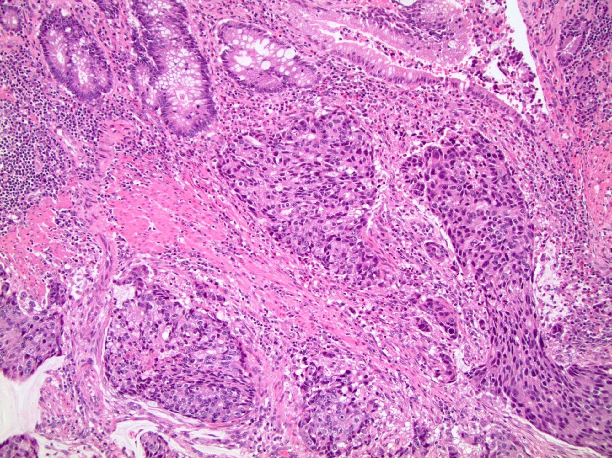
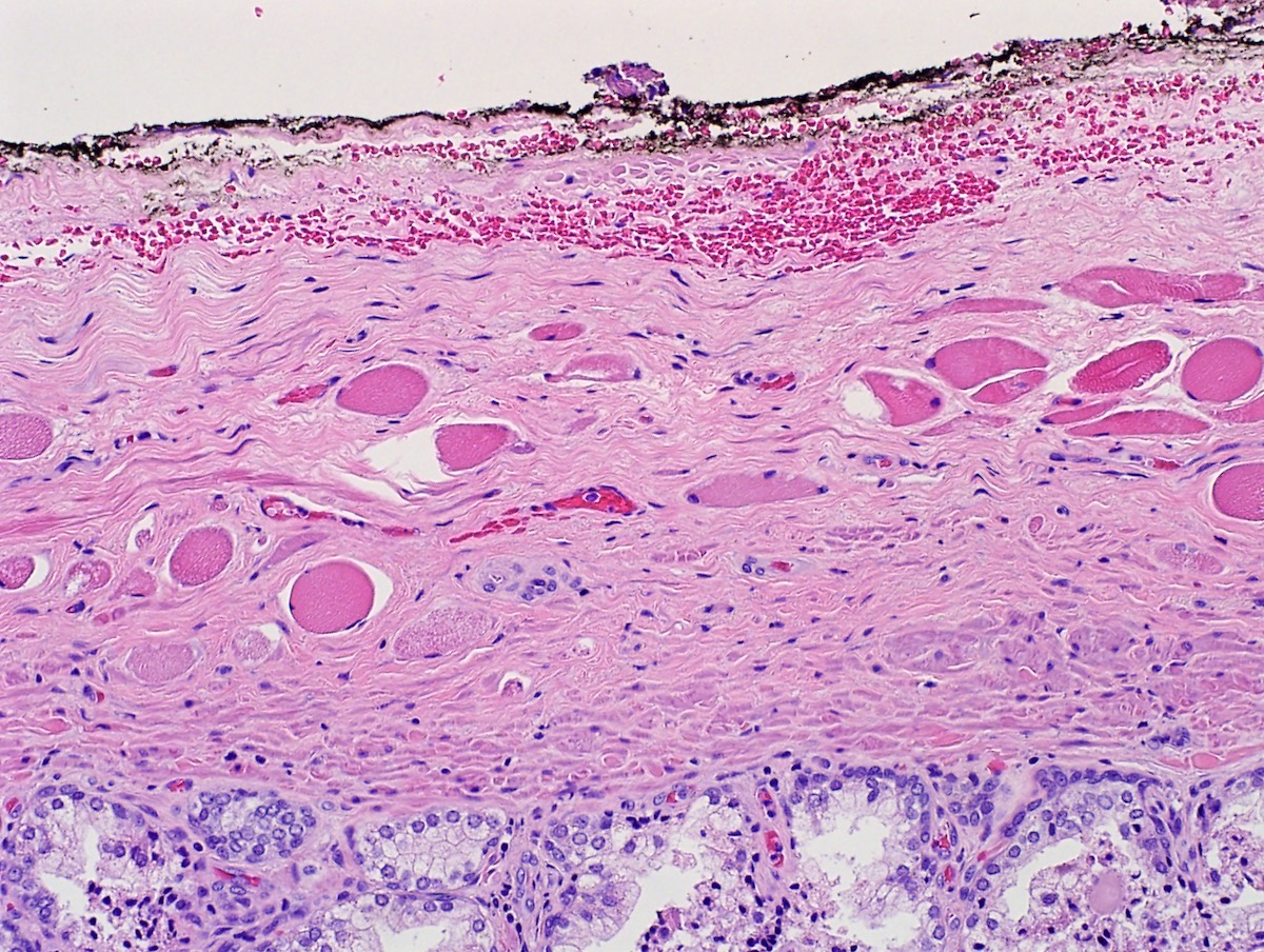
  • pT3a: extraprostatic extension or microscopic invasion of bladder neck
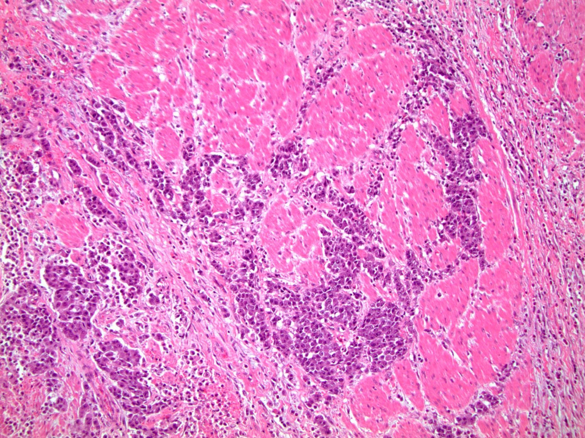
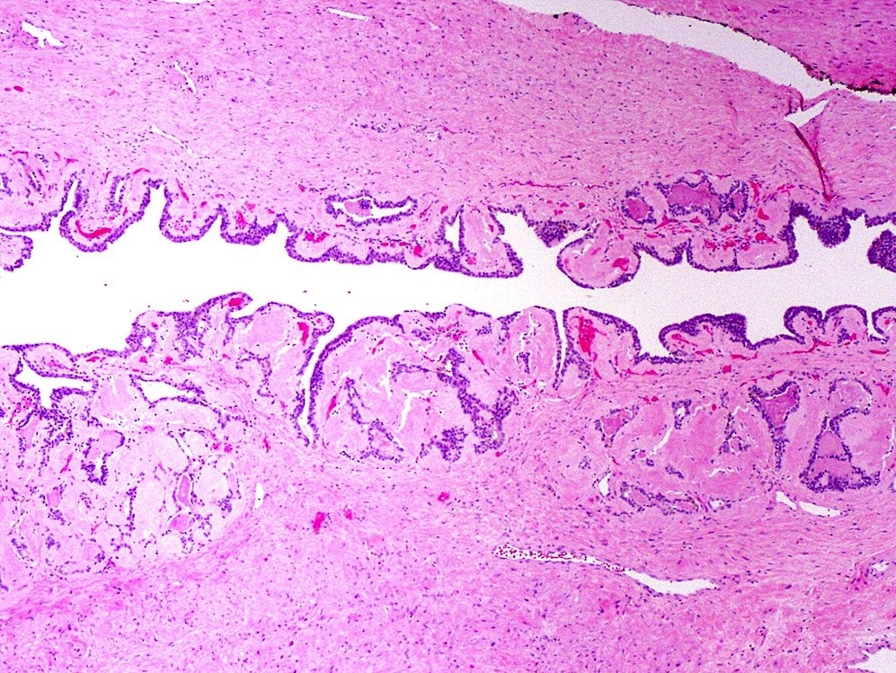
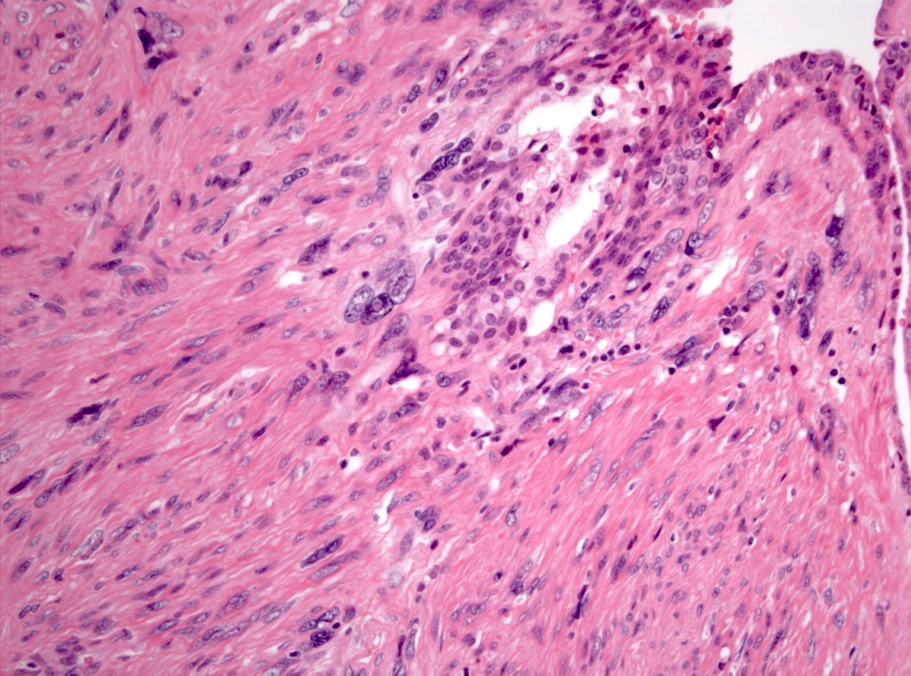
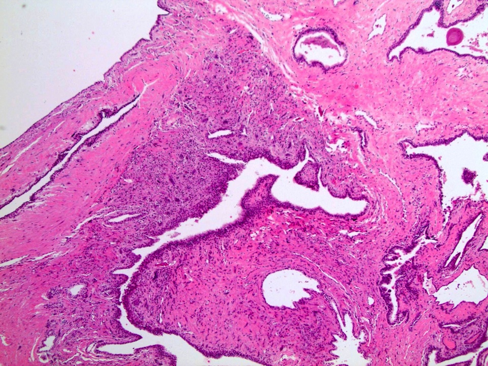
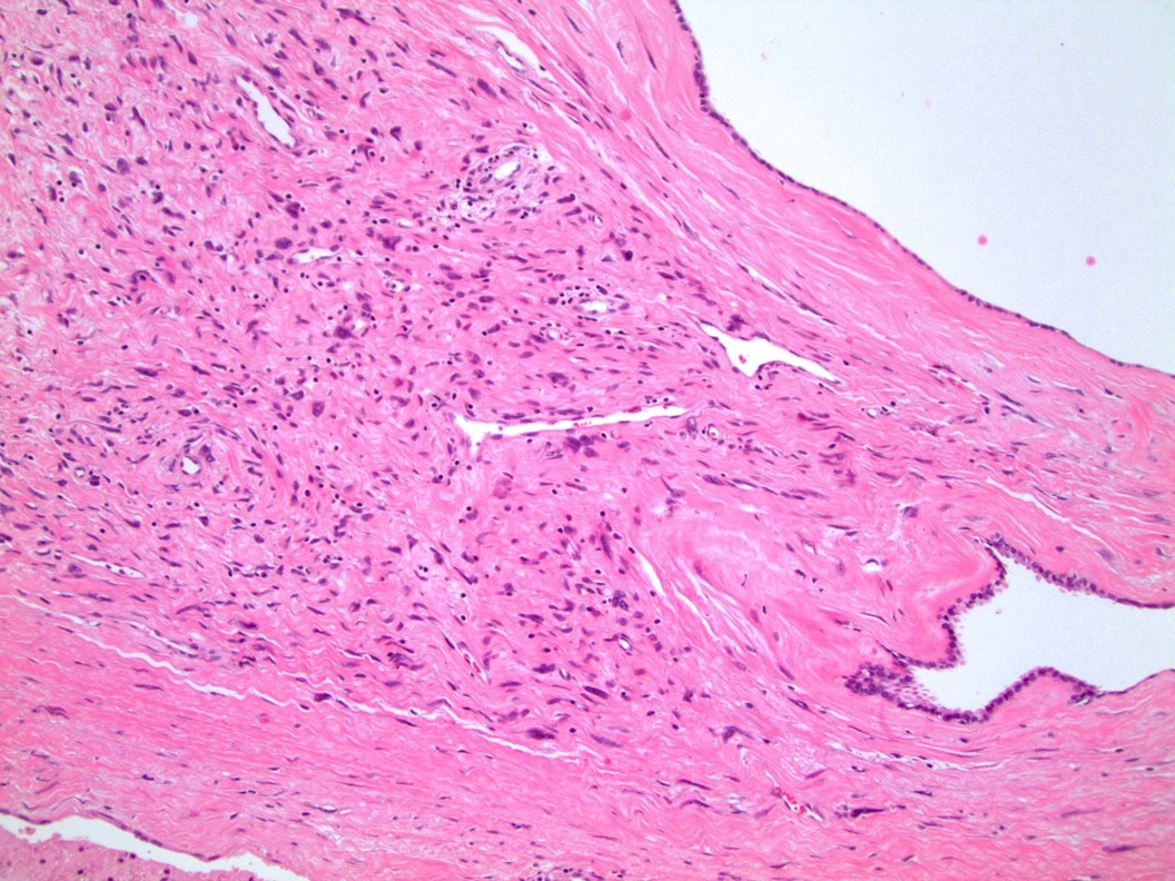
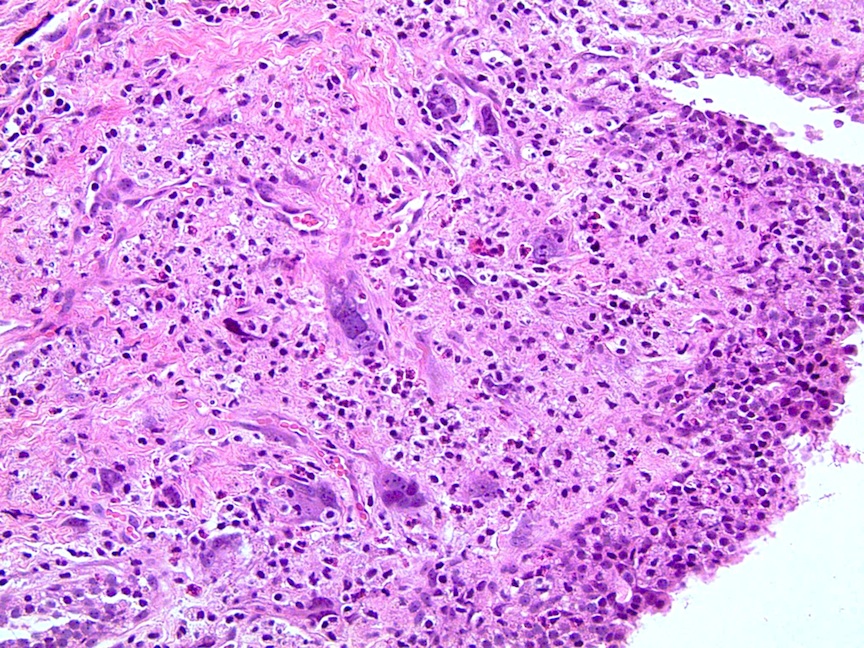
  • pT3b: seminal vesicle muscle invasion
  • pT4: fixed tumor or invasion of structures such as external sphincter, rectum, bladder, levator muscles or pelvic wall

Notes:
  • There is no pT1 classification
  • Note that cT1 is a part of clinical classification for clinically inapparent, nonpalpable tumor detected in specimens other than a prostatectomy such as a TURP or prostate biopsy with clinically inapparent, nonpalpable, incidental tumor in 5% or less of tissue defined as cT1a, clinically inapparent, nonpalpable, incidental tumor in > 5% defined as cT1b and clinically inapparent, nonpalpable tumor in a prostate needle biopsy defined as cT1c
  • Extraprostatic extension: usually this is dichotomized as focal (< 1 high power field in 1 - 2 slides) versus nonfocal / established (> 1 high power field in 1 - 2 slides)
  • Microscopic invasion of bladder neck: presence of tumor in thick muscle usually with no adjacent nonneoplastic glands
  • Seminal vesicle invasion: invasion of the extraprostatic seminal vesicle muscular wall
Regional lymph nodes (pN)
  • pNX: cannot be assessed
  • pN0: no regional lymph node metastasis
  • pN1: regional lymph node metastasis

Notes:
  • Regional lymph nodes include periprostatic, pelvic, hypogastric, obturator, internal iliac, external iliac, sacral
Distant metastasis (pM)
  • pM1a: metastasis in nonregional lymph node
  • pM1b: metastasis in bone
  • pM1c: metastasis in other distant site

Notes:
  • Nonregional lymph nodes include aortic, common iliac, deep / superficial inguinal, retroperitoneal
Prefixes
  • y: preoperative radiotherapy or chemotherapy
  • r: recurrent tumor stage
Histologic grade group (G)
Grade group Gleason score Gleason pattern
1 ≤ 6 ≤ 3+3
2 7 3+4
3 7 4+3
4 8 4+4, 3+5, 5+3
5 9 - 10 4+5, 5+4, 5+5
AJCC prognostic stage groups (utilizing pT categorization)
Stage group I: pT2 N0 M0  PSA < 10 Grade group 1 Gleason score ≤ 3+3=6
Stage group IIA: pT2 N0 M0 PSA ≥ 10 - < 20 Grade group 1 Gleason score ≤ 3+3=6
Stage group IIB: pT2 N0 M0 PSA < 20 Grade group 2 Gleason score 3+4=7
Stage group IIC: pT2 N0 M0 PSA < 20 Grade group 3 - 4 Gleason score 4+3=7, 4+4=7, 3+5=8, 5+3=8
Stage group IIIA: pT2 N0 M0 PSA ≥ 20 Grade group 1 - 4 Gleason score ≤ 6 - 8
Stage group IIIB: pT3 - 4 N0 M0 Any PSA Grade group 1 - 4 Gleason score ≤ 6 - 8
Stage group IIIC: pT2 - 4 N0 M0 Any PSA Grade group 5 Gleason score 9 - 10
Stage group IVA: pT2 - 4 N1 M0 Any PSA Any grade groupAny Gleason score
Stage group IVB: pT2 - 4 N0 - 1 M1 Any PSA Any grade groupAny Gleason score
Registry data collection variables
  • Pretreatment serum PSA
  • Grade group
  • Gleason score
  • Gleason patterns
  • Tertiary Gleason pattern on prostatectomy
  • Number of cores examined
  • Number of cores positive
  • Needle core biopsies positive one on side, both sides or beyond prostate
  • Metastatic sites
Gross images

Contributed by Debra L. Zynger, M.D.
Extraprostatic extension, multifocal (pT3a) Extraprostatic extension, multifocal (pT3a)

Extraprostatic extension, multifocal (pT3a)

Seminal vesicle invasion (pT3b)

Microscopic (histologic) images

Contributed by Debra L. Zynger, M.D.

Extraprostatic extension (pT3a)

Bladder neck invasion (pT3a)

Seminal vesicle invasion (pT3b)

Rectal invasion (pT4)


Periprostatic lymph node metastasis (pN1)

Pelvic lymph node metastasis (pN1)

Neck lymph node metastasis (pM1a)

Bone metastasis (pM1b)

Lung metastasis (pM1c)

Brain metastasis (pM1c)

Brain metastasis (pM1c)

Board review style question #1

A radical prostatectomy was performed and a cross section of the seminal vesicle wall is shown above. There is involvement by high grade prostatic adenocarcinoma. Which of the following is the correct pT?

  1. pT2
  2. pT3a
  3. pT3b
  4. pT4
Board review style answer #1
C. pT3b

Comment Here

Reference: Prostate - Staging

Treatment effect
Definition / general
  • There is some overlap of histologic alterations and some divergence, caused by androgen deprivation therapy and by radiation, to both prostate cancer and benign epithelium
Essential features
  • Nucleolus poor adenocarcinoma with clear cytoplasm is the hallmark of androgen deprivation effect; also, there may be loss of cytologic atypia and shrinkage of gland size and cytoplasm as well as atrophy of benign acini
  • Radiation effect causes atypical features of residual cancer as well as benign acini, such as marked nuclear enlargement and nucleoli in basal cells
  • Dual immunostain with basal cell markers and P504S / racemase is recommended to discriminate cancer from benign glands after both types of treatment
  • Grading is not recommended after androgen deprivation due to gland distortion; although there is some controversy about grading cancer after radiotherapy, the bulk of the evidence is in favor of grading if significant radiation change is absent
  • References: Clin Prostate Cancer 2004;2:228, Urology 2005;65:76
Terminology
  • Radiation effect, androgen deprivation effect, marked atrophy
ICD coding
  • ICD-10: N42.89 - other specified disorders of prostate (atrophy of prostate, infarct of prostate)
Epidemiology
  • History should be searched for radiotherapy to the pelvis for prostate or other cancers or for prior androgen deprivation (dutasteride or newer agents like enzalutamide and abiraterone)
Pathophysiology
Diagnosis
  • After androgen deprivation: do not grade
  • After radiation treatment: only assign a grade if marked radiation changes are absent
    • Certain immunostains are helpful to distinguish residual cancer (see Positive stains)
    • Gleason grade can and should still be assigned unless treatment effect is significant, in contrast to post androgen deprivation prostate (Mol Urol 1999;3:193, Urology 2005;65:76, Cancer 1997;79:81)
    • Therapy effect should be noted as a biopsy with marked radiation change to tumor may have a similar prognosis as a negative biopsy
Laboratory
  • Elevated serum PSA usually favors residual carcinoma; rarely, salivary ductal adenocarcinoma can produce PSA and demonstrate androgen receptor sensitivity; history of head and neck cancer should be assessed (Mayo Clin Proc Innov Qual Outcomes 2020;4:601)
    Prognostic factors
    • Adjuvant androgen deprivation with radiotherapy has benefits for progression free survival (J Clin Oncol 2021 Jan;39:136)
    • Grading of radiation effect on a 0 - 6 scale is highly predictive of local failure (Cancer 1997;79:81)
      • A system proposed in 1997 takes the sum of cytoplasmic changes (0 - 3) and nuclear changes (0 - 3):
        • Cytoplasmic change grades are (1) swelling and microvesicles, (2) extensive vacuolation and voluminous cytoplasm and (3) dilated glands plus single cells without lumens
        • Nuclear change grades are (1) some swelling or smudging but nucleoli identified, (2) large bizarre nuclei and (3) pyknotic condensed nuclei
      • In the above study, local failure was 55% for 0 - 2 grade effect, 30% for grade 3 - 4 and 0% for grade 5 - 6
    • There is no evidence for tumor dedifferentiation after radiotherapy (Cancer 1997;79:81)
    Case reports
    • Radiotherapy - case reports mainly deal with later complications:
    • Androgen deprivation
      • 66 year old man diagnosed with prostate cancer metastasis received, over the next 17 years, various forms of androgen deprivation, including use of newer agents such as abiraterone and MDV 3100 (Clin Endocrinol Metab 2012;97:360)
      • 72 and 80 year old men who developed sarcomatoid carcinoma after androgen deprivation, which is androgen independent (World J Clin Cases 2021;9:1668)
      • 83 year old man whose urethral prostatic adenocarcinoma metastasis resolved after androgen deprivation therapy (Urology 2019;129:e4)
    Treatment
    • Androgen deprivation:
      • Used for patients with advanced cancer or who are not surgical candidates but also can be used as neoadjuvant treatment prior to surgery
      • Newer agents, such as enzalutamide and abiraterone, have been used in addition to traditional LHRH agonists (J Clin Oncol 2019;37:923)
      • Antagonists are used, including leuprolide, goserelin, triptorelin, histrelin, buserelin and degarelix; they achieve the same effect as surgical castration
    • Radiotherapy:
      • This alternative to surgery has comparable long term outcome
      • External beam radiation therapy (EBRT) uses beams of radiation from outside the body, while brachytherapy uses radioactive seed implants to the prostate, allowing delivery of higher doses of radiation to a smaller area; the two may be combined for higher risk patients (Int J Radiat Oncol Biol Phys 2004;58:25)
      • Radiotherapy may be undertaken prior to prostatectomy, in which case the latter is termed salvage prostatectomy; histologic effects on the prostate are identical
    • Cryotherapy: this alternative to surgery uses a probe to freeze part of the prostate into an ice ball, thereby devitalizing cancer and adjacent stroma
    Microscopic (histologic) description
    • After androgen deprivation: do not grade
      • Nucleolus poor adenocarcinoma is the hallmark; also shows cytoplasmic clearing / vacuolization, reduced cytoplasmic quantity, nuclear pyknosis, reduced gland diameter, mucinous breakdown
      • Difficult to evaluate margin status after androgen deprivation
      • Current recommendations are to not assign a Gleason grade to androgen deprivation treatment specimens (Clin Prostate Cancer 2004;2:228)
      • There is a tendency toward greater neuroendocrine differentiation at prostatectomy after neoadjuvant androgen deprivation and a molecular basis for promotion of this has been described (Eur Urol 2004;45:586, Cancer Lett 2019;440-441:35)
      • High grade prostatic intraepithelial neoplasia (PIN) persists after endocrine treatment; tufted PIN may be replaced by flat high grade PIN (Hum Pathol 1999;30:1503)
      • Normal prostate shows atrophy, basal cell hyperplasia and prominence
    • After radiation treatment: assign a grade only if significant radiation change is absent
    • After cryotherapy:
      • The main finding is gland disappearance and washed out blue gray stroma
    Microscopic (histologic) images

    Contributed by Kenneth Iczkowski, M.D. and Debra L. Zynger, M.D.
    Androgen deprivation, benign

    Androgen deprivation, benign

    Cancer, androgen deprivation Androgen deprivation, cancer

    Androgen deprivation, cancer

    Radiotherapy, benign Radiotherapy, benign

    Radiotherapy, benign


    Radiotherapy, benign

    Radiotherapy, benign

    Cryotherapy, benign

    Cryotherapy, benign

    Cryotherapy, cancer

    Cryotherapy, cancer

    Suspicious for cancer

    Suspicious for cancer

    Shrunken benign acinus

    Shrunken benign acinus


    Radiation change, carcinoma Radiation change, carcinoma Radiation change, carcinoma Radiation change, carcinoma

    Radiation change, carcinoma

    Positive stains
    Negative stains
    Sample pathology report
    • Prostate, left apex, needle biopsies:
      • Prostatic adenocarcinoma with cytologic changes of androgen deprivation effect, not gradable
    • Prostate, left apex, needle biopsies:
      • Prostatic adenocarcinoma, Gleason 3 + 4 (score = 7, grade group 2) or
      • Prostatic adenocarcinoma with severe radiotherapy effect, not gradable
    Differential diagnosis
    • Atrophy (without therapeutic cause):
      • Exceedingly common finding in prostatic biopsies but there usually are some admixed nonatrophic glands
      • In the presence of 100% of glands being atrophic, history of treatment effect should be sought, particularly androgen deprivation
      • If atrophy is accompanied by basal cell hyperplasia, seek a history of radiotherapy
    • Xanthoma:
      • Contains cells with foamy cytoplasm but atypia would be absent and immunostain would reveal positivity for CD68 and negativity for cytokeratins
    • Foamy gland adenocarcinoma:
      • Has abundant foamy cytoplasm and less cytologic atypia than other cancers; this may mimic radiation effect but radiation effect creates vacuoles with both nuclear and cytoplasmic enlargement, which are different findings
    Board review style question #1
    Which immunostain may be falsely positive in benign glands after radiotherapy?

    1. Basal cell cytokeratin
    2. GATA3
    3. P504S
    4. p63
    5. PSA
    Board review style answer #1
    B. GATA3 may be falsely positive and mimic urothelial carcinoma in benign glands after radiotherapy. P504S should be positive in cancer and negative in benign glands. Basal cell cytokeratin and p63 should be negative in cancer after therapy. PSA reactivity is usually retained so expected to be positive.

    Comment Here

    Reference: Treatment effect - prostate
    Board review style question #2

    A 65 year old man with a history of prostate cancer diagnosed 8 years ago has a prostatectomy. Carcinoma from his prostatectomy specimen is shown. Which of the following is the best diagnosis?

    1. Prostatic adenocarcinoma 3+3=6
    2. Prostatic adenocarcinoma 3+4=7
    3. Prostatic adenocarcinoma 4+4=8
    4. Prostatic adenocarcinoma 4+5=9
    5. Prostatic adenocarcinoma with treatment effect
    Board review style answer #2
    E. Prostatic adenocarcinoma with treatment effect. In this case the patient was treated with brachytherapy. Radiation change is seen consisting of atrophic glands with nuclei that touch the cell membrane and gland retraction. A Gleason score is not rendered for tumors with marked therapy effect such as this case.

    Comment Here

    Reference: Treatment effect - prostate

    Treatment related neuroendocrine prostatic carcinoma (pending)
    [Pending]

    Urethral polyp
    Definition / general
    Essential features
    • Prostatic type urethral polyp is characterized by delicate papillae with true fibrovascular cores lined with prostatic epithelium with focal benign urothelial cells; prostatic acini are often present in the polyp (Am J Surg Pathol 1983;7:351)
    • Fibroepithelial urethral polyp is composed of cloverleaf-like and club-like dense fibrovascular cores covered by benign urothelium (Am J Surg Pathol 2005;29:460)
    Terminology
    • Prostatic type urethral polyp, also known as prostatic urethral polyp and prostatic type epithelial polyp of the urethra (J Urol 2015;193:2095)
    • Fibroepithelial urethral polyp, also known as fibroepithelial congenital urethral polyp (J Pediatr Surg 2014;49:835)
    ICD coding
    • ICD-10:
      • N36.9 - urethral disorder, unspecified
      • D30.4 - benign neoplasm of urethra
    Epidemiology
    • Prostatic type urethral polyp:
    • Fibroepithelial urethral polyp:
      • Age range 17 - 70 years, mean of 47 years (Am J Surg Pathol 2005;29:460)
      • Commonly occurs in pediatric males, usually 3 - 9 years of age (Can J Urol 2013;20:6974)
      • Rare patients present during infancy or adulthood
      • Rarely occurs in pediatric female patients
    Sites
    • Commonly occurs in posterior prostatic urethra adjacent to verumontanum or the bladder neck (J Urol 2015;193:2095)
    • Anterior urethra in rare case reports
    Pathophysiology
    • Ectopic prostatic acinar epithelium presents focally in urothelial tract, commonly in posterior urethra, rarely in bladder neck and penile urethra
    • Prostatic type urethral polyp likely caused by hyperplastic proliferations of the prostatic acinar epithelium results in overgrowth of the overlying urothelium (Am J Surg Pathol 1984;8:833)
    Etiology
    Clinical features
    • Common symptoms and signs: hematuria, dysuria, hemospermia
    • Urethrocystoscopic examination: a polypoid mass protruding from the posterior urethral wall (most common) or from anterior urethral wall (Urology 2020;143:238)
    Diagnosis
    • Ultrasonography reveals a protruding hyperechogenic polypoid mass protruding from the posterior (most common) urethral wall (Urology 2020;143:238)
    • Filling defect on voiding cystourethrogram (Clin Imaging 2018;51:164)
    • Appearance of a benign polyp on urethrocystoscopy followed by histopathological examination of the excised polyp
    Radiology description
    • Abdominal sonogram of the urinary bladder shows a hyperechogenic polypoid mass protruding to the bladder neck (Urol J 2020;18:86)
    • Filling defect is present at the bladder neck of a distended bladder (Clin Imaging 2018;51:164)
    Radiology images

    Images hosted on other servers:

    Polypoid mass

    Filling defect

    Prognostic factors
    • Excellent prognosis for prostatic type urethral polyp and fibroepithelial urethral polyp
    • Recurrence is unusual (J Urol 2015;193:2095)
    Case reports
    Treatment
    Clinical images

    Images hosted on other servers:

    Urethrocystoscopic view of polyp

    Gross description
    • Prostatic type urethral polyp (J Urol 2015;193:2095)
      • Pink-tan, exophytic papillary, filiform to rarely sessile structure
      • Single or multiple polyps
      • Usually < 1 cm
    • Fibroepithelial urethral polyp (Clin Imaging 2018;51:164)
      • Pink-tan, polypoid mass with narrow stalk
      • Usually < 4 cm
    Gross images

    Images hosted on other servers:

    Brown-tan knob-like polyp

    Microscopic (histologic) description
    • Prostatic type urethral polyp:
      • Delicate papillae with true fibrovascular cores lined by an outer layer of columnar cells and an underlying flattened to cuboidal basal cells (Am J Surg Pathol 1983;7:351)
      • Focal benign urothelium is present in the benign prostatic epithelium (J Urol 2015;193:2095)
      • Benign prostatic acini are present in the papillae and in adjacent fibromuscular stroma (J Urol 2015;193:2095)
    • Fibroepithelial urethral polyp (Am J Surg Pathol 2005;29:460):
      • Pattern 1: most common pattern includes the following features
        • Broad cloverleaf-like and club-like projections covered by normal urothelium and composed of dense fibrovascular stroma with florid cytitis cystica et glandularis (most common morphologic variant)
        • Back to back glands present in the stalk
        • Anastomosing nests of benign urothelial cells resembling inverted papilloma
        • Dilated cysts with intracystic papillary contents
        • Degenerative reactive atypia of stromal cells
      • Pattern 2: numerous small papillae with dense fibrous cores and areas of glandular differentiation
      • Pattern 3: urothelial lined broad edematous papillae mimicking polypoid urethritis; urothelial lined broad fibrous papillae with subepithelial edema
    Microscopic (histologic) images

    Contributed by Y. Albert Yeh, M.D., Ph.D.

    Fibroepithelial polyp
    Missing Image

    Polypoid mass

    Missing Image

    Cloverleaf-like and club-like projections

    Missing Image

    Broad based papillae

    Missing Image

    Cloverleaf-like projections

    Missing Image

    Club-like projections


    Missing Image

    Broad based finger-like papillae

    Missing Image Missing Image

    Urethritis cystica and glandularis

    Missing Image

    Polypoid and club-like projections

    Missing Image

    Focal calcifications

    Cytology description
    Cytology images

    Images hosted on other servers:

    Benign papillary cluster

    Positive stains
    Negative stains
    Sample pathology report
    • Urethral lesion, prostatic, excision:
      • Urethral polyp with prostatic type epithelium and changes, consistent with prostatic type urethral polyp (see comment)
      • Comment: The urethral lesion shows a polypoid and papillary growth lined by prostatic type epithelium and composed of benign prostatic acini. PSA and PSAP immunohistochemical stains reveal positive cytoplasmic staining in the prostatic glandular cells. These features are consistent with prostatic urethral polyp.
    • Urethral lesion, prostatic, excision:
      • Cloverleaf-like and club-like projections lined by normal urothelium, consistent with fibroepithelial urethral polyp (see comment)
      • Comment: The urethral lesion shows papillary projections arranged in cloverleaf-like and club-like pattern. The papillary structures are composed of broad fibrovascular cores covered by unremarkable urothelial cells. Neither urothelial proliferation nor epithelial atypia is noted. These features are consistent with fibroepithelial urethral polyp.
    Differential diagnosis
    Board review style question #1

    A 50 year old man presented with dysuria and hematuria for 3 months. Cystoscopic examination showed a polypoid mass protruded from the posterior wall of the prostatic urethra. Cystoscopic excision of the polyp and histopathological examination were performed. The microphotograph of the urethral polyp is shown above. What is the diagnosis?

    1. Fibroepthelial urethral polyp
    2. Polypoid urethritis
    3. Prostatic type urethral polyp
    4. Urethral caruncle
    Board review style answer #1
    A. Fibroepithelial urethral polyp

    Comment here

    Reference: Urethral polyp
    Board review style question #2
    A 40 year old man presented with dysuria for 2 months. During cystoscopy, a polypoid mass protruding from the posterior urethra was detected. Surgical excision of the polyp was performed and microscopic examination showed a papillary growth lined by tall benign columnar cells with pale eosinophilic cytoplasm. In the stroma are some glands lined by benign columnar cells with underlying flattened epithelial cells. What is the diagnosis?

    1. Fibroepithelial polyp
    2. Polypoid urethritis
    3. Prostatic type urethral polyp
    4. Urethral caruncle
    Board review style answer #2
    C. Prostatic type urethral polyp

    Comment here

    Reference: Urethral polyp

    Vanishing cancer
    Definition / general
    • First described in 1995, vanishing cancer indicates no residual tumor found in radical prostatectomy specimen despite confirmed tumor in needle biopsy (Am J Surg Pathol 1995;19:1002)
    • Sometimes, only high grade prostatic intraepithelial neoplasia (HGPIN) or atypical small acinar proliferation suspicious for but not diagnostic of cancer (ASAP) is found
    Essential features
    • Contemporary incidence of vanishing cancer is 0.2% of radical prostatectomy specimens; it was higher a decade or two ago when prostatectomy was being performed for smaller / lower grade cancers
    • Incidence is higher in large prostates or in preoperatively treated ones
    • Resolution of dilemma involves complete tissue submission, cancer specific immunostains, use of second opinion, consider flipping blocks to other surface; some residual cancers simply cannot be detected after the above efforts
    ICD coding
    • ICD-10: C61 - malignant neoplasm of prostate
    Epidemiology
    Etiology
    • Most often, the phenomenon is attributed to the fact that routine histologic sections cannot evaluate every cubic millimeter of prostate volume, so small cancer foci remain in the paraffin block
    • Rarely, prostate cancer may have been ablated by the biopsy or transurethral resection procedure (BJU Int 2004;94:939)
    • Currently, more patients are receiving preoperative androgen deprivation, such as enzalutamide, that shrinks the cancer (Urology 2005;65:76, Clin Cancer Res 2017;23:2169)
    • It is also proposed that a minute cancer focus at the edge of the prostate may be inadvertently left behind in the overzealous attempt to perform a nerve sparing procedure (Indian J Cancer 2013;50:170)
    Diagnosis
    • Diagnostic resolution should include these steps in the following order (Urol Oncol 2019;37:696):
      • If the prostatectomy tissue has been partially submitted, complete submission should be pursued
      • Get a second pathologist, particularly a urologic pathologist, to reread the resection slides
      • Review the biopsy that was diagnostic of cancer, if slides are available
      • Immunostains (see below)
      • Flip tissue in some or all of the paraffin blocks and have the opposite surfaces of the tissues cut
      • DNA identity analysis can be performed if specimen switching is suspected (see below)
    Laboratory
    Gross description
    Gross images

    Images hosted on other servers:
    Missing Image

    Paraffin blocks
    before and
    after block
    flipping

    Positive stains
    • P504S racemase is positive in cancer; get immunostains on deeper levels, on areas initially suspicious for cancer (ASAP)
    Negative stains
    Molecular / cytogenetics description
    • Microsatellite analysis can be performed on the prostatectomy and biopsy specimens to confirm specimen identity; this very rarely shows a specimen switch (1 in 10 cases of vanishing cancer was attributed to specimen mixup) (Am J Surg Pathol 2005;29:467)
    • This is particularly indicated if there is high grade or high volume cancer on the biopsy (Am J Surg Pathol 2005;29:467)
    Sample pathology report
    • Prostate, radical prostatectomy:
      • No residual cancer (see comment)
      • Comment: This diagnosis is reached after complete submission and microscopy of all prostatic tissue. The tissue in the blocks was also reembedded and the reverse sides were cut into microscopic slides. The prostatic basal cell / P504S immunostain was performed to interrogate block X and it was confirmatory. The original diagnosis of cancer was confirmed by rereviewing the biopsy specimen. Dr. X agrees with this interpretation.
    Differential diagnosis
    • Biopsy specimen switch:
      • Specimen mixup with another patient must be excluded if the biopsy showed high grade or high volume cancer and no cancer is seen in the resection
      • Molecular analysis can be performed (Am J Surg Pathol 2005;29:467)
    • Biopsy false positive:
      • Correct diagnosis of cancer on the prior biopsy should be confirmed
      • Immunostains (AMACR+ / p63, HMWK-) can be used if not previously performed
    Board review style question #1
    A 70 year old man undergoes radical prostatectomy with an organ weight of 90 g. What is the least likely contributor to finding no residual cancer?

    1. Partial sampling of every other tissue slice (as is commonly done for > 50 g prostates)
    2. Preoperative course of enzalutamide ablated any residual cancer
    3. Prior prostate biopsy specimen switch
    4. Residual cancer remains in the paraffin, even after flipping the blocks
    Board review style answer #1
    C. A specimen switch of the prior prostate biopsy is the least likely explanation and could be detected molecularly. If initial partial sampling was done, complete sampling of the tissue should be pursued; if cancer is not found, flipping the blocks is recommended; use of immunostains may help if any foci are suspicious. Finally, it is possible that a preoperative course of enzalutamide has ablated any residual cancer.

    Comment Here

    Reference: Vanishing cancer

    Verumontanum mucosal hyperplasia
    Definition / general
    • Well circumscribed small acinar proliferation in or adjacent to verumontanum and posterior prostatic urethra
    Sites
    • The verumontanum is located in the posterior aspect of the prostatic urethra
    • It is the area where the utricle and the ejaculatory ducts merge with the urethra
    Clinical features
    • The presence of verumontanum mucosa gland hyperplasia is an asymptomatic morphologic variant of the glands underlying the posterior wall of the prostatic urethra
    Diagnosis
    • Can be identified in TURP specimens or in needle biopsies of the prostate
    Case reports
    Treatment
    • Not required
    Microscopic (histologic) description
    • Crowded small acinar proliferation, usually arranged in a well circumscribed nodule
    • The glands can have intraluminal concretions that vary in color from orange to gray or green
    • May be seen underlying the urethral urothelium
    Positive stains
    • The immunophenotype is identical to normal prostate glands, including basal cell markers
    Differential diagnosis
    • Verumontanum mucosal gland hyperplasia (VMGH), when present in a prostate needle biopsy, could be mistaken for low grade prostatic adenocarcinoma (Gleason pattern 3)
    • The lobular architecture and the presence of basal cells are features to differentiate it from Gleason pattern 3 cancer
    • Another helpful feature is the presence of corpora amylacea or the orange-grey-green concretions that are features of VMGH and not of prostatic carcinoma
    • Occasionally, it can present with a papillary architecture simulating prostatic duct adenocarcinoma
    • In contrast to prostatic duct carcinoma, the cells in VMGH are more cuboidal and without nuclear atypia

    Well differentiated neuroendocrine tumor
    Definition / general
    • Very rare tumor of prostate
    • Also called low grade neuroendocrine carcinoma
    • May metastasize
    Case reports
    Microscopic (histologic) images

    Images hosted on other servers:

    Primary carcinoid tumor of the verumontanum


    WHO classification
    Definition / general
    • The World Health Organization (WHO) 2022 Classification of Urinary and Male Genital Tumors (5th edition) replaces the previous WHO 2016 Classification
    Major updates
    • Low grade prostatic intraepithelial neoplasia is no longer recognized as a reportable entity as it is not possible to distinguish from benign glandular hyperplasia
    • Cribriform subtype of high grade prostatic intraepithelial neoplasia (HGPIN) is no longer regarded as a distinct entity
      • The term atypical intraductal proliferation is preferred to denote intraductal proliferations that are more complex than HGPIN but less complex than intraductal carcinoma; however, consensus on the terminology has not been reached (Pathology 2018;50:60, Arch Pathol Lab Med 2021;145:461)
    • Intraductal carcinoma of the prostate (IDC-P) may represent 2 distinct entities
      • Small subset of IDC-P lesions is thought to have progressed from HGPIN precursor and are truly in situ
      • Majority of IDC-P lesions represent late events associated with high grade invasive carcinoma colonizing nonneoplastic prostatic ducts and acini
    • Whether to include (or exclude) IDC-P into Gleason grading remains controversial with diverging recommendations by leading societies (Am J Surg Pathol 2020;44:e87, Arch Pathol Lab Med 2021;145:461)
    • Prostatic intraepithelial neoplasia-like adenocarcinoma has been moved and is discussed as a subtype of acinar adenocarcinoma (Am J Surg Pathol 2018;42:1693, Histopathology 2021;78:327)
    • Ductal adenocarcinoma remains separate from acinar adenocarcinoma, although there is consideration to reclassify it as a subtype of acinar adenocarcinoma (Mod Pathol 2009;22:359, Prostate 2015;75:1610, Prostate 2017;77:1242, Eur Urol Focus 2019;5:433, JCO Precis Oncol 2019;3:PO.18.00327)
    • Adenoid cystic (basal cell) carcinoma of the prostate is the preferred diagnostic terminology, rather than basal cell carcinoma, to avoid confusion with the more common skin tumor
    • The designation of subtypes has replaced variants for distinct clinical or morphologic categories within a tumor type; the term variant is used for genomic alterations
      • Examples of subtypes of prostatic acinar adenocarcinoma include
        • Prostatic intraepithelial neoplasia-like adenocarcinoma
        • Signet ring-like cell acinar adenocarcinoma
        • Sarcomatoid acinar adenocarcinoma
        • Pleomorphic giant cell acinar adenocarcinoma
    • Neuroendocrine, mesenchymal, hematolymphoid, melanocytic, metastatic and genetic syndrome related tumors are now discussed in separate chapters
      • Exceptions include
        • Mesenchymal tumors thought to arise from the prostatic stroma: prostatic stromal tumor of uncertain malignant potential and prostatic stromal sarcoma
        • Treatment related neuroendocrine prostatic carcinomas that remain under glandular neoplasms of the prostate
    • Urothelial carcinoma of the prostate and prostatic urethra has been moved to the urinary tract chapter
    WHO (2022)
    Tumors of the prostate ICD-O ICD-11
    • Epithelial tumors of the prostate
    • Glandular neoplasms of the prostate
    8440/0 2F34 & XH5RJ2
    8148/2 2E67.5
    8148/2 2E67.5
    8140/3 C61
    8500/3 2C822.0 & XH7KH3
    8574/3 2C82.Y & XH0U20
    • Squamous neoplasms of the prostate
    8560/3 2C82.Y & XH7873
    8070/3 2C82.Y & XH0945
    8147/3 2C82.Y & XH4ZC3
    • Mesenchymal tumors unique to the prostate
    • Stromal tumors of the prostate
    8935/1 2F77 & XH8747
    8935/3 2C82.Y & XH49Y5
    Diagrams / tables
    None
    Microscopic (histologic) images

    Contributed by Alcino Pires Gama, M.D. and Bonnie Choy, M.D.
    Prostatic ductal adenocarcinoma

    Prostatic ductal adenocarcinoma

    Adenoid cystic (basal cell) carcinoma of the prostate

    Adenoid cystic (basal cell) carcinoma of the prostate

    Treatment related neuroendocrine carcinoma Treatment related neuroendocrine carcinoma

    Treatment related neuroendocrine carcinoma

    Intraductal carcinoma of the prostate Intraductal carcinoma of the prostate

    Intraductal carcinoma of the prostate

    Board review style question #1
    Which of the following is most accurate according to the WHO 2022 Classification of Urinary and Male Genital Tumors (5th edition)?

    1. Cribriform subtype of high grade prostatic intraepithelial neoplasia (HGPIN) is no longer a distinct entity
    2. Ductal adenocarcinoma has been incorporated as a subtype of acinar adenocarcinoma
    3. Intraductal carcinoma should be graded and incorporated in the final Gleason score
    4. PIN-like adenocarcinoma remains a subtype of ductal adenocarcinoma
    Board review style answer #1
    A. Cribriform subtype of high grade prostatic intraepithelial neoplasia (HGPIN) is no longer recognized as a distinct entity as it belongs to a group of intraductal proliferations that are more complex than HGPIN but less complex than intraductal carcinoma. Answer B is incorrect because ductal adenocarcinoma remains separate from acinar adenocarcinoma, although there have been discussions to reclassify it as a subtype of acinar adenocarcinoma. Answer C is incorrect because the inclusion / exclusion of intraductal carcinoma into Gleason grading remains controversial; additional studies are required to compare the 2 approaches from a prognostic perspective. Answer D is incorrect because PIN-like adenocarcinoma has been moved to the chapter on acinar adenocarcinoma.

    Comment Here

    Reference: Prostate gland & seminal vesicles - WHO classification
    Board review style question #2
    Which of the following is an update from the WHO 2022 Classification of Urinary and Male Genital Tumors (5th edition)?

    1. Basal cell markers are useful in distinguishing adenoid cystic carcinoma of the prostate from basal cell carcinoma
    2. Low grade intraepithelial neoplasia remains a distinct entity
    3. The descriptors subtypes and variants can be used interchangeably
    4. Treatment related neuroendocrine carcinomas of the prostate remain under glandular neoplasms of the prostate
    Board review style answer #2
    D. Treatment related neuroendocrine carcinomas of the prostate remain under glandular neoplasms of the prostate. Given the distinct risk factors, prognosis and management, treatment related neuroendocrine carcinomas of the prostate remain under glandular neoplasms of the prostate. All other mesenchymal tumors were moved to a separate chapter. Answer A is incorrect because basal cell carcinoma of the prostate has been renamed as adenoid cystic (basal cell) carcinoma of the prostate. Answer B is incorrect because low grade prostatic intraepithelial neoplasia is no longer a distinct entity. Answer C is incorrect because the designation of subtypes has replaced variant for distinct clinical or morphologic categories within a tumor type.

    Comment Here

    Reference: Prostate gland & seminal vesicles - WHO classification

    WHO classification (pending)
    [Pending]
    Back to top
    Recent Prostate gland & seminal vesicles Pathology books

    Amin: 2022

    Cheng: 2019

    Epstein: 2021

    Epstein: 2020

    Epstein: 2020

    IARC: 2022

    Lopez-Beltran: 2018

    Matoso: 2020

    Robinson: 2018

    Shah: 2019

    VandenBussche: 2022

    Wobker: 2021

    Yang: 2020

    Zhou: 2022


    Find related Pathology books: cytopathology, GU/adrenal
    Image 01 Image 02