
25 October 2017 - Case of the Week #441
All cases are archived on our website. To view them sorted by case number, diagnosis or category, visit our main Case of the Week page. To subscribe or unsubscribe to Case of the Week or our other email lists, click here.
Thanks to Drs. Aysha Mubeen, Ahmad Alkhasawneh and Arun Gopinath, University of Florida College of Medicine, Jacksonville, Florida (USA) for contributing this case and Dr. Raul S. Gonzalez, Assistant Professor, University of Rochester Medical Center, New York (USA), for editing the discussion. To contribute a Case of the Week, first make sure that we are currently accepting cases, then follow the guidelines on our main Case of the Week page.

Advertisement
Website news:
(1) We are looking for highly qualified authors to keep our textbook up to date. More information is available at http://www.pathologyoutlines.com/authors.html and http://www.pathologyoutlines.com/Instructionsforauthors.html.
(2) We are now offering Sponsorships for 2018 which will support hiring additional pathologists as authors and editors. Click here for Sponsorship categories, here for why you should be a Sponsor, and here for a mockup of the new home page. Please contact us at (248) 646-0325 or PathOutAds@gmail.com for more information.
We would like to take the opportunity to thank LOXO Oncology and Cleveland Clinic for their 2018 Sponsorships!
(3) We now have regional totals by subspecialty on our jobs page, as well as jobs by location and subspecialty. At the top of the jobs page, under the heading Search Jobs, there is a subsection for subspecialties.
Visit and follow our Blog to see recent updates to the website.
Case of the Week #441
Clinical history:
A 59 year old man presented with abdominal bloating and vague abdominal pain. CT and PET scan showed ascites and findings suggestive of peritoneal carcinomatosis. However, no primary could be identified. A diagnostic laparoscopy with omental and peritoneal biopsies was performed. The patient underwent a debulking procedure. He died 6 months later.
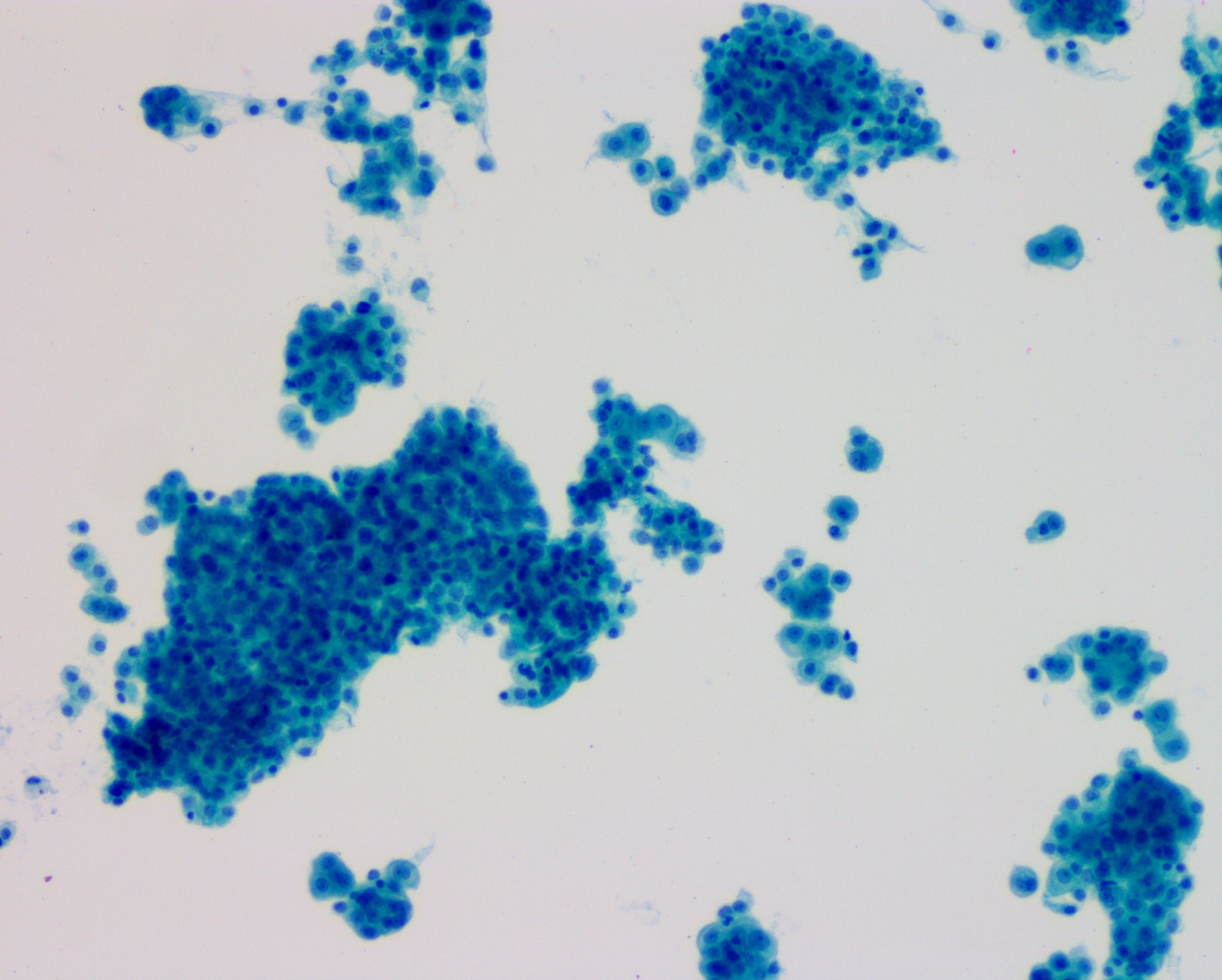
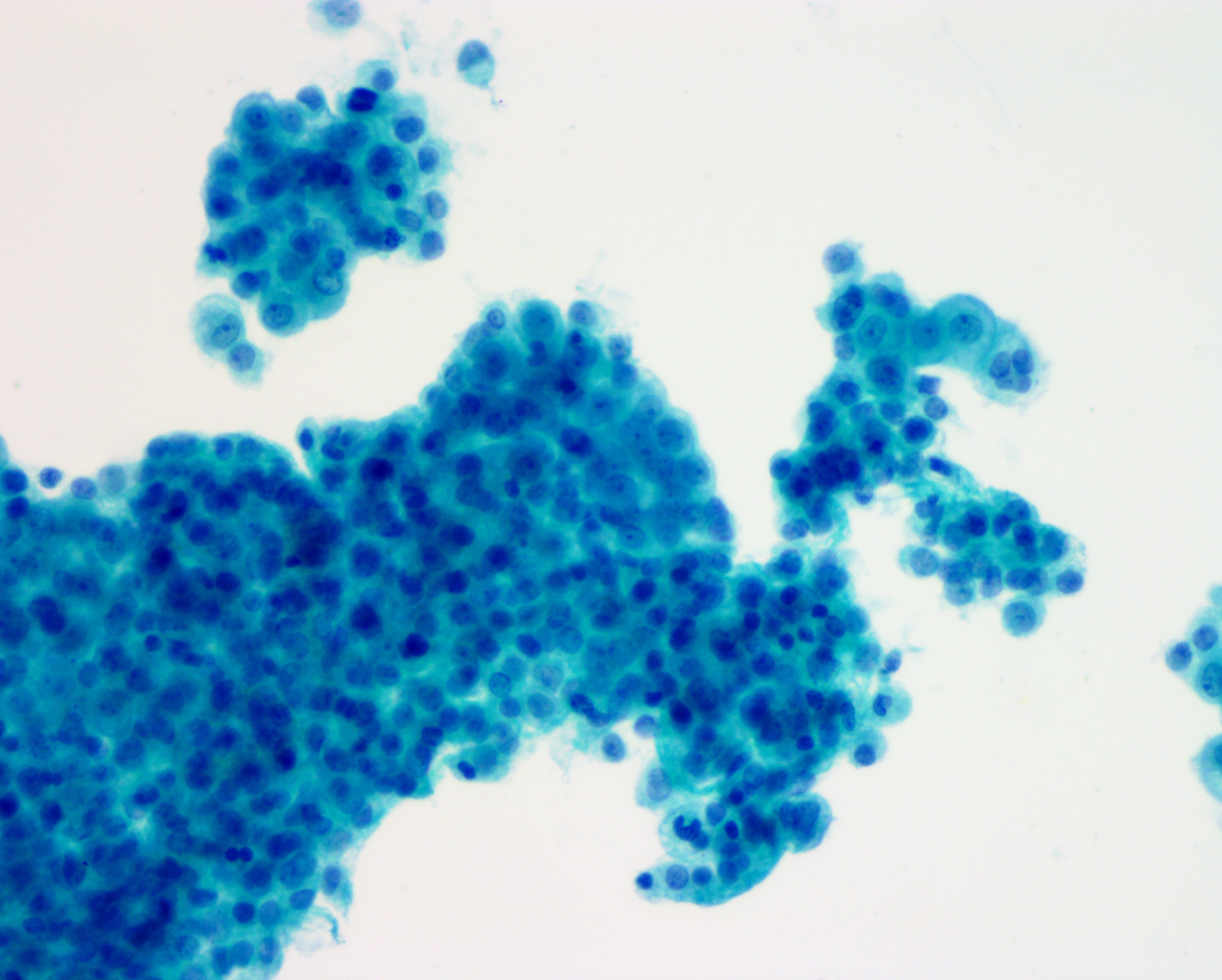
Cytology images:

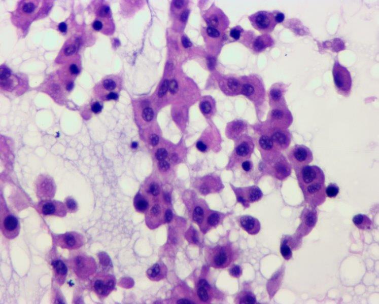
Histopathology images:


What is your diagnosis?
Diagnosis:
Myxoid variant of peritoneal epithelioid mesothelioma
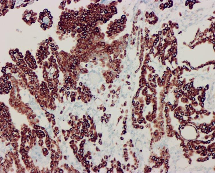
Special stains:

Test question (answer at the end):
Which of the following immunostains would be positive in peritoneal myxoid mesothelioma but not pseudomyxoma peritonei?
A. CDX2
B. Claudin4
C. D2-40
D. Pankeratin
Discussion:
Up to one third of malignant mesotheliomas arise in the peritoneum, and epithelioid mesothelioma is the most frequent histologic type of malignant mesothelioma (sarcomatoid and biphasic mesotheliomas are less common). There are several subtypes of mesothelioma, including common and rare variants. The myxoid variant of epithelioid mesothelioma is a rare subtype that retains the secretory properties of normal mesothelium. About two dozen cases have been reported in the literature, with only 4 of these arising in the peritoneum (Virchows Arch 2005;447:828, Cesk Patol 2014;50:149). There is no sex predilection. Patients can present at any stage, and about half of the published cases report asbestos exposure. Overall, myxoid features appear to confer a slightly improved prognosis in epithelioid mesothelioma.
Histologically, the tumor consists of dyscohesive medium sized to large epithelioid cells with a moderate to abundant amount of eosinophilic cytoplasm dispersed in a myxoid background. The nuclei show coarse chromatin with prominent nucleoli. Mitotic figures are usually inconspicuous. Immunohistochemically, the tumor cells show diffuse positivity for calretinin, D2-40, EMA, CK5 / CK6 and vimentin. MOC31, BerEP4 and Claudin4 are negative in tumor cells, supporting mesothelial differentiation (Am J Clin Pathol 2013;139:611).
The main differential diagnoses are mucinous adenocarcinoma and pseudomyxoma peritonei. All of these lesions are positive for keratins, but only mesothelioma is positive for the above mesothelial markers.
Test Question Answer:
C. D2-40
All cases are archived on our website. To view them sorted by case number, diagnosis or category, visit our main Case of the Week page. To subscribe or unsubscribe to Case of the Week or our other email lists, click here.
Thanks to Drs. Aysha Mubeen, Ahmad Alkhasawneh and Arun Gopinath, University of Florida College of Medicine, Jacksonville, Florida (USA) for contributing this case and Dr. Raul S. Gonzalez, Assistant Professor, University of Rochester Medical Center, New York (USA), for editing the discussion. To contribute a Case of the Week, first make sure that we are currently accepting cases, then follow the guidelines on our main Case of the Week page.
Advertisement
Website news:
(1) We are looking for highly qualified authors to keep our textbook up to date. More information is available at http://www.pathologyoutlines.com/authors.html and http://www.pathologyoutlines.com/Instructionsforauthors.html.
(2) We are now offering Sponsorships for 2018 which will support hiring additional pathologists as authors and editors. Click here for Sponsorship categories, here for why you should be a Sponsor, and here for a mockup of the new home page. Please contact us at (248) 646-0325 or PathOutAds@gmail.com for more information.
We would like to take the opportunity to thank LOXO Oncology and Cleveland Clinic for their 2018 Sponsorships!
(3) We now have regional totals by subspecialty on our jobs page, as well as jobs by location and subspecialty. At the top of the jobs page, under the heading Search Jobs, there is a subsection for subspecialties.
Visit and follow our Blog to see recent updates to the website.
Case of the Week #441
Clinical history:
A 59 year old man presented with abdominal bloating and vague abdominal pain. CT and PET scan showed ascites and findings suggestive of peritoneal carcinomatosis. However, no primary could be identified. A diagnostic laparoscopy with omental and peritoneal biopsies was performed. The patient underwent a debulking procedure. He died 6 months later.
Cytology images:
ThinPrep
Diff-Quik
Histopathology images:
2x
4x
40x
What is your diagnosis?
Diagnosis:
Myxoid variant of peritoneal epithelioid mesothelioma
Special stains:
Calretinin
MOC31
Test question (answer at the end):
Which of the following immunostains would be positive in peritoneal myxoid mesothelioma but not pseudomyxoma peritonei?
A. CDX2
B. Claudin4
C. D2-40
D. Pankeratin
Discussion:
Up to one third of malignant mesotheliomas arise in the peritoneum, and epithelioid mesothelioma is the most frequent histologic type of malignant mesothelioma (sarcomatoid and biphasic mesotheliomas are less common). There are several subtypes of mesothelioma, including common and rare variants. The myxoid variant of epithelioid mesothelioma is a rare subtype that retains the secretory properties of normal mesothelium. About two dozen cases have been reported in the literature, with only 4 of these arising in the peritoneum (Virchows Arch 2005;447:828, Cesk Patol 2014;50:149). There is no sex predilection. Patients can present at any stage, and about half of the published cases report asbestos exposure. Overall, myxoid features appear to confer a slightly improved prognosis in epithelioid mesothelioma.
Histologically, the tumor consists of dyscohesive medium sized to large epithelioid cells with a moderate to abundant amount of eosinophilic cytoplasm dispersed in a myxoid background. The nuclei show coarse chromatin with prominent nucleoli. Mitotic figures are usually inconspicuous. Immunohistochemically, the tumor cells show diffuse positivity for calretinin, D2-40, EMA, CK5 / CK6 and vimentin. MOC31, BerEP4 and Claudin4 are negative in tumor cells, supporting mesothelial differentiation (Am J Clin Pathol 2013;139:611).
The main differential diagnoses are mucinous adenocarcinoma and pseudomyxoma peritonei. All of these lesions are positive for keratins, but only mesothelioma is positive for the above mesothelial markers.
Test Question Answer:
C. D2-40
