
All cases are archived on our website. To view them sorted by case number, diagnosis or category, visit our main Case of the Week page. To subscribe or unsubscribe to Case of the Week or our other email lists, click here.
Thanks to Dr. Julie Guilmette and Dr. Olguta Gologan, University of Montreal Hospital Center (Canada) for contributing this case and discussion. To contribute a Case of the Week, follow the guidelines on our main Case of the Week page.

See how leading oncologists are visualizing tumor cells with PerkinElmer's Phenoptics™ Research Solution
To advance the understanding of disease mechanisms in cancer, it's critical that you see everything the tumor has to show you. With our Phenoptics research solution, you can visualize and measure tumor cells and multiple immune-cell phenotypes simultaneously in FFPE tissue. Phenoptics integrates multiplexed immunohistochemistry and imaging to quantitatively capture systems biology data with cellular detail. It reveals multi-parameter cellular expressions and interactions while retaining spatial context, offering insights into the complexity of immune-cancer interactions.
Learn how industry leaders are advancing their cancer research with our award winning Phenoptics Research Solution for Cancer Immunology and Immunotherapy by downloading our e-book now.
Click here to download.
Website news:
(1) Don't forget to subscribe to our 5 available email newsletters: (a) Case of the Week; (b) Commercial / Promotions email; (c) Jobs, Fellowships and Conferences; (d) Website News / New Books and (5) What's New in Pathology (new). Click here to learn more, and remember, you can unsubscribe at any time. If you are starting a new job or fellowship in July, please email us with your new email so you can continue to receive our newsletters.
(2) What do you think? Should we have a page for classified ads (buying and selling expensive pathology related equipment) at PathologyOutlines.com? We would charge $400 for an ad (prepaid only, perhaps a little less at the beginning), which could include 100 words and 3 images. Buyers / sellers would communicate with each other directly. We would do nothing other than post the ad and remove it after 90 days (or sooner, if requested by the seller). We could either create a separate Classifieds page on our website, or put this on our Facebook page. Is this a good idea / bad idea? Let us know.
(3) Our newest Commercial Email contest winner is Dr. Varsha Ingale, from Maharastra, India, who received a $50 Amazon gift card. Please sign up for and read our Commercial Emails for a chance to win future contests.
Visit and follow our Blog to see recent updates to the website.
Case of the Week #394
Clinical history:
A 71 year old man presented with a painless, right sided neck mass increasing in size for three months. He had mild dysphagia but was otherwise asymptomatic. Ultrasound revealed three hypoechoic thyroid nodules, all within the right lobe, measuring 4.2 cm, 3.0 cm and 1.0 cm. As fine needle aspiration was suspicious for neoplasia, he underwent a total thyroidectomy.
Gross description:
Gross examination showed 3 non encapsulated, well circumscribed white masses. No necrosis was seen.
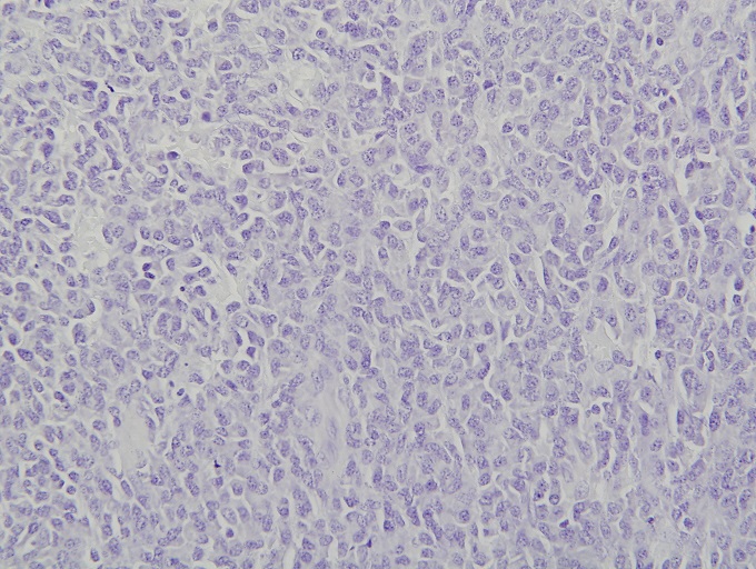
Micro images:
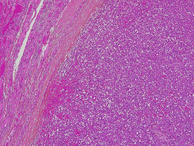
10x

20x

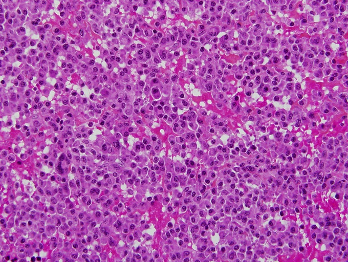
40x
What is your diagnosis?
Diagnosis:
Solitary extramedullary plasmacytoma of the thyroid
Discussion:
Immunohistochemistry:
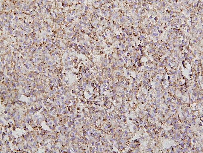
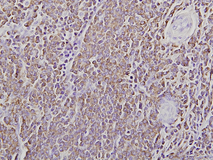
The neoplastic plasma cells were positive for CD138, CD79a and kappa light chain, but were negative for lambda light chain, as well as TTF1, thyroglobulin, synaptophysin, chromogranin and calcitonin.

CD79a
CD138
Kappa
Lambda
Micro description:
Plasma cell dyscrasias are separated into six distinct classes: plasmacytoma, multiple myeloma, lymphoplasmacytic lymphoma, heavy chain disease, primary or immunocyte associated amyloidosis and monoclonal gammopathy of undetermined significance (MGUS, Case Rep Otolaryngol 2012;2012:282784). Plasmacytoma is further divided into two subtypes, solitary plasmacytoma of the bone (SPB) and extramedullary plasmacytoma (EMP). EMP involves the soft tissue without systemic involvement (Blood Res 2014;49:280).
Solitary EMP is a rare neoplasm that occurs most frequently in the head and neck region, especially the upper aerodigestive tract. It occurs only rarely in the thyroid gland (Thyroid 2012;22:861, Acta Clin Belg 2015;70:133, J Cytol 2014;31:53). Avila et al., have documented a slight female predominance with a median age of 64 years (Ann Diagn Pathol 2009;13:119).
Specific diagnostic criteria for extramedullary plasmacytoma are required to ensure that multiple myeloma has been excluded. These include monoclonal plasma cell histology at tissue biopsy, plasma cell infiltration of less than 5% of all nucleated cells at bone marrow biopsy, a low serum M protein concentration, and the absence of osteolytic bone lesions, hypercalcemia, anemia and renal failure (Haematologica 2000;85:47).
Histopathology shows discohesive cells with few entrapped atrophic thyroid follicles. The neoplastic cells demonstrate plasma cell morphology, featuring eccentric, round nuclei with clumped, "cartwheel" chromatin and abundant basophilic cytoplasm with perinuclear clearing. Sporadic binucleated and multinucleated plasma cells may be noted. The thyroid parenchyma often shows significant lymphocytic infiltration with germinal centers (background of lymphocytic thyroiditis).
The histologic differential diagnosis includes thyroid medullary carcinoma, as well as involvement of the thyroid by multiple myeloma and plasma cell granuloma. Medullary carcinoma of the thyroid may present as a solid proliferation with plasmacytoid cells, but will have a different immunochemical profile (positive for synaptophysin, chromogranin and calcitonin, Diagn Cytopathol 2000;23:354). Differentiating EMP from secondary thyroid involvement by multiple myeloma requires additional clinical information (ScientificWorldJournal 2012;2012:895765). Similar to EMP, plasma cell granuloma may arise in a background of Hashimito thyroiditis, but unlike EMP, it is polyclonal on immunohistochemistry (Endocr Pract 2008;14:611).
The prognosis of extramedullary plasmacytoma is favorable. All sites included, the 10 year overall survival rate is 70%. Only 11% of cases progress to multiple myeloma over a 10 year period (Clin Oncol (R Coll Radiol) 2004;16:405).
Discussion edited by: Dr. Jennifer R. Kaley, University of Arkansas for Medical Sciences (USA).
