
11 February 2015 - Case #342
All cases are archived on our website. To view them sorted by case number, diagnosis or category, visit our main Case of the Month page. To subscribe or unsubscribe to Case of the Month or our other email lists, click here.
Thanks to Dr. Cristina Aguilar, The Guthrie Clinic, Pennsylvania (USA), for contributing this case.
Advertisement
Case #342
Clinical history:
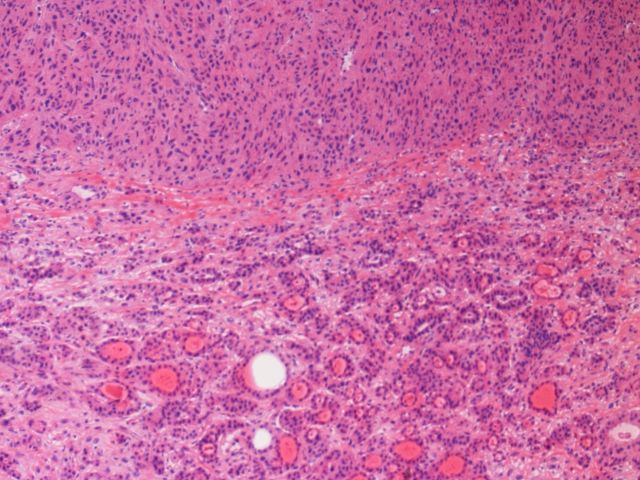
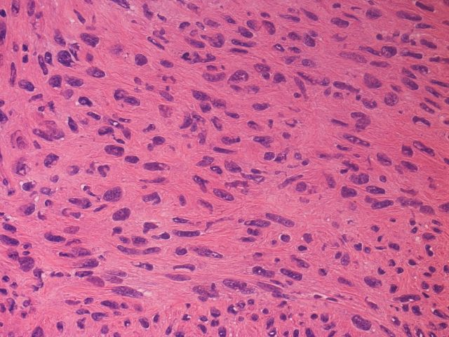
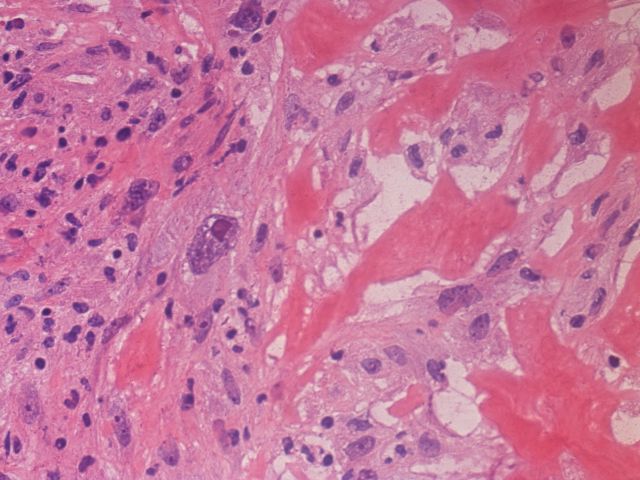
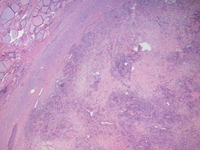
A 54 year old woman had 2 firm thyroid nodules, which were excised.
Microscopic images:

What is your diagnosis?
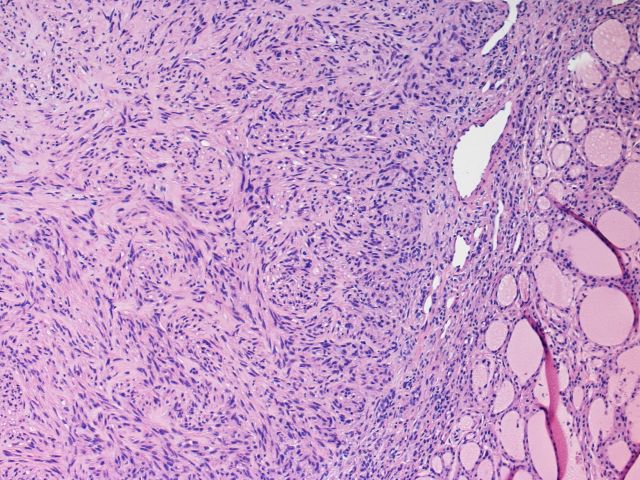
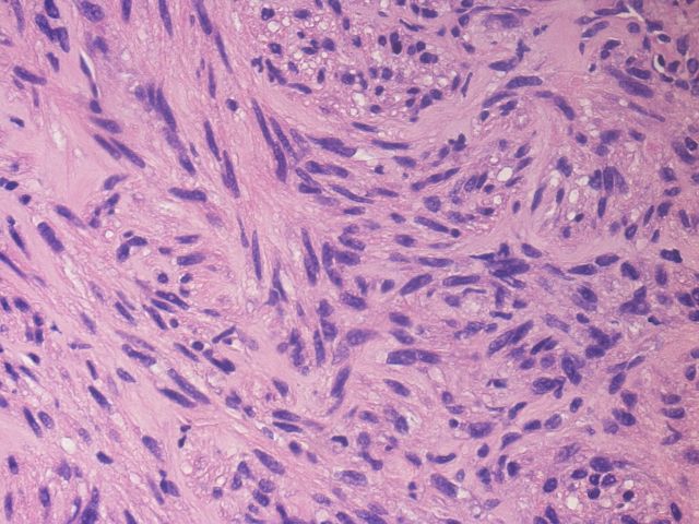
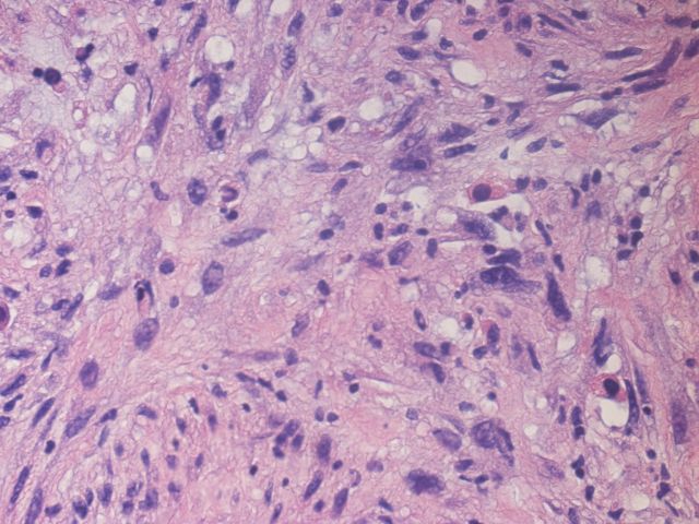
Diagnosis: Leiomyosarcoma (metastatic)
Discussion:
Tumor cells were immunoreactive (images not provided) for actin, desmin, caldesmon and vimentin, weakly positive for CD68 and had a proliferation rate of 28% with Ki67. They were negative for pan-keratin, TTF1, CD117/c-kit, S100, PAX8, CD34 and Factor VIII.
The tumor is composed of hypercellular fascicles of spindle cells resembling smooth muscle cells, with moderate to occasionally severe pleomorphism. Although mitotic figures are not numerous in the images, Ki67 shows a high proliferation rate of 28%. These histologic features, together with immunoreactivity for smooth muscle markers (actin, desmin, caldesmon) and the high Ki67 is characteristic of leiomyosarcoma. In this case, the patient was reported to have a uterine leiomyosarcoma removed 2 years prior.
Although the thyroid gland is very vascular, clinical metastases to this gland are uncommon, accounting for only 1 - 3% of thyroid malignancies, usually from kidney, lung, breast, esophagus or uterus (Head Neck Pathol 2009;3:217, Cancer 1997;79:574). In patients dying of cancer, 10 - 24% have metastases at autopsy in the thyroid gland. The differential diagnosis also includes direct extension of tumors from the pharynx, larynx, trachea, esophagus and neck, which are usually squamous cell carcinomas.
Metastatic leiomyosarcoma to the thyroid gland is very rare (Thyroid 2007;17:1295, Obstet Gynecol 2002;100:1122). The differential diagnosis includes the more common anaplastic carcinoma (Cancer 1988;62:2558). Excision of isolated metastases may be effective treatment.
All cases are archived on our website. To view them sorted by case number, diagnosis or category, visit our main Case of the Month page. To subscribe or unsubscribe to Case of the Month or our other email lists, click here.
Thanks to Dr. Cristina Aguilar, The Guthrie Clinic, Pennsylvania (USA), for contributing this case.
Advertisement
Website news:
(1) We have now posted the top 20 books sold at Amazon.com through PathologyOutlines.com from July to December 2014 on our Top Books page, which is also accessed through our Books page.
Visit Amazon.com for great shopping deals and support PathologyOutlines.com at the same time with any purchase (if you visit Amazon through a link from our website, Amazon will pay us part of their profits on any items purchased, with no extra cost to you)!
(2) Our Feature page for February is Microscopes / Microscopy Products and highlights our advertiser OPTRONICS. It also contains an original short article, "Ultrastructural Microscopy: Beyond Light and Cells", by Jaleh Mansouri, M.D.
(3) Thanks for the great response to Case of the Week submissions but it has been too great! We have decided to put new Case of the Week submissions on hold, since we now have over 40 cases in the queue. Read more.
(4) In January 2015, we broke new records on traffic to our Jobs and Conference pages. We had 45,766 visits to our Jobs pages and 10,106 visits to our Conference page! Traffic to our Fellowship pages was the highest it's been since the record in January 2013 (6,843 visits), with 6,648 visits. Thanks again for making us the "go to" place to find Jobs, Fellowships and Conferences.
(5) Typically, we respond to emails, particularly relating to ads, within 4 business hours or 24 hours at the most. If you don't get a response, please resend. We make these emails our highest priority but emails occasionally get lost.
Visit and follow our Blog to see recent updates to the website.
Case #342
Clinical history:
A 54 year old woman had 2 firm thyroid nodules, which were excised.
Microscopic images:
Frozen section slides
Permanent sections
What is your diagnosis?
Click here for diagnosis and discussion:
Diagnosis: Leiomyosarcoma (metastatic)
Discussion:
Tumor cells were immunoreactive (images not provided) for actin, desmin, caldesmon and vimentin, weakly positive for CD68 and had a proliferation rate of 28% with Ki67. They were negative for pan-keratin, TTF1, CD117/c-kit, S100, PAX8, CD34 and Factor VIII.
The tumor is composed of hypercellular fascicles of spindle cells resembling smooth muscle cells, with moderate to occasionally severe pleomorphism. Although mitotic figures are not numerous in the images, Ki67 shows a high proliferation rate of 28%. These histologic features, together with immunoreactivity for smooth muscle markers (actin, desmin, caldesmon) and the high Ki67 is characteristic of leiomyosarcoma. In this case, the patient was reported to have a uterine leiomyosarcoma removed 2 years prior.
Although the thyroid gland is very vascular, clinical metastases to this gland are uncommon, accounting for only 1 - 3% of thyroid malignancies, usually from kidney, lung, breast, esophagus or uterus (Head Neck Pathol 2009;3:217, Cancer 1997;79:574). In patients dying of cancer, 10 - 24% have metastases at autopsy in the thyroid gland. The differential diagnosis also includes direct extension of tumors from the pharynx, larynx, trachea, esophagus and neck, which are usually squamous cell carcinomas.
Metastatic leiomyosarcoma to the thyroid gland is very rare (Thyroid 2007;17:1295, Obstet Gynecol 2002;100:1122). The differential diagnosis includes the more common anaplastic carcinoma (Cancer 1988;62:2558). Excision of isolated metastases may be effective treatment.
