
All cases are archived on our website. To view them sorted by case number, diagnosis or category, visit our main Case of the Month page. To subscribe or unsubscribe to Case of the Month or our other email lists, click here.
Thanks to Dr. Margaret Lawless, University of Washington (USA), for contributing this case.

Establishing the spatial / cellular context of gene expression and validating NGS gene discoveries using
RNA in situ Hybridization
Pronin et al. used RNA-seq to sequence the entire
transcriptome of the mouse cornea and identified
~10,000 actively transcribed genes. Among them
there were 194 GPCRs, of which 96 were putative
olfactory receptors (Olfrs). They went on to validate
the 20 Olfrs by RT-PCR and RNA in situ hybridization
(ISH) via RNAscope® technology.
Join a webinar on Tues, Jan 20th, 2015 at 11 am
(Pacific US Time), 2 pm (Eastern US Time) and
understand new molecular techniques to:
- Validate RNA-Seq results
- Get spatial and quantitative gene expression information in complex tissues
- Visualize RNA targets in the full context of
tissue architecture
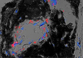
Expression of Olfr558 in vessels of the choroid and retina (Pronin et al., 2014). In situ hybridization of the mouse choroid slices with Olfr558 probe (red dots). Nuclei staining is DAPI (blue dots).
(1) Our Feature page for January is Billing / Consulting / Management and highlights our advertisers AdvantEdge Healthcare Solutions (AHS), Physicians Independent Management Services (PIMS) and Vachette Pathology. It also contains an original short article, "Medical Billing In Pathology: Billing and Consulting Firms", by Jaleh Mansouri, M.D.
(2) Now that our new Home Page has been completed, we will work on a new template for the Topic and Chapter pages. It will be similar but more consistent and with a cleaner look. After that, we will work on templates for mobile devices, to improve our website's usability for mobile.
(3) Are you shopping at Amazon.com? Before you do, please click on any Amazon.com link from our Home Page, our Books pages, or here. Purchases at Amazon.com through a link from our website support PathologyOutlines.com, without costing you anything!
Visit and follow our Blog to see recent updates to the website.
Case #338
Clinical history:
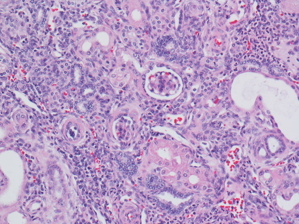
2 week old female infant presents with nephrotic syndrome and renal failure. A renal biopsy was perfomed.
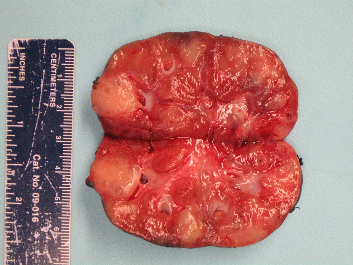
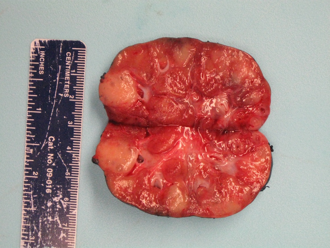
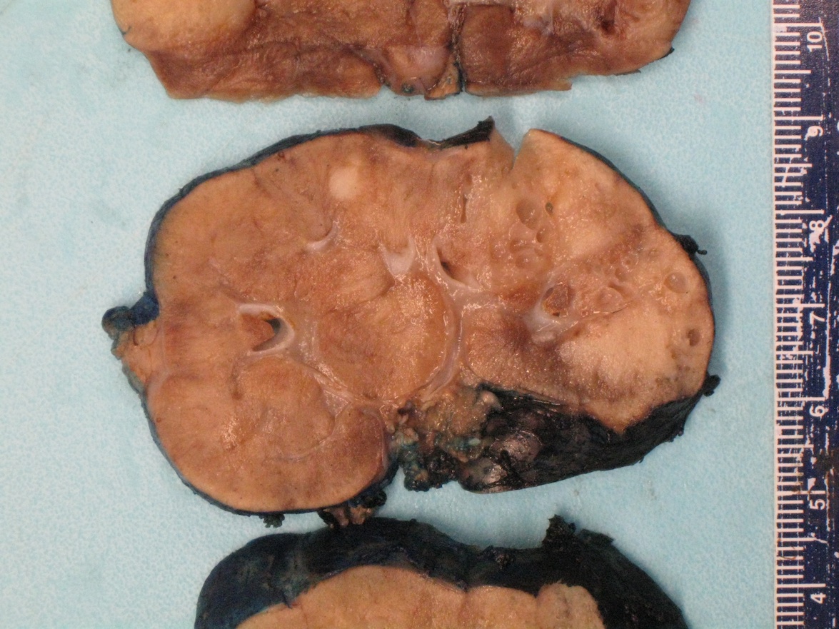
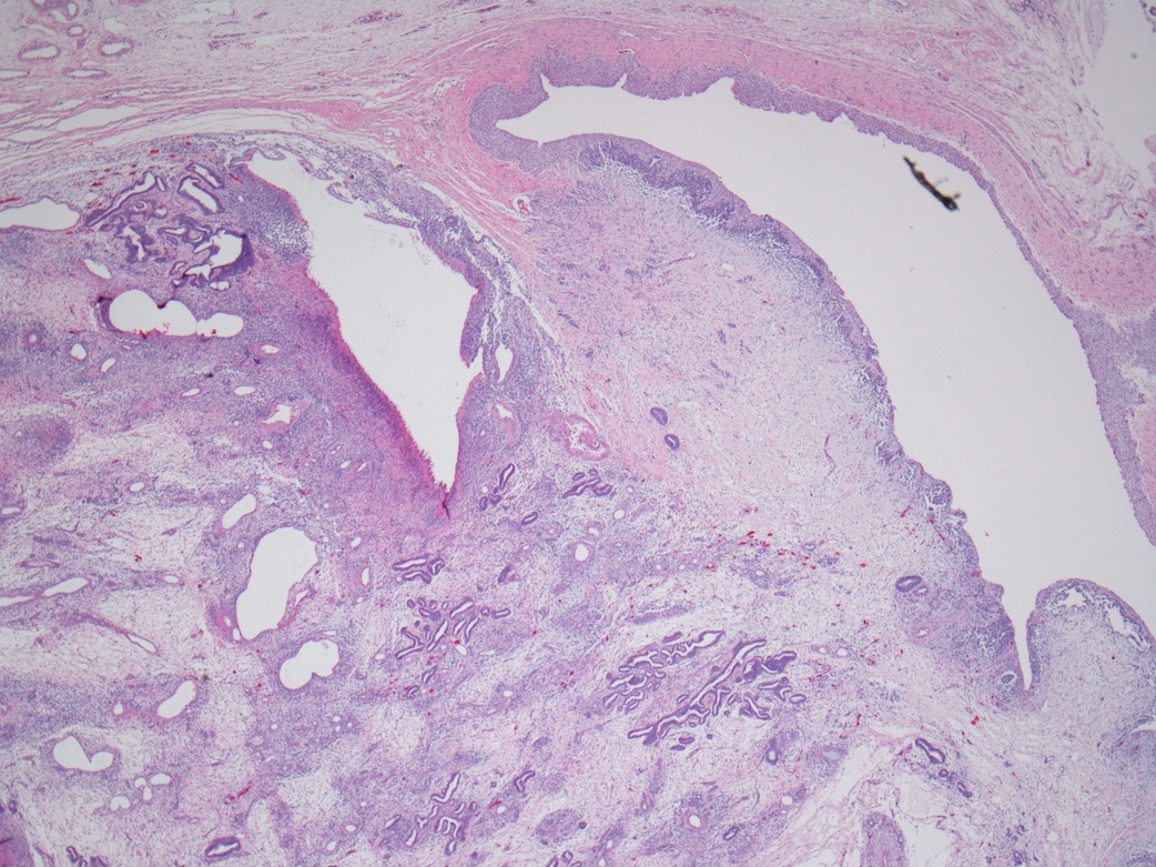
Due to the patient's syndrome (discussed below), genetic testing was performed, which revealed a 46XY karyotype and WT1 mutation. Bilateral nephrectomies were performed, which showed multiple, ill defined, firm, tan nodules distributed throughout the cortex and medulla.
Gross images:

Microscopic images:


What is your diagnosis?
Syndrome: Denys-Drash syndrome
Diagnosis: Multifocal hyperplastic intralobar rests
Discussion:
The renal biopsy shows diffuse mesangial sclerosis, a component of Denys-Drash syndrome, which is characterized by a germline WT1 mutation, male pseudohermaphroditism, diffuse mesangial sclerosis and subsequent Wilm tumor (see Wilms' tumor of children). The nephrogenic rests in the nephrectomy specimens are persistent foci of embryonal cells seen after normal nephrogenesis. When diffuse or multifocal, they are termed nephroblastomatosis. WT1 is a transcriptional regulator that mediates the mesenchymal-epithelial transition and differentiation during kidney and gonad morphogenesis. It normally represses genes that encode cell proliferation factors and activates genes that encode epithelial cell differentiation (eMedicine: Denys-Drash Syndrome [Accessed 27 December 2023]). WT1 point mutations cause result in loss of its regulatory function and the abnormalities in glomerular formation and gonadal differentiation indicated above.
Nephrogenic rests may be inapparent on gross examination but may also appear as nodules or foci of capsular thickening / scar beneath the capsule or in the deep cortex or medulla. Microscopically, they are tightly packed nests or diffuse sheets of primitive but non-anaplastic blastemal / primitive epithelial cells with scanty stroma. There is no cartilage or primitive mesenchyme. Patterns include intralobar (randomly distributed throughout cortex and medulla with irregular margins, more stroma than blastema or tubules) and perilobar (peripheral with sharply demarcated margins, composed of blastema and tubules with scanty or sclerotic stroma, often solitary). Intralobar rests have an increased rate of progression to Wilm tumor and are more commonly associated with WT1 mutations, Denys-Drash syndrome and WAGR syndrome. Perilobar rests are seen in sporadic tumors and are associated with idiopathic hemihypertrophy and Beckwith-Wiedemann syndrome (Adv Anat Pathol 2014;21:166).
Nephrogenic rests are seen in 1% of neonatal kidneys and if incidental, a wait and see policy is appropriate, because they spontaneously disappear (J Pediatr Hematol Oncol 2007;29:361). In Denys-Drash syndrome patients, most patients undergo bilateral nephrectomy, as they are otherwise destined to develop Wilm tumor.
