
10 December 2014 - Case #336
All cases are archived on our website. To view them sorted by case number, diagnosis or category, visit our main Case of the Month page. To subscribe or unsubscribe to Case of the Month or our other email lists, click here.
Thanks to Dr. Cristina Aguilar, The Guthrie Clinic, Pennsylvania (USA), for contributing this case.
Advertisement
Case #336
Clinical history:
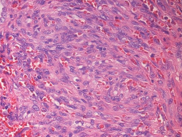
A 42 year old man with HIV and Hepatitis B virus had six months of coughing / congestion. He underwent a video assisted thoracoscopy with wedge biopsy.
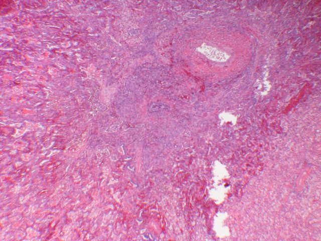
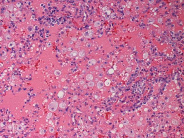
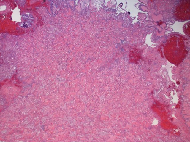
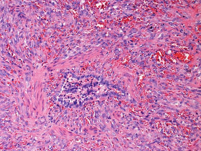
Microscopic images:

What is your diagnosis?
Diagnosis: Kaposi sarcoma (multiple foci)
Discussion:
Kaposi sarcoma of the lung or soft tissue is a vascular proliferative disorder mediated by inflammatory cytokines and angiogenic growth factors in patients with HHV8 / KHSV (Kaposi sarcoma associated herpesvirus) infection, influenced by immune status. It may originate from a cell type capable of undergoing lymphatic differentiation, based on D2-40 staining (Mod Pathol 2002;15:434). It is usually associated with AIDS but occasionally is present in HIV negative patients with other immunocompromise or organ transplant. Kaposi sarcoma is usually limited to skin but may involve mucus membranes, lymph nodes and visceral organs such as the lung.
In the lung, radiologic and clinical findings include pleural effusion, solitary lung nodules, diffuse reticulonodular pulmonary infiltrates and peribronchovascular and interlobular septal thickening (Orphanet J Rare Dis 2009 Jul 14;4:18). Grossly, tumor follows lymphatic channels and appears as discrete, dark red hemorrhagic nodules with growth along septa and infiltration of small airways, pulmonary arteries and veins.
In this case (reviewed by Dr. Thomas Colby, Mayo Clinic), the biopsy showed alveoli with foamy macrophages, proteinaceous material and inflammatory debris. However, no fungal elements or pneumocystis organisms were identified on GMS / Grocott methenamine-silver staining (stain not shown). The nodules were composed of a fascicle-like pattern of mitotically active, pleomorphic spindle cells in a background of inflammatory cells and red blood cells. Small vessels and slitlike spaces with hyaline droplets are often present. The spindle cells are positive for CD34 and HHV8 (immunostains not shown). HHV8 immunostaining is also present in primary effusion lymphoma, multicentric Castleman disease and multicentric Castleman disease associated plasmablastic lymphoma. In HIV+ patients, HHV8 may also be present in hemangiomas (Mod Pathol 2005;18:463).
Management consists of HAART / highly active antiretroviral therapy, followed by systemic chemotherapy (Mayo Clin Proc 2012;87:e77).
All cases are archived on our website. To view them sorted by case number, diagnosis or category, visit our main Case of the Month page. To subscribe or unsubscribe to Case of the Month or our other email lists, click here.
Thanks to Dr. Cristina Aguilar, The Guthrie Clinic, Pennsylvania (USA), for contributing this case.

PathConsulting was founded to meet the business needs of pathologists, pathology managers and laboratories to provide access to high quality, professional management and advisory assistance. PathConsulting is a subsidiary of the American Pathology Foundation (APF) and has adopted the philosophy and ethics built by the APF over the past 50 years. PathConsulting serves ONLY the pathology and laboratory industry, with the scope of engagements provided by some of the most highly regarded duly respected individuals in the business. PathConsulting delivers advantages in areas of:
• Financial Management • Operations Management • Human Resources • Compliance
To learn more about PathConsulting, visit www.pathconsulting.org.
Website news:
(1) Are you shopping at Amazon.com? Be sure to click on any Amazon.com link from our Home Page, our Books pages or here. It helps support our website, without costing you anything!
(2) Our Feature page for December is Grossing Equipment / Workstations and highlights our advertisers EXAKT Technologies, Inc., Leica Biosystems, Milestone Medical, MOPEC and Photodyne Technologies. It also contains an original short article, "Pathology Grossing Tools of the Trade: Blades and Scalpels", by Jaleh Mansouri, M.D.
(3) Get ready for our new Home Page, to arrive by the end of the year. All of the old links will be there but they will be rearranged to make them a little easier to access and to provide a cleaner look. At this time, ONLY the Home page is being changed. Underlying the new Home Page is a state of the art Content Management System ("computer infrastructure"), which will create more uniformity on the pages and allow for a Topic Search in the future.
(4) We are seeking experts to serve on our Editorial Board to provide a secondary review of topics as they are updated. Editorial Board members are listed on the Authors page, on the relevant Chapter page and on all topics that they review. For more information on joining our Editorial Board, please click here or email Dr. Pernick at NatPernick@hotmail.com.
Visit and follow our Blog to see recent updates to the website.
Case #336
Clinical history:
A 42 year old man with HIV and Hepatitis B virus had six months of coughing / congestion. He underwent a video assisted thoracoscopy with wedge biopsy.
Microscopic images:
What is your diagnosis?
Click here for diagnosis and discussion:
Diagnosis: Kaposi sarcoma (multiple foci)
Discussion:
Kaposi sarcoma of the lung or soft tissue is a vascular proliferative disorder mediated by inflammatory cytokines and angiogenic growth factors in patients with HHV8 / KHSV (Kaposi sarcoma associated herpesvirus) infection, influenced by immune status. It may originate from a cell type capable of undergoing lymphatic differentiation, based on D2-40 staining (Mod Pathol 2002;15:434). It is usually associated with AIDS but occasionally is present in HIV negative patients with other immunocompromise or organ transplant. Kaposi sarcoma is usually limited to skin but may involve mucus membranes, lymph nodes and visceral organs such as the lung.
In the lung, radiologic and clinical findings include pleural effusion, solitary lung nodules, diffuse reticulonodular pulmonary infiltrates and peribronchovascular and interlobular septal thickening (Orphanet J Rare Dis 2009 Jul 14;4:18). Grossly, tumor follows lymphatic channels and appears as discrete, dark red hemorrhagic nodules with growth along septa and infiltration of small airways, pulmonary arteries and veins.
In this case (reviewed by Dr. Thomas Colby, Mayo Clinic), the biopsy showed alveoli with foamy macrophages, proteinaceous material and inflammatory debris. However, no fungal elements or pneumocystis organisms were identified on GMS / Grocott methenamine-silver staining (stain not shown). The nodules were composed of a fascicle-like pattern of mitotically active, pleomorphic spindle cells in a background of inflammatory cells and red blood cells. Small vessels and slitlike spaces with hyaline droplets are often present. The spindle cells are positive for CD34 and HHV8 (immunostains not shown). HHV8 immunostaining is also present in primary effusion lymphoma, multicentric Castleman disease and multicentric Castleman disease associated plasmablastic lymphoma. In HIV+ patients, HHV8 may also be present in hemangiomas (Mod Pathol 2005;18:463).
Management consists of HAART / highly active antiretroviral therapy, followed by systemic chemotherapy (Mayo Clin Proc 2012;87:e77).
