
27 August 2014 - Case #324
All cases are archived on our website. To view them sorted by case number, diagnosis or category, visit our main Case of the Month page. To subscribe or unsubscribe to Case of the Month or our other email lists, click here.
Thanks to Dr. Govindaswamy Koteeswaran, Mahatma Gandhi Medical College and Research Institute (India), for contributing this case and discussion.
Advertisement
Case #324
Clinical history:
A 3 month old child presented with a painful nodule on the left little finger, present since birth, which was excised.
Gross examination showed a 2.0 x 1.5 cm skin covered nodule. The cut section showed a homogenous dark brown appearance.
Gross images:

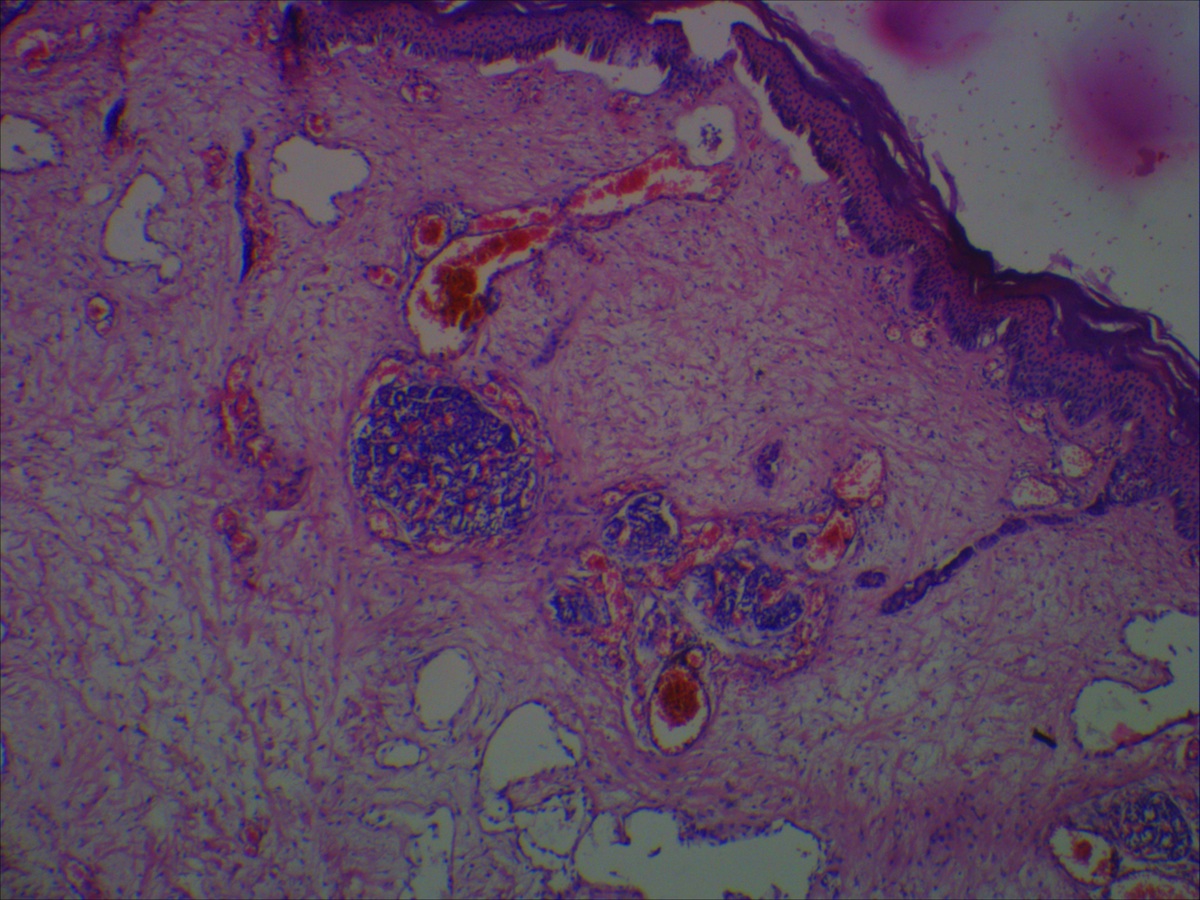
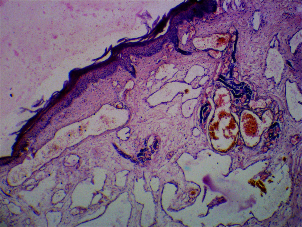
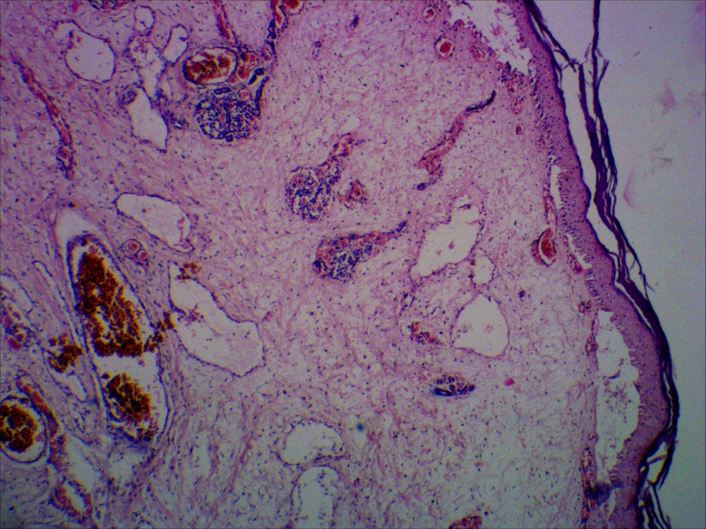
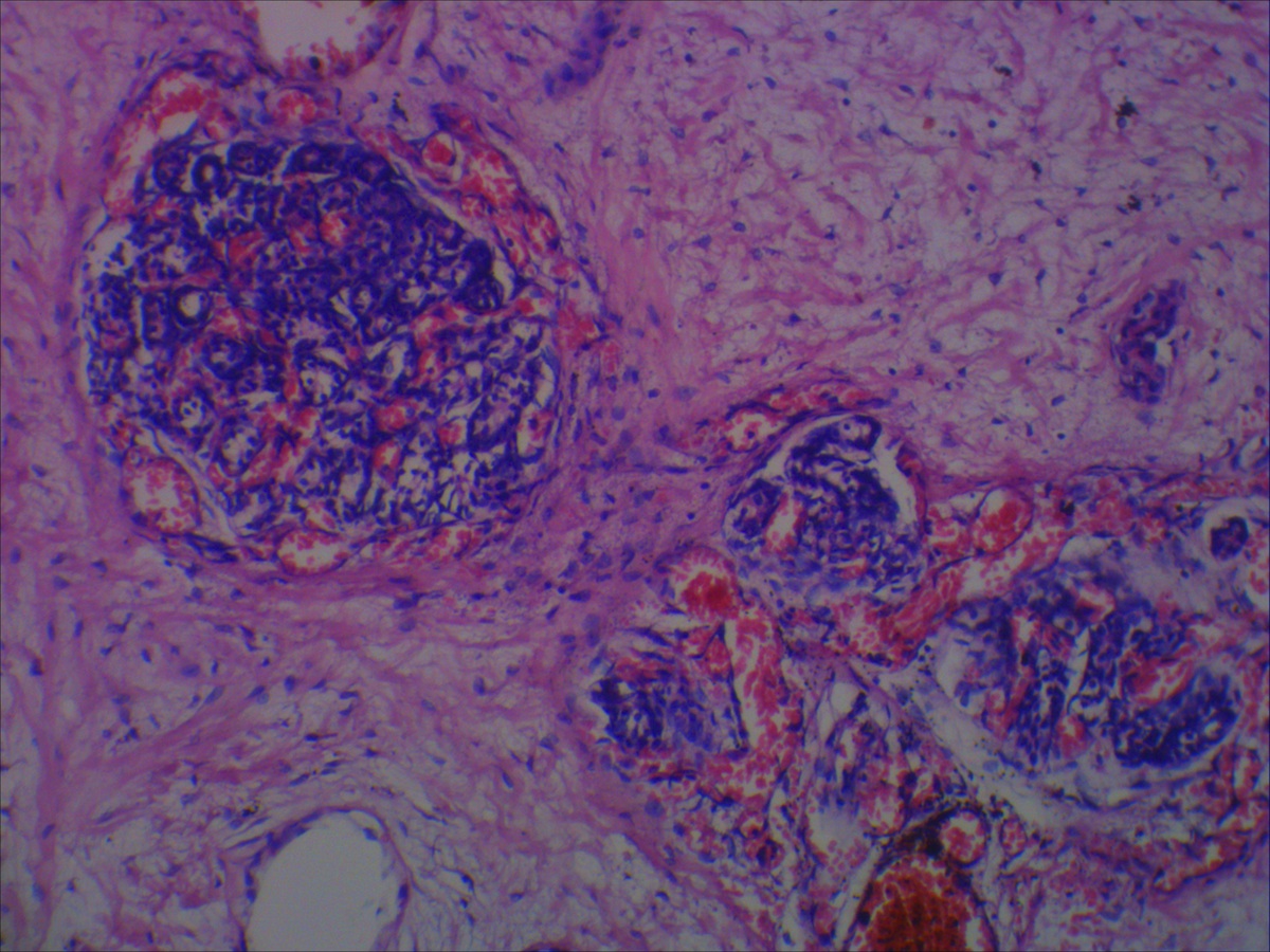
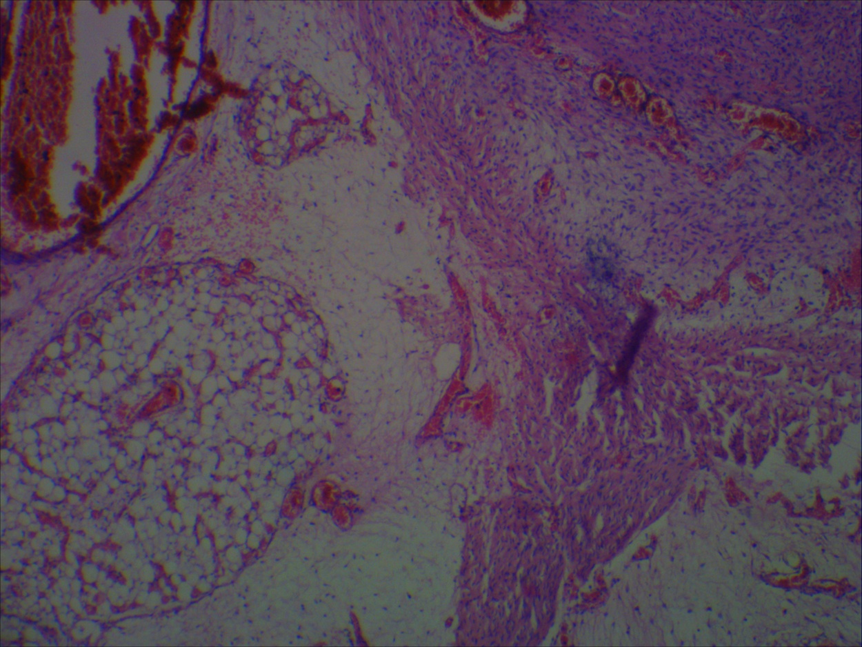
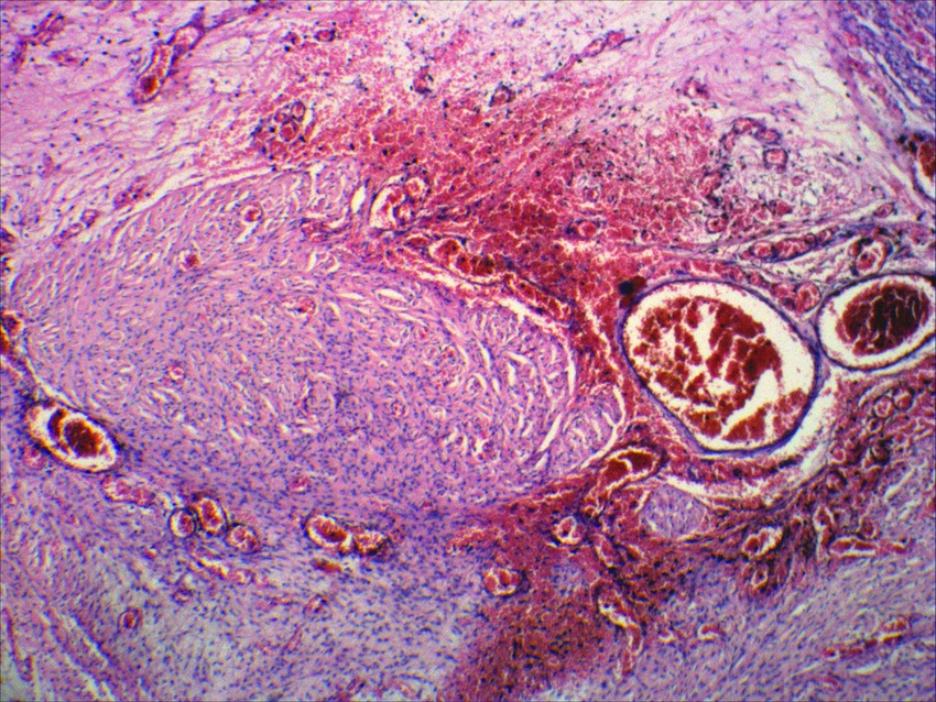
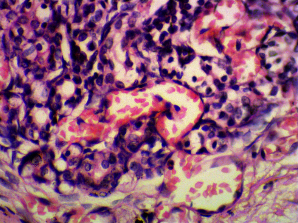
Microscopic images:
What is your diagnosis?
Diagnosis: Congenital eccrine angiomatous hamartoma
Discussion:
Microscopic examination showed an increased number of eccrine glands in the superficial dermis, with blood vessels between and around the glands and ducts. The vessels varied in size, but were predominantly small and lined by endothelial cells. Other areas of dermis showed smooth muscle bundles, fat cells and focal areas of myxoid change.
Congenital eccrine angiomatous hamartoma (EAH) is a rare benign hamartoma of eccrine glands and blood vessels that often occurs in the extremities (Ann Dermatol 2013;25:208). It may be present at birth or develop during childhood (Dermatology Online Journal 2009;15:10). It may be painful and be associated with hyperhidrosis, which can be helpful in diagnosis.
EAH must be differentiated from similar conditions including angiokeratoma, eccrine nevus, glomus tumor, hemorrhage, macular telangiectatic mastocytosis, nevus flammeus and smooth muscle hamartoma (Ann Dermatol 2011;23 Suppl 1:S84).
EAH is typically slow growing with benign behavior. Simple excision is usually curative, but is reserved for painful and cosmetically disfiguring lesions (Ann Dermatol Venereol 1997;124:623, Pediatr Dermatol 2009;26:316).
All cases are archived on our website. To view them sorted by case number, diagnosis or category, visit our main Case of the Month page. To subscribe or unsubscribe to Case of the Month or our other email lists, click here.
Thanks to Dr. Govindaswamy Koteeswaran, Mahatma Gandhi Medical College and Research Institute (India), for contributing this case and discussion.

Scientific Symposiums International Fall CME Series
Essentials of Molecular and Surgical Pathology in the Diagnosis of Lymphomas, Leukemias and CNS, ENT and Endocrine Lesions
October 6-9, 2014
The Hapuna Beach Prince Hotel, Mauna Kea Resort, Big Island of Hawaii (USA)
Register for our upcoming Surgical Pathology Course course being held this October in order to learn the essentials of molecular and surgical pathology in the diagnosis of lymphomas, leukemias and CNS, ENT and Endocrine Lesions.
Our world-renowned faculty includes Gregory Fuller, M.D., Ph.D., Adam Bagg, M.D., Thomas J. Giordano, M.D., Ph.D., and Bruce M. Wenig, M.D.
Website news:
(1) We are now adding thumbnails and links of new books to the chapter pages each month, to help visitors learn about new offerings. Remember, if you click on any of these links, and then buy that book or anything else from Amazon.com, you are helping to support our website.
(2) We hope you will stop by our booth this year at CAP '14 in Chicago! We are booth #623. Please let us know how we can make the website more useful to you. We can also take your picture for our Facebook page.
(3) Some of our readers may have already noticed, but the thumbnail links to many of the Webpathology.com images are no longer loading. We are finding that many Mac users are having difficulty connecting and loading the Webpathology pages while using Firefox or Chrome browsers, with a slight delay using Safari. From what we understand, this problem is not being experienced by PC users. We are still continuing to add those images and are working with the owner of Webpathology to resolve these issues.
Visit and follow our Blog to see recent updates to the website.
Case #324
Clinical history:
A 3 month old child presented with a painful nodule on the left little finger, present since birth, which was excised.
Gross examination showed a 2.0 x 1.5 cm skin covered nodule. The cut section showed a homogenous dark brown appearance.
Gross images:
Microscopic images:
What is your diagnosis?
Click here for diagnosis and discussion:
Diagnosis: Congenital eccrine angiomatous hamartoma
Discussion:
Microscopic examination showed an increased number of eccrine glands in the superficial dermis, with blood vessels between and around the glands and ducts. The vessels varied in size, but were predominantly small and lined by endothelial cells. Other areas of dermis showed smooth muscle bundles, fat cells and focal areas of myxoid change.
Congenital eccrine angiomatous hamartoma (EAH) is a rare benign hamartoma of eccrine glands and blood vessels that often occurs in the extremities (Ann Dermatol 2013;25:208). It may be present at birth or develop during childhood (Dermatology Online Journal 2009;15:10). It may be painful and be associated with hyperhidrosis, which can be helpful in diagnosis.
EAH must be differentiated from similar conditions including angiokeratoma, eccrine nevus, glomus tumor, hemorrhage, macular telangiectatic mastocytosis, nevus flammeus and smooth muscle hamartoma (Ann Dermatol 2011;23 Suppl 1:S84).
EAH is typically slow growing with benign behavior. Simple excision is usually curative, but is reserved for painful and cosmetically disfiguring lesions (Ann Dermatol Venereol 1997;124:623, Pediatr Dermatol 2009;26:316).
