
13 August 2014 - Case #322
All cases are archived on our website. To view them sorted by case number, diagnosis or category, visit our main Case of the Month page. To subscribe or unsubscribe to Case of the Month or our other email lists, click here.
Thanks to Dr. Divya Sharma, University of Cincinnati Medical Center (USA), for contributing this case and discussion.

Scientific Symposiums International Fall CME Series
Essentials of Molecular and Surgical Pathology in the Diagnosis of Lymphomas, Leukemias and CNS, ENT and Endocrine Lesions
October 6-9, 2014
The Hapuna Beach Prince Hotel, Mauna Kea Resort, Big Island of Hawaii (USA)
Register for our upcoming Surgical Pathology Course course being held this October in order to learn the essentials of molecular and surgical pathology in the diagnosis of lymphomas, leukemias and CNS, ENT and Endocrine Lesions.
Our world-renowned faculty includes Gregory Fuller, M.D., Ph.D., Adam Bagg, M.D., Thomas J. Giordano, M.D., Ph.D., and Bruce M. Wenig, M.D.
This course is one not to be missed!
Advertisement
Case #322
Clinical history:
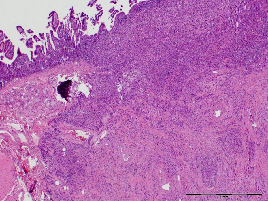
A 50 year old man with obstructive jaundice had an ampullary mass. A biopsy was followed by the Whipple procedure (pancreaticoduodenectomy).
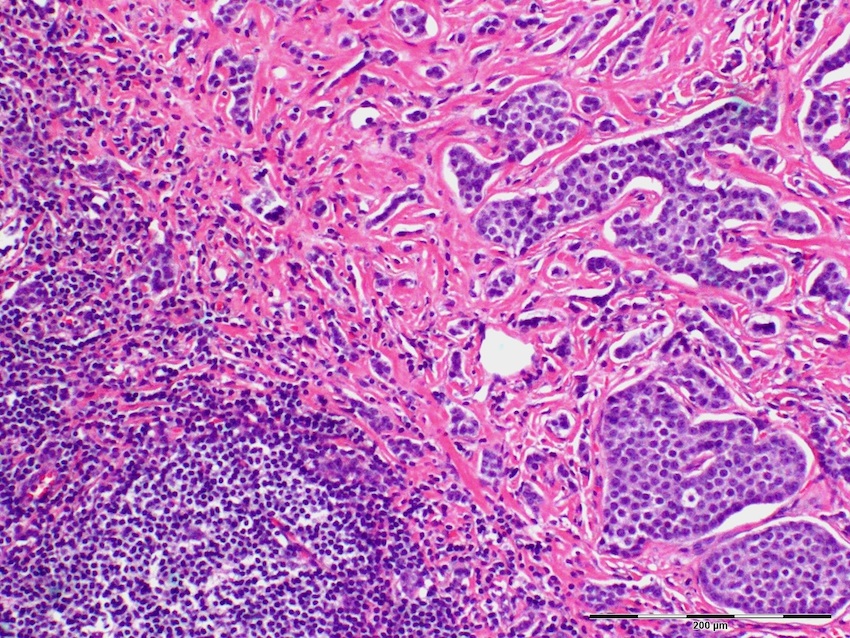
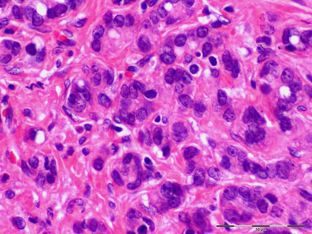
Microscopic images:

What is your diagnosis?
Diagnosis: Collision tumor mixed adenoneuroendocrine carcinoma of the duodenum (> 30% each component)
Immunostains:


Discussion:
In addition, many cells are positive for CK20, occasional cells are positive for CK7.
Collision tumor is a well documented but uncommon phenomenon characterized by (a) 2 distinctive and spatially independent tumor components in the same location and (b) distinct and well demarcated morphology and immunoexpression patterns between the 2 tumors microscopically, with no intervening intermediate cell populations. This entity is distinct from neoplasms demonstrating heterologous or mixed elements. Collision tumors in general, and those involving adenocarcinoma and neuroendocrine carcinomas in particular, are extremely rare in the duodenum, ampulla and stomach (Rare Tumors 2012;4:e20, Gut 2001;48:853, Rev Esp Enferm Dig 2007;99:235, HPB Surg 1997;10:241, Case Rep Gastroenterol 2014;8:89).
The differential diagnosis includes adenocarcinoma with neuroendocrine features.
All cases are archived on our website. To view them sorted by case number, diagnosis or category, visit our main Case of the Month page. To subscribe or unsubscribe to Case of the Month or our other email lists, click here.
Thanks to Dr. Divya Sharma, University of Cincinnati Medical Center (USA), for contributing this case and discussion.
Scientific Symposiums International Fall CME Series
Essentials of Molecular and Surgical Pathology in the Diagnosis of Lymphomas, Leukemias and CNS, ENT and Endocrine Lesions
October 6-9, 2014
The Hapuna Beach Prince Hotel, Mauna Kea Resort, Big Island of Hawaii (USA)
Register for our upcoming Surgical Pathology Course course being held this October in order to learn the essentials of molecular and surgical pathology in the diagnosis of lymphomas, leukemias and CNS, ENT and Endocrine Lesions.
Our world-renowned faculty includes Gregory Fuller, M.D., Ph.D., Adam Bagg, M.D., Thomas J. Giordano, M.D., Ph.D., and Bruce M. Wenig, M.D.
This course is one not to be missed!
Website news:
(1) We continue to add more surveys to our Surveys and Buyer's Guide pages. Check out what is new in products and services for pathologists.
(2) We have now posted the top books sold at Amazon.com through PathologyOutlines.com for the first half of 2014, click here or on the Top Books link from the Books page. Buy your books through PathologyOutlines.com - we are the only place where you can see all the books that have been recently published!
(3) Our Feature page for August is Books & Journals, and highlights the publishers ARP Press, ASCP, CAP, LWW and WHO Press. It also contains an original short article, "Pathology Books and Journals: A Multidisciplinary Approach", by Jaleh Mansouri, M.D.
Visit and follow our Blog to see recent updates to the website.
Case #322
Clinical history:
A 50 year old man with obstructive jaundice had an ampullary mass. A biopsy was followed by the Whipple procedure (pancreaticoduodenectomy).
Microscopic images:
What is your diagnosis?
Click here for diagnosis and discussion:
Diagnosis: Collision tumor mixed adenoneuroendocrine carcinoma of the duodenum (> 30% each component)
Immunostains:
CDX2
Chromogranin
Synaptophysin
Discussion:
In addition, many cells are positive for CK20, occasional cells are positive for CK7.
Collision tumor is a well documented but uncommon phenomenon characterized by (a) 2 distinctive and spatially independent tumor components in the same location and (b) distinct and well demarcated morphology and immunoexpression patterns between the 2 tumors microscopically, with no intervening intermediate cell populations. This entity is distinct from neoplasms demonstrating heterologous or mixed elements. Collision tumors in general, and those involving adenocarcinoma and neuroendocrine carcinomas in particular, are extremely rare in the duodenum, ampulla and stomach (Rare Tumors 2012;4:e20, Gut 2001;48:853, Rev Esp Enferm Dig 2007;99:235, HPB Surg 1997;10:241, Case Rep Gastroenterol 2014;8:89).
The differential diagnosis includes adenocarcinoma with neuroendocrine features.
