
2 July 2014 - Case #318
All cases are archived on our website. To view them sorted by case number, diagnosis or category, visit our main Case of the Month page. To subscribe or unsubscribe to Case of the Month or our other email lists, click here.
Thanks to Dr. Nasir Uddin, Aga Khan University Hospital (Pakistan), for contributing this case.
Case #318
Clinical history:
A 50 year old man presented with a right lung tumor, which as biopsied. Grossly, multiple fragments of tumor measured 5 cm in aggregate.
Radiology images:

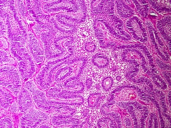
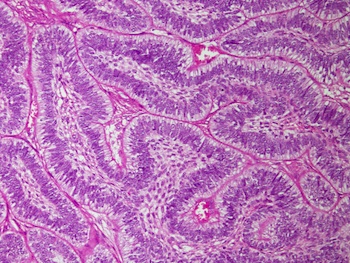
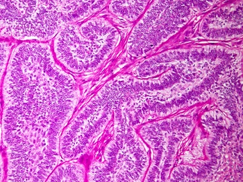
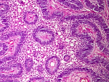
Microscopic images:
What is your diagnosis?
Diagnosis: Ameloblastoma metastasis to lung
Immunostains:

Discussion:
No normal lung tissue was seen in the biopsy. The cells have a benign appearance, arranged as back to back elongated nests with peripheral palisading, central loose reticulum, minimal hyperchromatism and rare mitotic activity. The tumor cells were positive for CK AE1 / AE3 and p63 and negative for BerEP4 and TTF1. The MIB1 index was low. The referring clinician subsequently reported that the patient had a left mandible tumor resected 10 years ago which was reported as ameloblastoma. There was no histology report but a recent Xray of the mandible was obtained, which showed a previous tumor excision site.
Ameloblastoma, previously called adamantinoma, represents 1% of jaw tumors and cysts. It occurs in men and women of all ages, with a mean age of 39 years. The tumor arises from remnants of ameloblast, dental lamina, dentigerous cysts or the basal layer of oral mucosa. Clinically, it is slow growing and locally aggressive, with a recurrence rate of 25 - 35%. Metastases to lungs or CNS, as in this case, are rare, and are associated with tumor of long duration, multiple surgical procedures and radiation therapy (Rom J Morphol Embryol 2014;55:183, J Craniomaxillofac Surg 2014;42:e301, Int J Surg Pathol 2013;22:343, J Craniomaxillofac Surg 2012;40:e470). Metastases may be treated successfully by carboplatin / paclitaxel chemotherapy (Ecancermedicalscience 2013;7:323).
The differential diagnosis for pulmonary metastases includes primary squamous cell carcinoma or a basaloid variant (Korean J Thorac Cardiovasc Surg 2014;47:63).
All cases are archived on our website. To view them sorted by case number, diagnosis or category, visit our main Case of the Month page. To subscribe or unsubscribe to Case of the Month or our other email lists, click here.
Thanks to Dr. Nasir Uddin, Aga Khan University Hospital (Pakistan), for contributing this case.
Case #318
Clinical history:
A 50 year old man presented with a right lung tumor, which as biopsied. Grossly, multiple fragments of tumor measured 5 cm in aggregate.
Radiology images:
Microscopic images:
What is your diagnosis?
Click here for diagnosis and discussion:
Diagnosis: Ameloblastoma metastasis to lung
Immunostains:
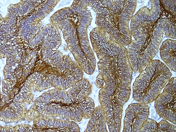
CK AE1 / AE3
p63
Discussion:
No normal lung tissue was seen in the biopsy. The cells have a benign appearance, arranged as back to back elongated nests with peripheral palisading, central loose reticulum, minimal hyperchromatism and rare mitotic activity. The tumor cells were positive for CK AE1 / AE3 and p63 and negative for BerEP4 and TTF1. The MIB1 index was low. The referring clinician subsequently reported that the patient had a left mandible tumor resected 10 years ago which was reported as ameloblastoma. There was no histology report but a recent Xray of the mandible was obtained, which showed a previous tumor excision site.
Ameloblastoma, previously called adamantinoma, represents 1% of jaw tumors and cysts. It occurs in men and women of all ages, with a mean age of 39 years. The tumor arises from remnants of ameloblast, dental lamina, dentigerous cysts or the basal layer of oral mucosa. Clinically, it is slow growing and locally aggressive, with a recurrence rate of 25 - 35%. Metastases to lungs or CNS, as in this case, are rare, and are associated with tumor of long duration, multiple surgical procedures and radiation therapy (Rom J Morphol Embryol 2014;55:183, J Craniomaxillofac Surg 2014;42:e301, Int J Surg Pathol 2013;22:343, J Craniomaxillofac Surg 2012;40:e470). Metastases may be treated successfully by carboplatin / paclitaxel chemotherapy (Ecancermedicalscience 2013;7:323).
The differential diagnosis for pulmonary metastases includes primary squamous cell carcinoma or a basaloid variant (Korean J Thorac Cardiovasc Surg 2014;47:63).
