
11 June 2014 - Case #315
All cases are archived on our website. To view them sorted by case number, diagnosis or category, visit our main Case of the Month page. To subscribe or unsubscribe to Case of the Month or our other email lists, click here.
Thanks to Dr. Nasir Uddin, Aga Khan University Hospital (Pakistan), for contributing this case.
Advertisement
Case #315
Clinical history:
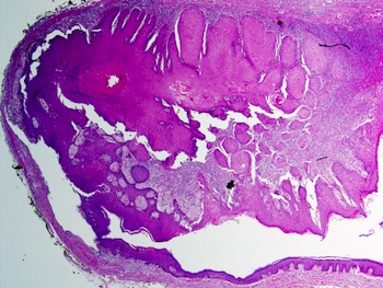
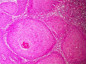
A 53 year old man presented with a chest wall sebaceous cyst which was excised. The gross specimen was 2.5 cm of excised skin with subcutaneous tissue containing a 1.5 cm cyst.
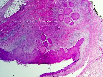
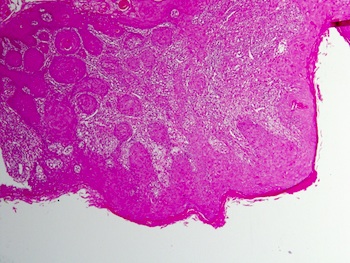
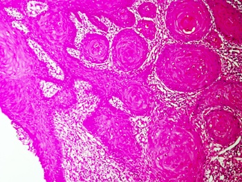
Microscopic images:


What is your diagnosis?
Diagnosis: Squamous cell carcinoma arising in an epidermal inclusion cyst
Discussion:
Epidermoid cysts are the most common cutaneous cysts, typically appearing on the face, scalp, neck and trunk (eMedicine: Epidermal Inclusion Cyst [Accessed 21 March 2024]). Most lesions originate from the follicular infundibulum, so the clinical term sebaceous cyst is inaccurate. Histologically, these lesions are lined by keratinized epithelium with a distinct granular layer. The cyst contains lamellated keratin but no calcification.
Although benign, premalignant and malignant lesions have been identified in their walls, often as incidental findings, illustrating the importance of routine histology in these lesions (J Plast Reconstr Aesthet Surg 2012;65:1267, Ann Acad Med Singapore 2000;29:757, N Am J Med Sci 2010;2:46).
Treatment consists of excision and possibly retinoids for chemoprevention (Dermatol Ther 2014;27:94).
All cases are archived on our website. To view them sorted by case number, diagnosis or category, visit our main Case of the Month page. To subscribe or unsubscribe to Case of the Month or our other email lists, click here.
Thanks to Dr. Nasir Uddin, Aga Khan University Hospital (Pakistan), for contributing this case.


Leica Biosystems releases two new customer apps for Immunohistochemistry (IHC) Education and Reference for Path Lead and IHC Antibody Catalog.
- PathLead is a mobile educational tool and pathology resource application, designed for use by healthcare professionals in the field of surgical pathology and diagnostic immunohistochemistry (IHC).
- IHC Antibody Catalog is an application for Laboratory Managers and Pathologists looking for both routine and innovative IHC antibodies and consumables.
- Both are currently available for download at the Apple App Store.
Click here for more information!
Website news:
(1) There is now an easier way to post images on our Flickr page. Just email the pictures to part85keep@photos.flickr.com and send a CC to CommentsPathout@gmail.com. You can use the subject line to give the image a title and the body to add a description, which should include the diagnosis and the contributor's name.
(2) The links to Webpathology.com do work, but we are in the process of updating the thumbnails, which may take some time since there are so many.
(3) In May 2014, we had record traffic of 623,482 visits, with a daily record of 25,244 visits on May 14.
(4) Our Feature page for June is Diagnostic Testing / Reagents, and highlights Advanced Cell Diagnostics, Inc. (ACD), bioTheranostics, Cell Marque, Leica Biosystems and Ventana Medical. It also contains an original short article, "Diagnostic Reagents: The Potential Role of microRNAs", by Jaleh Mansouri, M.D.
Visit and follow our Blog to see recent updates to the website.
Case #315
Clinical history:
A 53 year old man presented with a chest wall sebaceous cyst which was excised. The gross specimen was 2.5 cm of excised skin with subcutaneous tissue containing a 1.5 cm cyst.
Microscopic images:
What is your diagnosis?
Click here for diagnosis and discussion:
Diagnosis: Squamous cell carcinoma arising in an epidermal inclusion cyst
Discussion:
Epidermoid cysts are the most common cutaneous cysts, typically appearing on the face, scalp, neck and trunk (eMedicine: Epidermal Inclusion Cyst [Accessed 21 March 2024]). Most lesions originate from the follicular infundibulum, so the clinical term sebaceous cyst is inaccurate. Histologically, these lesions are lined by keratinized epithelium with a distinct granular layer. The cyst contains lamellated keratin but no calcification.
Although benign, premalignant and malignant lesions have been identified in their walls, often as incidental findings, illustrating the importance of routine histology in these lesions (J Plast Reconstr Aesthet Surg 2012;65:1267, Ann Acad Med Singapore 2000;29:757, N Am J Med Sci 2010;2:46).
Treatment consists of excision and possibly retinoids for chemoprevention (Dermatol Ther 2014;27:94).
