
23 April 2014 - Case #309
All cases are archived on our website. To view them sorted by case number, diagnosis or category, visit our main Case of the Month page. To subscribe or unsubscribe to Case of the Month or our other email lists, click here.
Thanks to Dr. Joseph Fullmer, SUNY Upstate Medical University (USA) and Dr. Wei Huang, University of Wisconsin Hospital and Clinics (USA), for contributing this case.
Case #309
Clinical history:
A 16 year old boy presented with a chronic left testicular mass, with no recent change in size. Serum tumor markers were normal. Imaging revealed a homogenous, 3.6 cm mass adjacent to the left testis. It did not have the classic appearance of testicular cancer and may have represented polyorchidism, but a malignant rhabdomyosarcoma or an infectious cause could not be excluded without biopsy or excision. A left radical orchiectomy was performed.
Microscopic images:

What is your diagnosis?
Diagnosis: Splenogonadal fusion syndrome
Discussion:
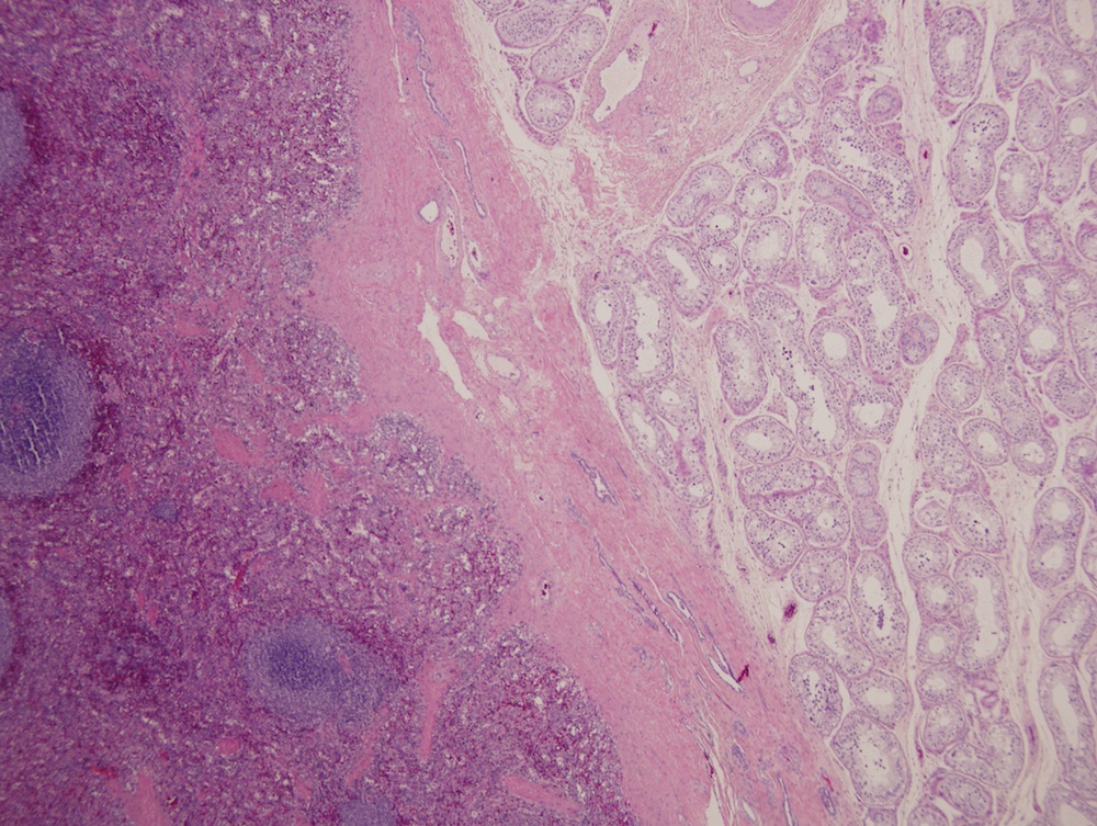
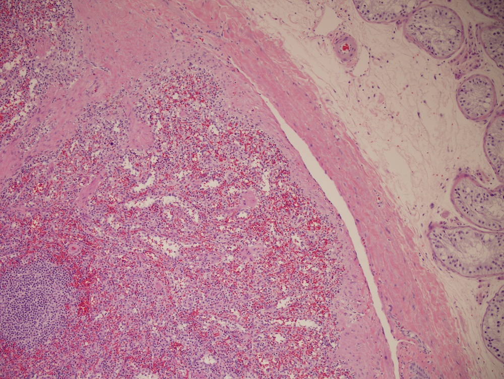
The microscopic sections show histologically normal splenic tissue adherent to testicular parenchyma. Splenogonal fusion syndrome is a rare congenital condition, usually presenting before age 20, with ≥ 50% of cases presenting before age 10. In men, it occurs only in the left testis. It can also occur in women.
The fusion is termed either continuous (attaching to spleen) or discontinuous (intrascrotal splenic nodules attached to testis, spermatic cord, epididymis, appendix of testis or appendix of epididymis). The continuous form is associated with limb-bud anomalies such as peromelia (severe congenital anomalies of extremities identical to thalidomide embryopathy) and micrognatia (small jaw) (Urology 1988;32:521, OMIM: 183300 [Accessed 22 March 2024]). The discontinuous type is rarely associated with cardiac defects.
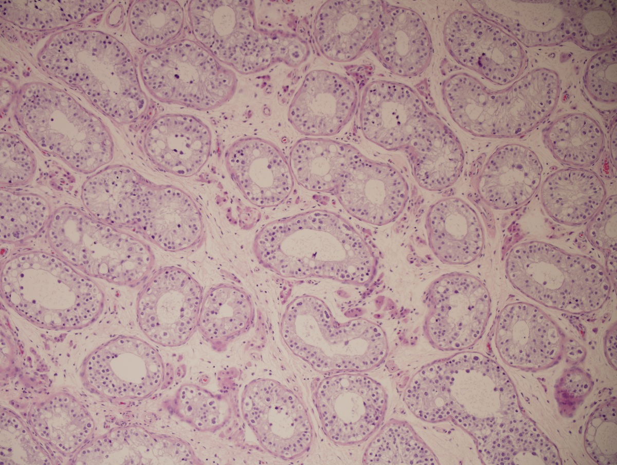
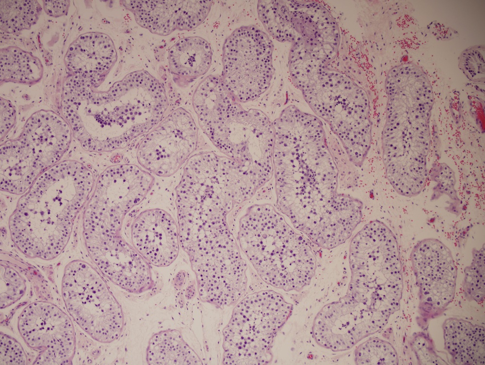
Gross examination shows splenic tissue well demarcated from the gonad / testis. Microscopically, as in this case, normal splenic parenchyma is identified, but there may also be fibrosis, thrombi, calcification, fatty degeneration and hemosiderin. The testicular tissue may be normal, or have atrophy or fibrosis of seminiferous tubules, increased Leydig cells or thrombosis of spermatic vessels.
The differential diagnosis includes lymphoma or a germ cell tumor.
Optimal treatment is excision of the ectopic splenic tissue only, to prevent testicular atrophy, torsion or infarction and preserve fertility (Ann R Coll Surg Engl 2006;88:163, Exp Ther Med 2013;6:816).
All cases are archived on our website. To view them sorted by case number, diagnosis or category, visit our main Case of the Month page. To subscribe or unsubscribe to Case of the Month or our other email lists, click here.
Thanks to Dr. Joseph Fullmer, SUNY Upstate Medical University (USA) and Dr. Wei Huang, University of Wisconsin Hospital and Clinics (USA), for contributing this case.
Website news:
(1) Huy Phu Pham, M.D. has written 3 new Apharesis topics for the Transfusion Medicine chapter: LDL apheresis, leukocytapheresis and plateletpheresis.
(2) Here are the ten most popular books sold through our website and Amazon.com, from January to March 2014:
Visit and follow our Blog to see recent updates to the website.
(1) Huy Phu Pham, M.D. has written 3 new Apharesis topics for the Transfusion Medicine chapter: LDL apheresis, leukocytapheresis and plateletpheresis.
(2) Here are the ten most popular books sold through our website and Amazon.com, from January to March 2014:
- Patil: McGraw-Hill Specialty Board Review Anatomic Pathology
- Dabbs: Diagnostic Immunohistochemistry
- Clement: Atlas of Gynecologic Surgical Pathology
- Amin: Urological Pathology
- Lakhani: WHO Classification of Tumours of the Breast
- Montgomery: Biopsy Interpretation of theGastrointestinal Tract Mucosa: Volume 1: Non-Neoplastic
- Montgomery: Biopsy Interpretation of theGastrointestinal Tract Mucosa: Volume 2: Neoplastic
- Fletcher: WHO Classification of Tumours of Soft Tissue and Bone
- Fang: Cytopathology Review
- Bostwick: Urologic Surgical Pathology
Visit and follow our Blog to see recent updates to the website.
Case #309
Clinical history:
A 16 year old boy presented with a chronic left testicular mass, with no recent change in size. Serum tumor markers were normal. Imaging revealed a homogenous, 3.6 cm mass adjacent to the left testis. It did not have the classic appearance of testicular cancer and may have represented polyorchidism, but a malignant rhabdomyosarcoma or an infectious cause could not be excluded without biopsy or excision. A left radical orchiectomy was performed.
Microscopic images:
What is your diagnosis?
Click here for diagnosis and discussion:
Diagnosis: Splenogonadal fusion syndrome
Discussion:
The microscopic sections show histologically normal splenic tissue adherent to testicular parenchyma. Splenogonal fusion syndrome is a rare congenital condition, usually presenting before age 20, with ≥ 50% of cases presenting before age 10. In men, it occurs only in the left testis. It can also occur in women.
The fusion is termed either continuous (attaching to spleen) or discontinuous (intrascrotal splenic nodules attached to testis, spermatic cord, epididymis, appendix of testis or appendix of epididymis). The continuous form is associated with limb-bud anomalies such as peromelia (severe congenital anomalies of extremities identical to thalidomide embryopathy) and micrognatia (small jaw) (Urology 1988;32:521, OMIM: 183300 [Accessed 22 March 2024]). The discontinuous type is rarely associated with cardiac defects.
Gross examination shows splenic tissue well demarcated from the gonad / testis. Microscopically, as in this case, normal splenic parenchyma is identified, but there may also be fibrosis, thrombi, calcification, fatty degeneration and hemosiderin. The testicular tissue may be normal, or have atrophy or fibrosis of seminiferous tubules, increased Leydig cells or thrombosis of spermatic vessels.
The differential diagnosis includes lymphoma or a germ cell tumor.
Optimal treatment is excision of the ectopic splenic tissue only, to prevent testicular atrophy, torsion or infarction and preserve fertility (Ann R Coll Surg Engl 2006;88:163, Exp Ther Med 2013;6:816).
