
16 April 2014 - Case #308
All cases are archived on our website. To view them sorted by case number, diagnosis or category, visit our main Case of the Month page. To subscribe or unsubscribe to Case of the Month or our other email lists, click here.
Thanks to Drs. Akash Jain and Sushama Desai, Krishna Institute of Medical Sciences (India), for contributing this case.
Advertisement
Case #308
Clinical history:
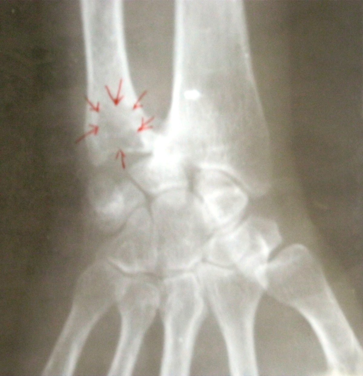
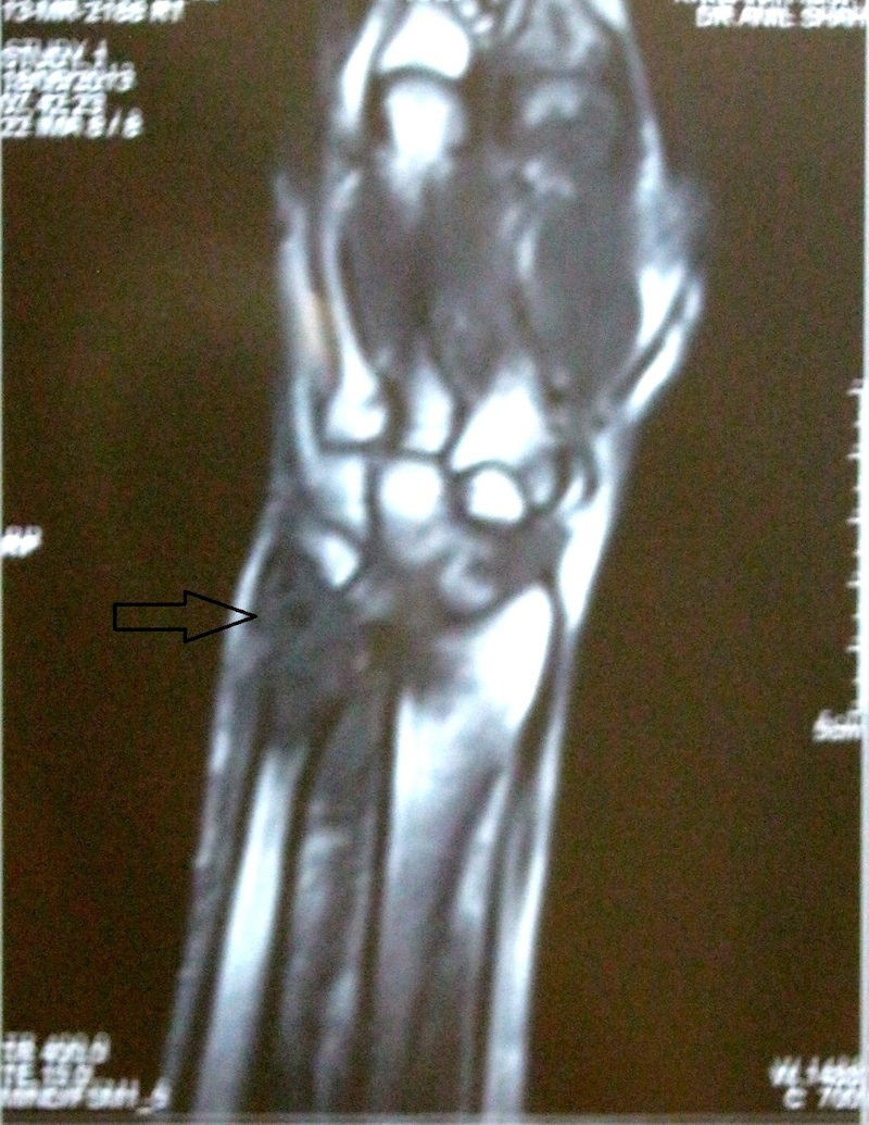
A 52 year old woman had pain in the right wrist joint for 6 months. The Xray was suggestive of aggressive giant cell tumor. A biopsy was obtained.
Radiology images:
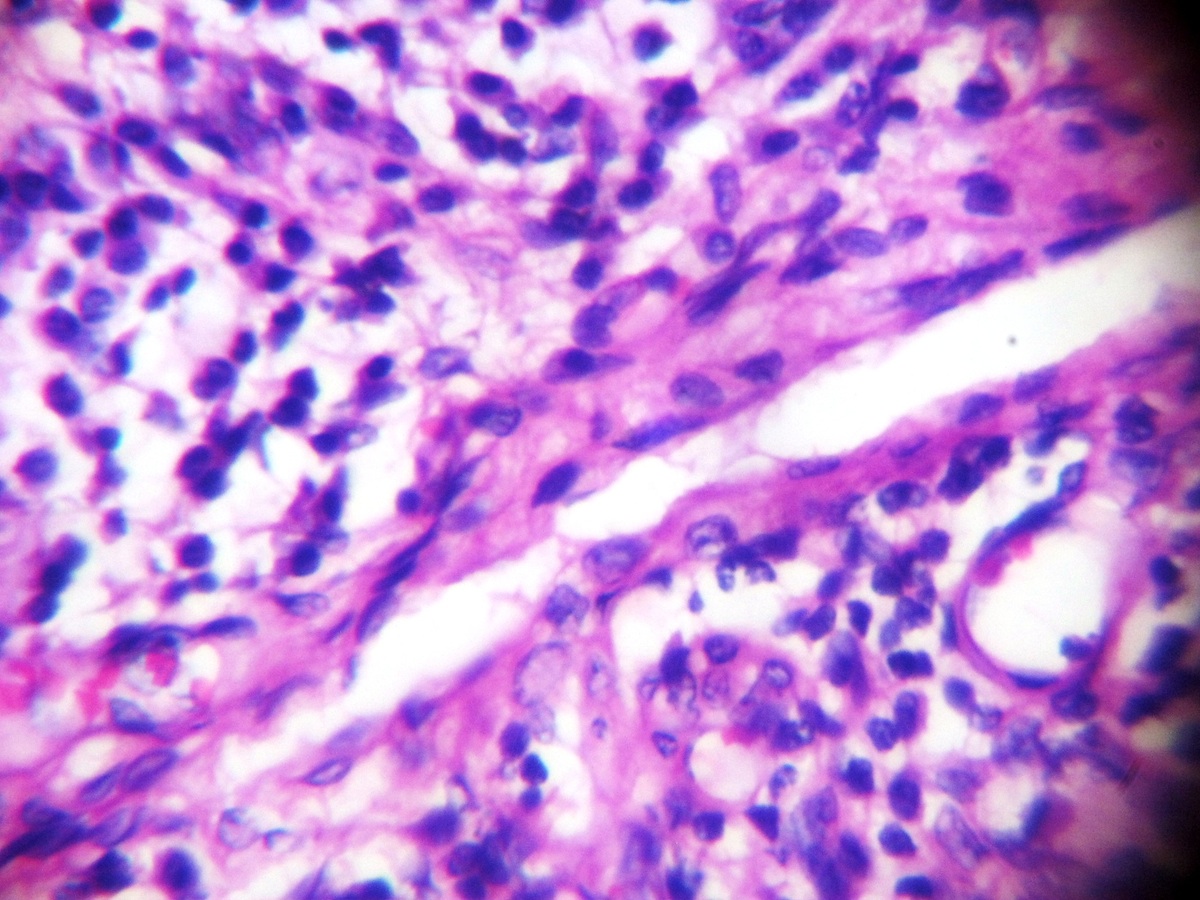
Microscopic images:

What is your diagnosis?
Diagnosis: Monoarticular erosive rheumatoid arthritis
Discussion:
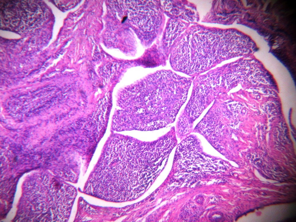
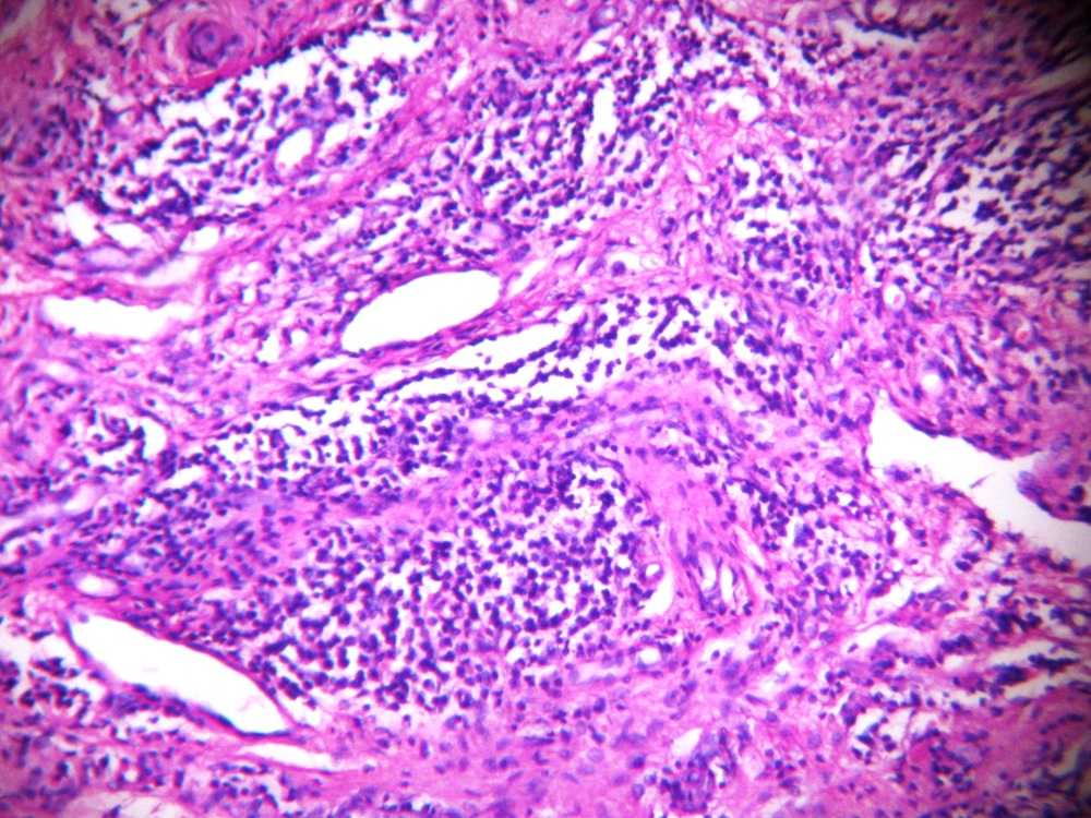
Histology showed a pannus (abnormal layer of fibrovascular or granulation tissue) with a significant papillary proliferation of capillaries and mononuclear cell infiltrate. Serology showed that the RA factor was strongly positive and anti-CCP antibodies were present at > 300 U/mL (Wikipedia: Anti-citrullinated protein antibody [Accessed 26 January 2022]). These features are consistent with rheumatoid arthritis.
Arthroscopic synovial biopsy may give conclusive diagnosis where clinical diagnosis is equivocal (Int J Appl Basic Med Res 2012;2:102). Biopsies of rheumatoid arthritis affected joints show a proliferative synovitis with synovial cell hyperplasia and hypertrophy. There is a dense perivascular infiltrate of T cells, plasma cells (often with eosinophilic cytoplasmic inclusions called Russell bodies) and macrophages. There are variable germinal centers, necrobiotic nodules and fibrosis. The inflammation may extend to the subchondral bone, which is relatively specific for rheumatoid arthritis.
Later findings include organizing fibrin floating in joint space as rice bodies, pannus formation progressing to fibrous ankylosis, then ossifying to form bony ankylosis. There is typically minimal evidence of repair (proliferative cartilage, sclerotic bone or osteophytes).
All cases are archived on our website. To view them sorted by case number, diagnosis or category, visit our main Case of the Month page. To subscribe or unsubscribe to Case of the Month or our other email lists, click here.
Thanks to Drs. Akash Jain and Sushama Desai, Krishna Institute of Medical Sciences (India), for contributing this case.
Advertisement

Case #308
Clinical history:
A 52 year old woman had pain in the right wrist joint for 6 months. The Xray was suggestive of aggressive giant cell tumor. A biopsy was obtained.
Radiology images:
Bony lesion
MRI of bony lesion
Microscopic images:
What is your diagnosis?
Click here for diagnosis and discussion:
Diagnosis: Monoarticular erosive rheumatoid arthritis
Discussion:
Histology showed a pannus (abnormal layer of fibrovascular or granulation tissue) with a significant papillary proliferation of capillaries and mononuclear cell infiltrate. Serology showed that the RA factor was strongly positive and anti-CCP antibodies were present at > 300 U/mL (Wikipedia: Anti-citrullinated protein antibody [Accessed 26 January 2022]). These features are consistent with rheumatoid arthritis.
Arthroscopic synovial biopsy may give conclusive diagnosis where clinical diagnosis is equivocal (Int J Appl Basic Med Res 2012;2:102). Biopsies of rheumatoid arthritis affected joints show a proliferative synovitis with synovial cell hyperplasia and hypertrophy. There is a dense perivascular infiltrate of T cells, plasma cells (often with eosinophilic cytoplasmic inclusions called Russell bodies) and macrophages. There are variable germinal centers, necrobiotic nodules and fibrosis. The inflammation may extend to the subchondral bone, which is relatively specific for rheumatoid arthritis.
Later findings include organizing fibrin floating in joint space as rice bodies, pannus formation progressing to fibrous ankylosis, then ossifying to form bony ankylosis. There is typically minimal evidence of repair (proliferative cartilage, sclerotic bone or osteophytes).
