
21 February 2014 - Case #301
All cases are archived on our website. To view them sorted by case number, diagnosis or category, visit our main Case of the Month page. To subscribe or unsubscribe to Case of the Month or our other email lists, click here.
Thanks to Dr. Saroona Haroon and Dr. Muhammad Usman, The Aga Khan University Hospital (Pakistan), for contributing this case.
Advertisement
Case #301
Clinical history:
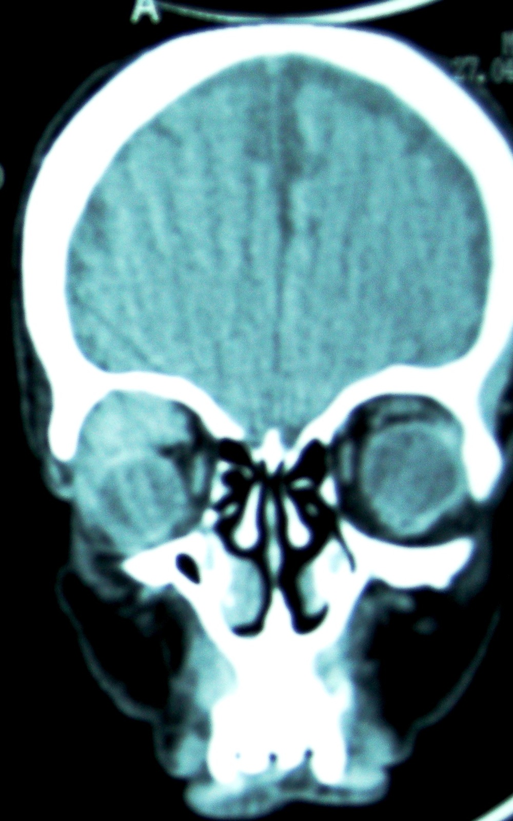
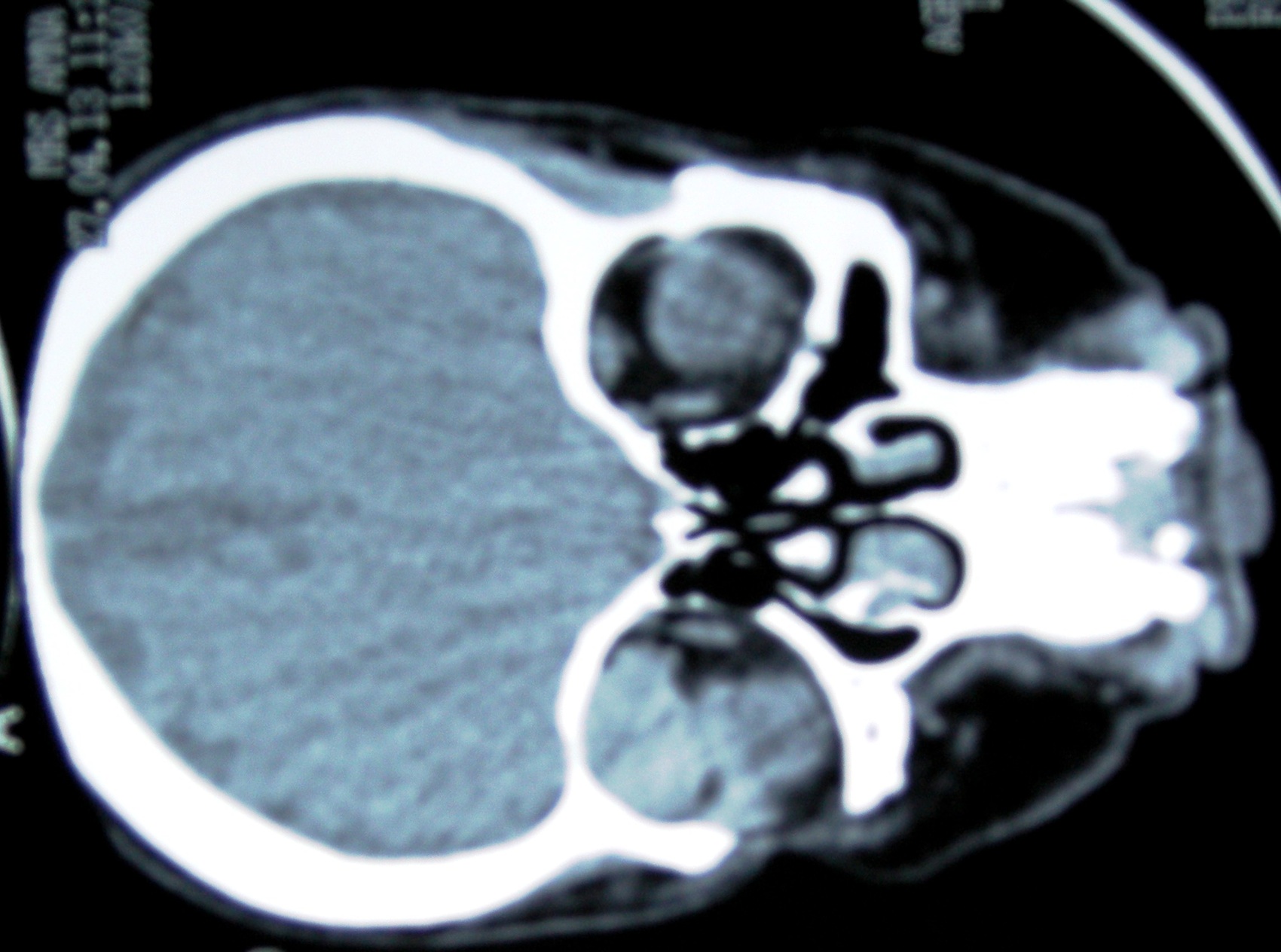
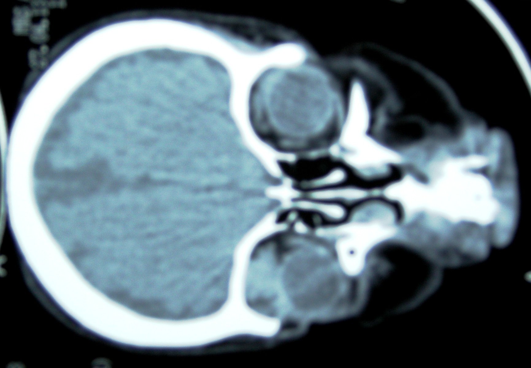
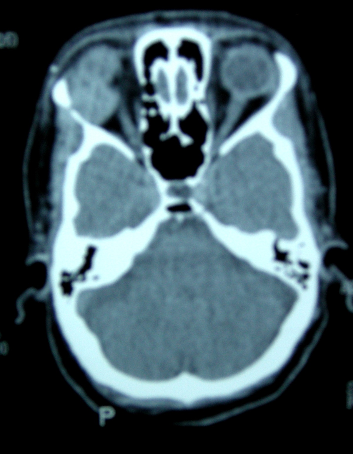
A 45 year old woman presented with a 6 month history of blurry vision and proptosis of the right eye. Clinically, there was a soft tissue swelling confined to the orbit.
Radiology images:
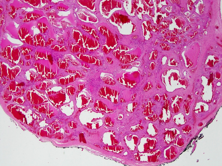
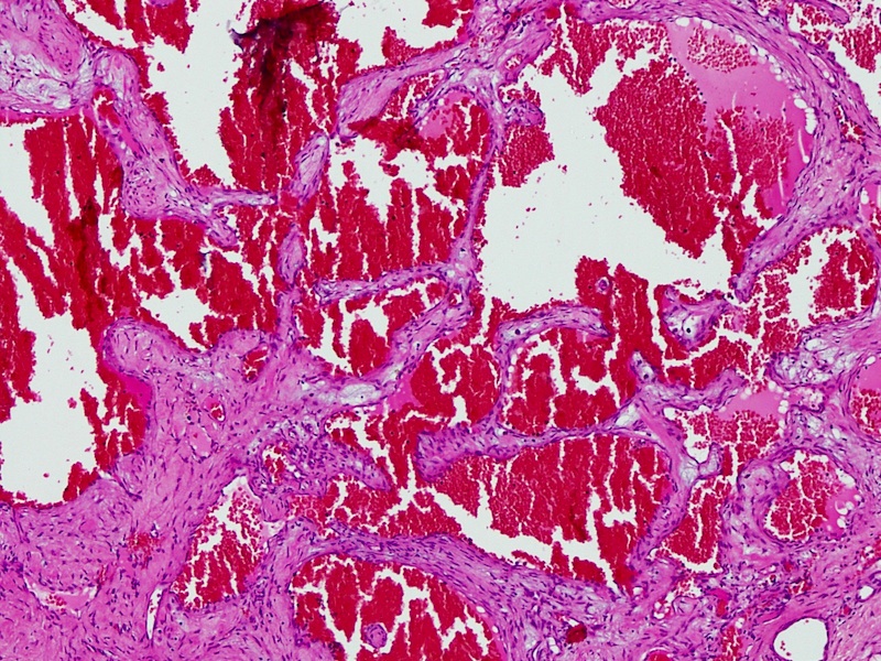
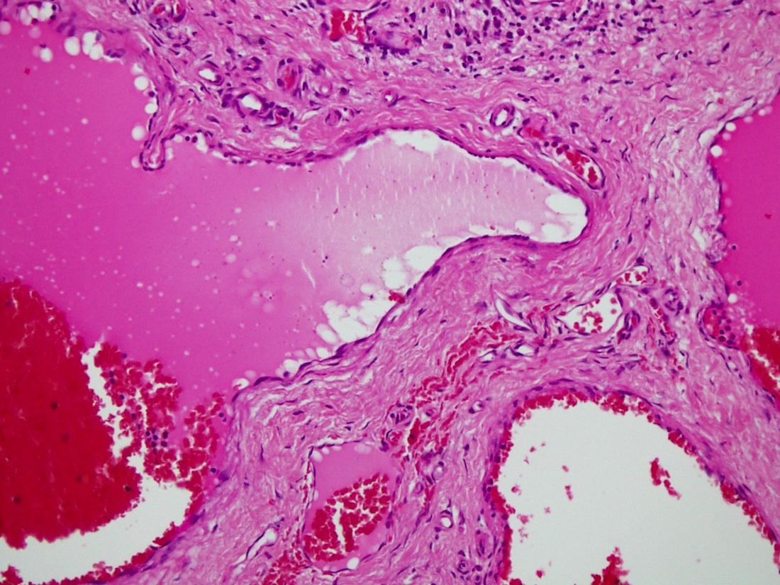
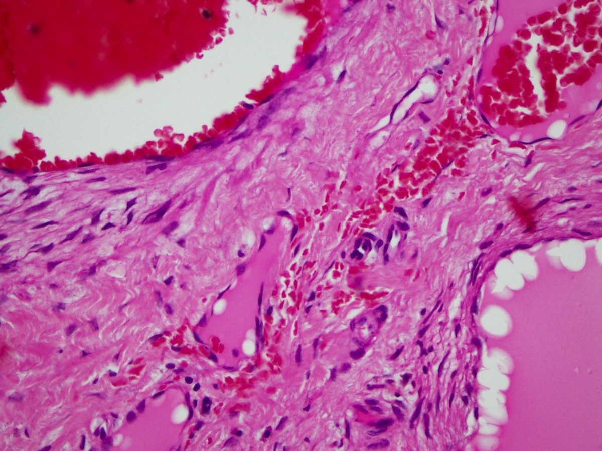
Microscopic images:

What is your diagnosis?
Diagnosis: Orbital cavernous hemangioma
Discussion:
Hemangioma of the orbit is a common lesion of the orbit and occurs more frequently than lymphangioma (Radiopaedia: Orbital Cavernous Haemangioma [Accessed 25 March 2024], EyeWiki: Cavernous Hemangioma [Accessed 25 March 2024]). Most cases occur in adults and most tumors are cavernous (Cases J 2009;2:6912).
CT and MRI scans often show a round mass with well defined margins in the extraocular muscles (Int J Ophthalmol 2011;4:195). Moderate to severe adhesion is frequent. In these cases, CT / MRI may show an irregular or ovoid mass filling the orbital apex or distorted / spiky margins in the posterior region of the mass.
Microscopically, these tumors are large and encapsulated, with cavernous vascular channels or spaces separated by scant connective tissue stroma. The vascular spaces are lined by a single layer of bland endothelial cells with variable smooth muscle in their walls. The differential diagnosis includes other vascular tumors which show mitotic activity, pleomorphism or atypia.
Hemangioma of the orbit is benign but may be slowly progressive.
All cases are archived on our website. To view them sorted by case number, diagnosis or category, visit our main Case of the Month page. To subscribe or unsubscribe to Case of the Month or our other email lists, click here.
Thanks to Dr. Saroona Haroon and Dr. Muhammad Usman, The Aga Khan University Hospital (Pakistan), for contributing this case.

June 16-19, 2014
The Sea Pines Resort
Hilton Head Island, South Carolina
Diagnosing Lymphomas and Leukemias:
Challenges for the Surgical Pathologist
Website
Hotel
Faculty:
Steven H. Swerdlow, M.D., Course Director, University of Pittsburgh School of Medicine, Pittsburgh, PA;
Kathy Foucar, M.D., University of New Mexico, Albuquerque, NM;
Marsha C. Kinney, M.D., University of Texas Health Sciences Center, San Antonio, TX;
Lawrence M. Weiss, M.D., Clarient, Inc., Aliso Viejo, CA.
Website news:
(1) On 19 February 2014, we had record traffic of 23,252 visits.
(2) We are always looking for reviewers for our 6700+ topics. Reviewers should have expertise in the topics reviewed. They can be 3rd/4th year residents or fellows, if working under the direction of a staff.
(3) We are starting to add these images:
Visit and follow our Blog to see recent updates to the website.
(1) On 19 February 2014, we had record traffic of 23,252 visits.
(2) We are always looking for reviewers for our 6700+ topics. Reviewers should have expertise in the topics reviewed. They can be 3rd/4th year residents or fellows, if working under the direction of a staff.
(3) We are starting to add these images:
- Submitted by contributors by email
- Posted to our Flickr page
- AFIP Third Fascicle
- Webpathology.com
- Contributed by Dr. Mark Wick
- From other Flickr pages
Visit and follow our Blog to see recent updates to the website.
Case #301
Clinical history:
A 45 year old woman presented with a 6 month history of blurry vision and proptosis of the right eye. Clinically, there was a soft tissue swelling confined to the orbit.
Radiology images:
Microscopic images:
What is your diagnosis?
Click here for diagnosis and discussion:
Diagnosis: Orbital cavernous hemangioma
Discussion:
Hemangioma of the orbit is a common lesion of the orbit and occurs more frequently than lymphangioma (Radiopaedia: Orbital Cavernous Haemangioma [Accessed 25 March 2024], EyeWiki: Cavernous Hemangioma [Accessed 25 March 2024]). Most cases occur in adults and most tumors are cavernous (Cases J 2009;2:6912).
CT and MRI scans often show a round mass with well defined margins in the extraocular muscles (Int J Ophthalmol 2011;4:195). Moderate to severe adhesion is frequent. In these cases, CT / MRI may show an irregular or ovoid mass filling the orbital apex or distorted / spiky margins in the posterior region of the mass.
Microscopically, these tumors are large and encapsulated, with cavernous vascular channels or spaces separated by scant connective tissue stroma. The vascular spaces are lined by a single layer of bland endothelial cells with variable smooth muscle in their walls. The differential diagnosis includes other vascular tumors which show mitotic activity, pleomorphism or atypia.
Hemangioma of the orbit is benign but may be slowly progressive.
