
11 September 2013 - Case #285
All cases are archived on our website. To view them sorted by case number, diagnosis or category, visit our main Case of the Month page. To subscribe or unsubscribe to Case of the Month or our other email lists, click here.
Thanks to Dr. Samir Ghani, Wexham Park Hospital (United Kingdom), for contributing this case and the discussion.

Advertisement
Case #285
Clinical history:
A 41 year old woman delivered a full term baby without complications and underwent tubal ligation and greater omentum sampling during a Caesarean section.
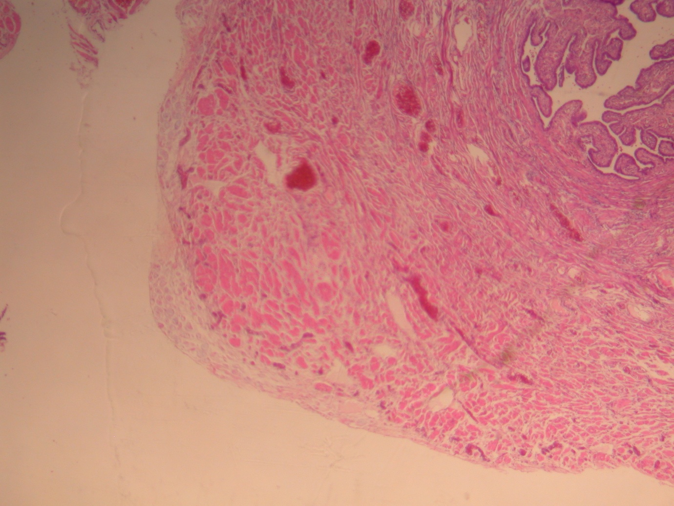
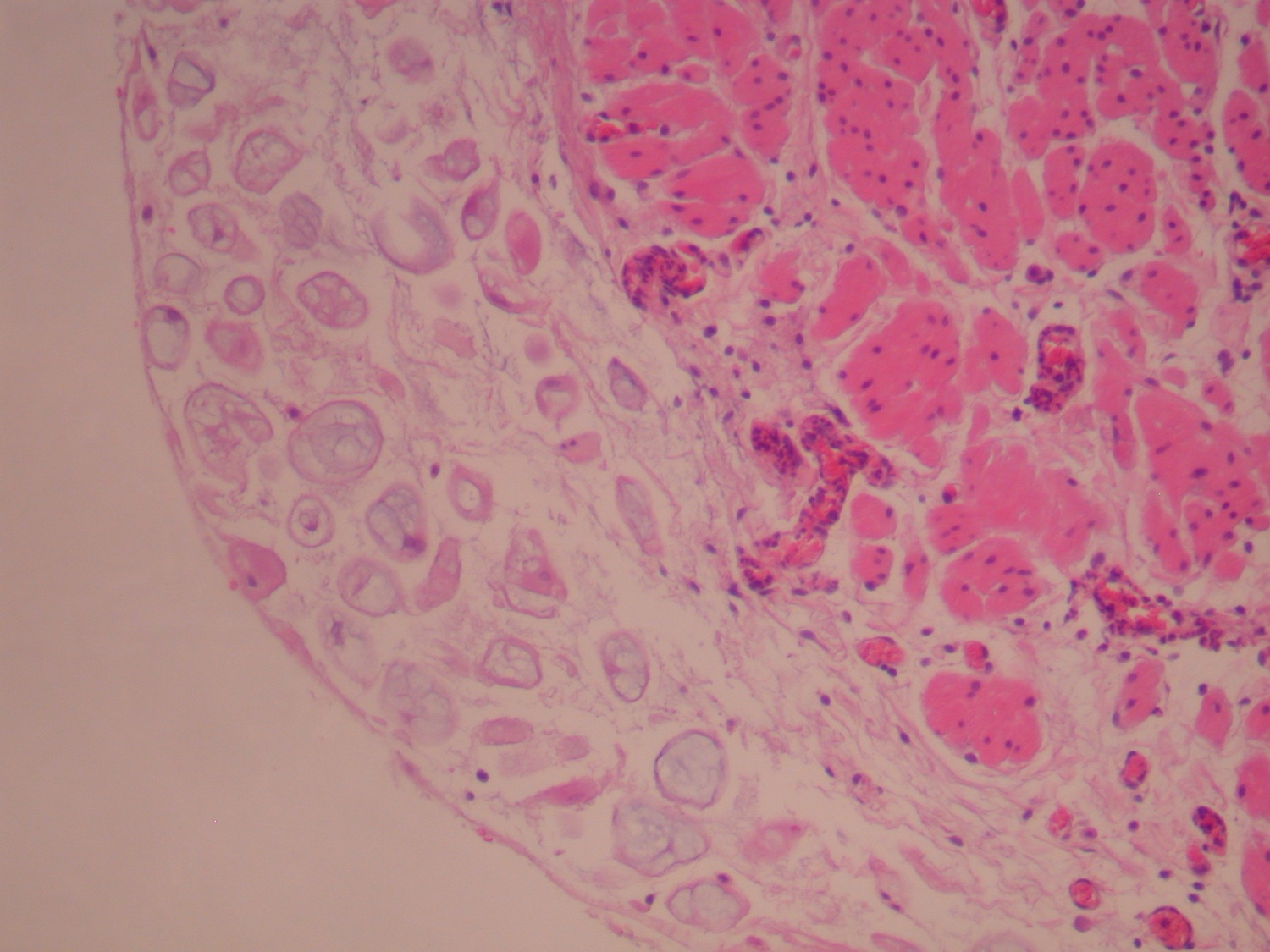
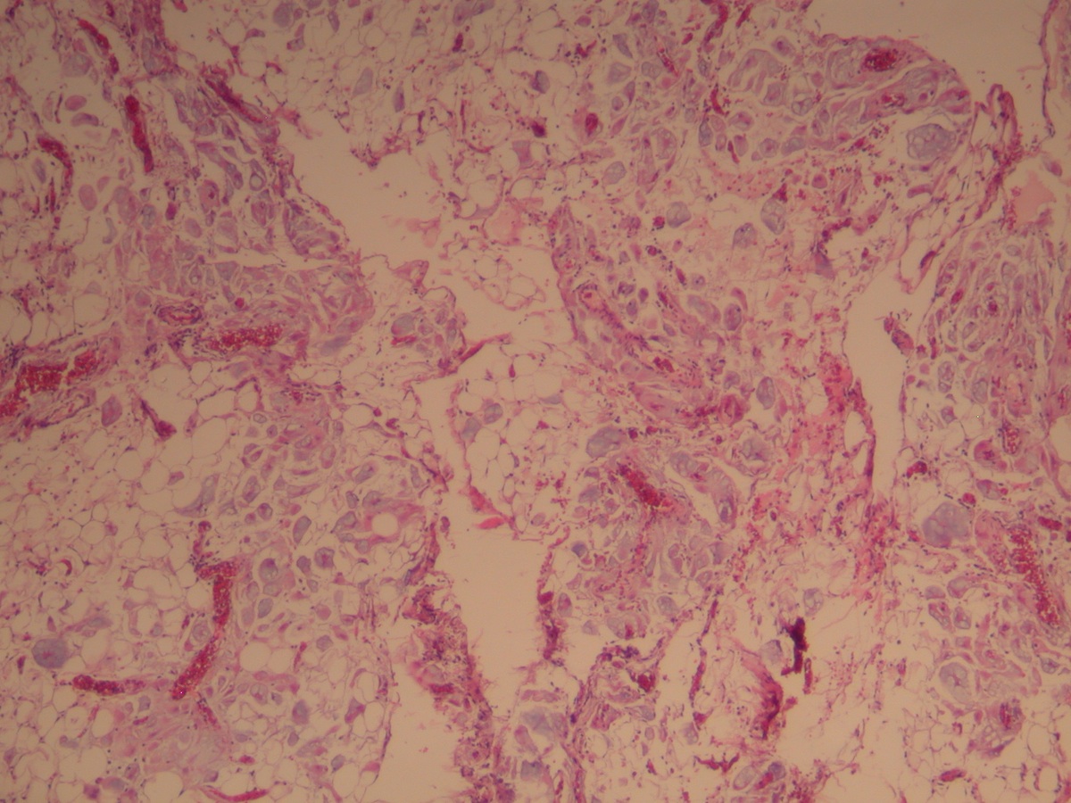
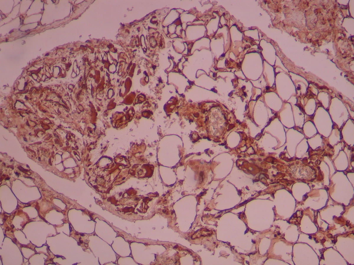
Microscopic images:
What is your diagnosis?
Diagnosis: Extrauterine deciduosis
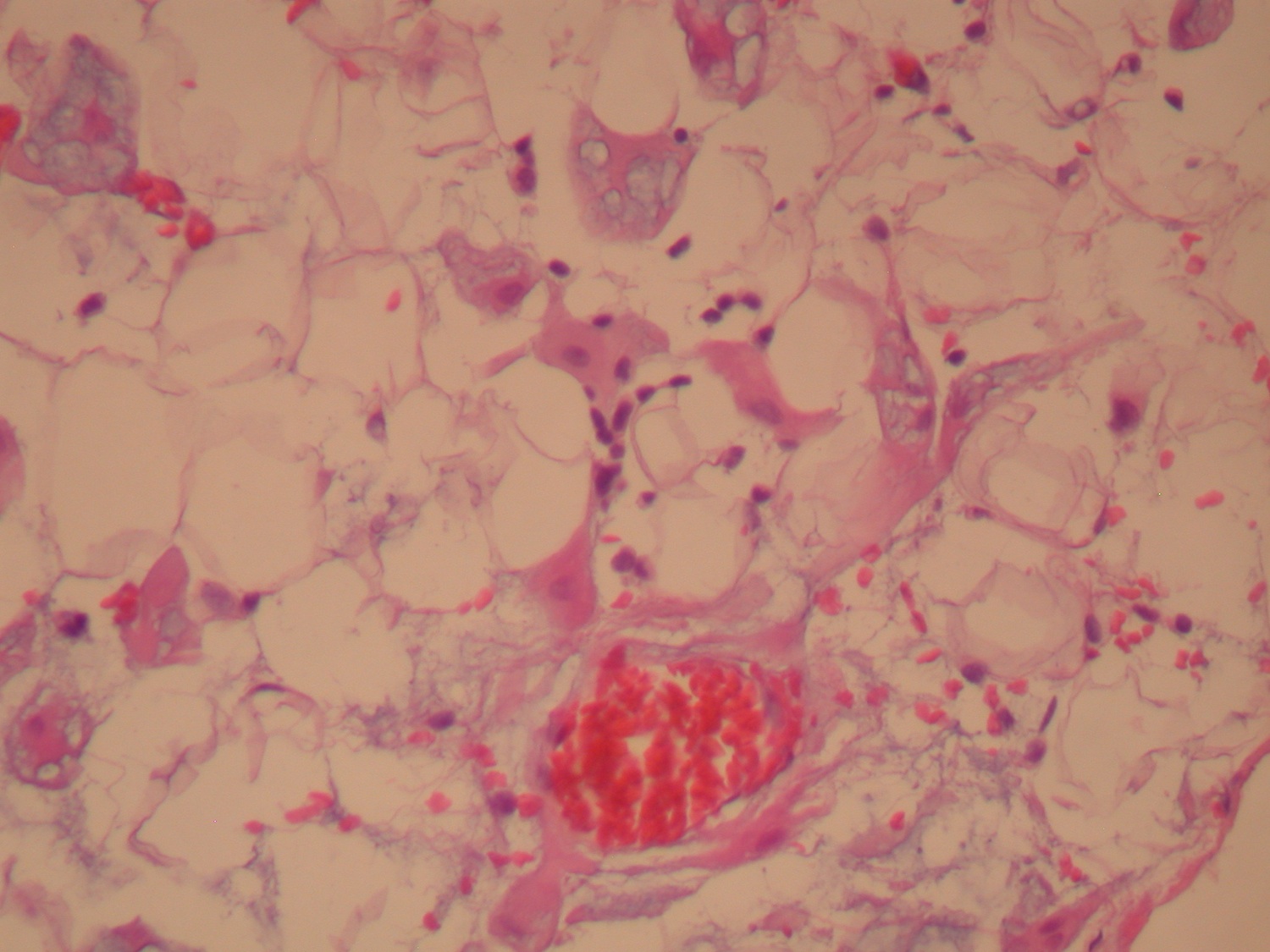
Immunostains:

Discussion:
The omentum and the peritoneal surface of the fallopian tube show exaggerated extrauterine deciduosis (EUD) / decidual reaction, with single / cell aggregates displaying vacuolated cytoplasm with mucoid-like material and bland nuclear morphology. No mitotic figures are seen. There are only a few cells with eosiophilic cytoplasm, which provide the clue to the nature of the lesion, in addition to the clinical presentation.
Immunohistochemistry showed strong progesterone receptor nuclear staining in the lesional cells. The vimentin stain was diffusely positive. Other markers such as EMA and calretinin were negative (not shown).
Extrauterine deciduosis (EUD) is a benign condition which can be mistaken macroscopically for malignancy and resembles peritoneal carcinomatosis. EUD can be discovered accidentally in pregnancy during caesarean sections and during pelvic surgery in women taking oral contraceptives (Am J Surg Pathol 1987;11:526).
Deposits are found mainly on the ovary and cervix, but also on abdominal serosal surfaces of the fallopian tubes, bowel, peritoneum and vagina, as well as lungs, pleura, retroperitoneal lymph nodes and rarely skin. Most lesions do not require further treatment and spontaneously involute within 4 - 6 weeks of delivery.
All cases are archived on our website. To view them sorted by case number, diagnosis or category, visit our main Case of the Month page. To subscribe or unsubscribe to Case of the Month or our other email lists, click here.
Thanks to Dr. Samir Ghani, Wexham Park Hospital (United Kingdom), for contributing this case and the discussion.
Advertisement
Website news:
(1) We are continously updating topics in our Stains and CD Markers chapters. However, there are over 600 stains / CD Marker topics, so we are seeking pathologists interested in reviewing 3-5 of these topics. If interested, please contact Kristina at editor.pathout@gmail.com.
(2) Our Case of the Week page now lists upcoming cases with the contributor's name and the number of cases pending in our queue. Currently, we have a 8-9 month turn around time from when we accept a case to when it is posted on our website. We now are only posting a maximum of 5 cases per contributor per calendar year.
(3) We have a new Microbiology chapter that is being written by Christopher Hale, M.D. Be sure to take a look!
Visit and follow our Blog to see recent updates to the website.
Case #285
Clinical history:
A 41 year old woman delivered a full term baby without complications and underwent tubal ligation and greater omentum sampling during a Caesarean section.
Microscopic images:
What is your diagnosis?
Click here for diagnosis and discussion:
Diagnosis: Extrauterine deciduosis
Immunostains:
Left: PR, right: vimentin
Discussion:
The omentum and the peritoneal surface of the fallopian tube show exaggerated extrauterine deciduosis (EUD) / decidual reaction, with single / cell aggregates displaying vacuolated cytoplasm with mucoid-like material and bland nuclear morphology. No mitotic figures are seen. There are only a few cells with eosiophilic cytoplasm, which provide the clue to the nature of the lesion, in addition to the clinical presentation.
Immunohistochemistry showed strong progesterone receptor nuclear staining in the lesional cells. The vimentin stain was diffusely positive. Other markers such as EMA and calretinin were negative (not shown).
Extrauterine deciduosis (EUD) is a benign condition which can be mistaken macroscopically for malignancy and resembles peritoneal carcinomatosis. EUD can be discovered accidentally in pregnancy during caesarean sections and during pelvic surgery in women taking oral contraceptives (Am J Surg Pathol 1987;11:526).
Deposits are found mainly on the ovary and cervix, but also on abdominal serosal surfaces of the fallopian tubes, bowel, peritoneum and vagina, as well as lungs, pleura, retroperitoneal lymph nodes and rarely skin. Most lesions do not require further treatment and spontaneously involute within 4 - 6 weeks of delivery.
