
All cases are archived on our website. To view them sorted by case number, diagnosis or category, visit our main Case of the Month page. To subscribe or unsubscribe to Case of the Month or our other email lists, click here.
Thanks to Dr. Carolina Martinez Ciarpaglini, University Clinic Hospital of Valencia (Spain), for contributing this case and the discussion and to Dr. Borislav Alexiev, Northwestern University, Chicago, Illinois (USA), for the Editorial Board review. To contribute a Case of the Month, follow the guidelines on our Case of the Month page.
Introducing 23 New Rabbit Monoclonal Antibodies: MSH2, CD25, RRM1, and others.
Epitomics is releasing 23 new EP Clones, high quality antibodies for anatomic pathology.
Our collection of EP Clones now includes new breast cancer markers: Aurora B and Mammaglobin; new Lymphoma markers CD25, RRM1, as well as various other Colon and Lung Cancer targets. Rabbit monoclonal antibodies (RabMAbs) are the product of true rabbit-rabbit hybridomas. The inherent qualities of the rabbit immune system allow us to select antibodies which include: 1) stronger binding affinity and 2) higher specificity.
Click here to view our new products or visit us at epitomics.com/diagnostics for our complete product listing.
Advertisement
Case of the Week #276
Clinical history:
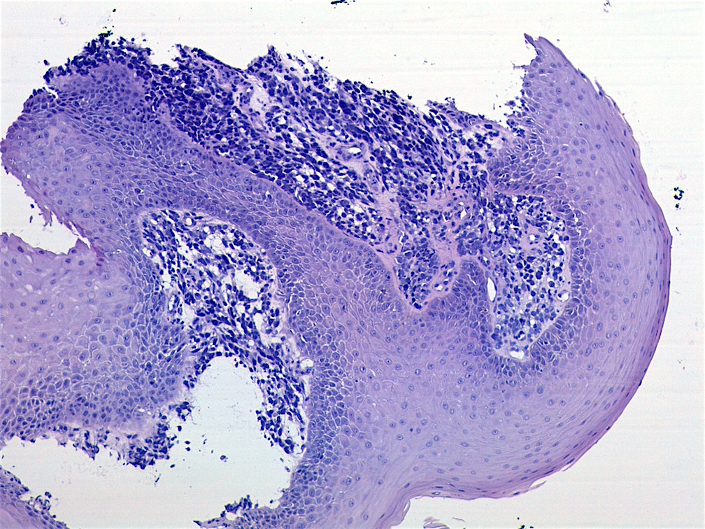
An 11 year old boy presented with a left tonsillar mass present for three weeks. A biopsy was taken.
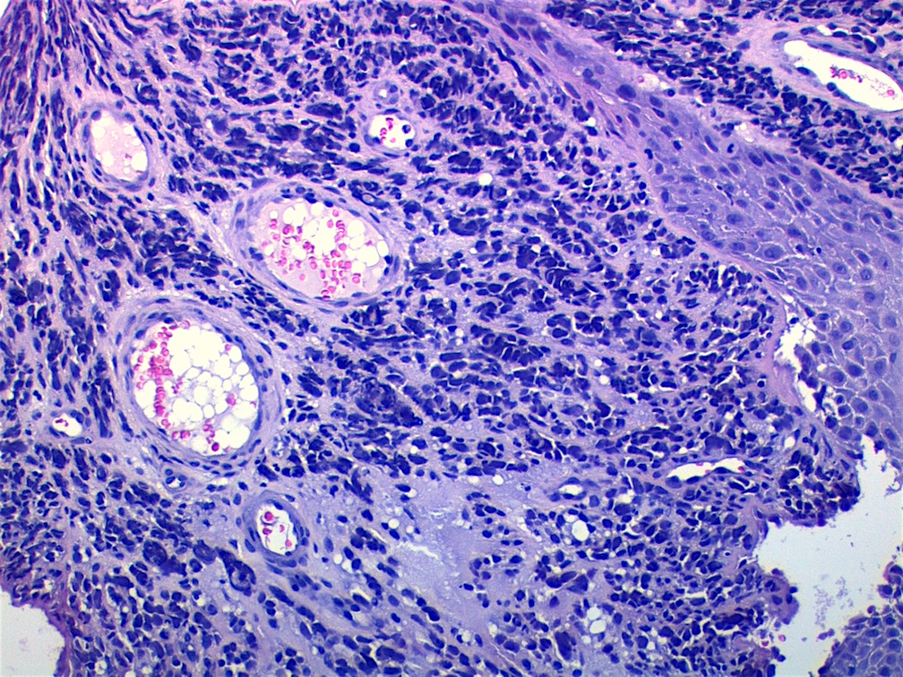
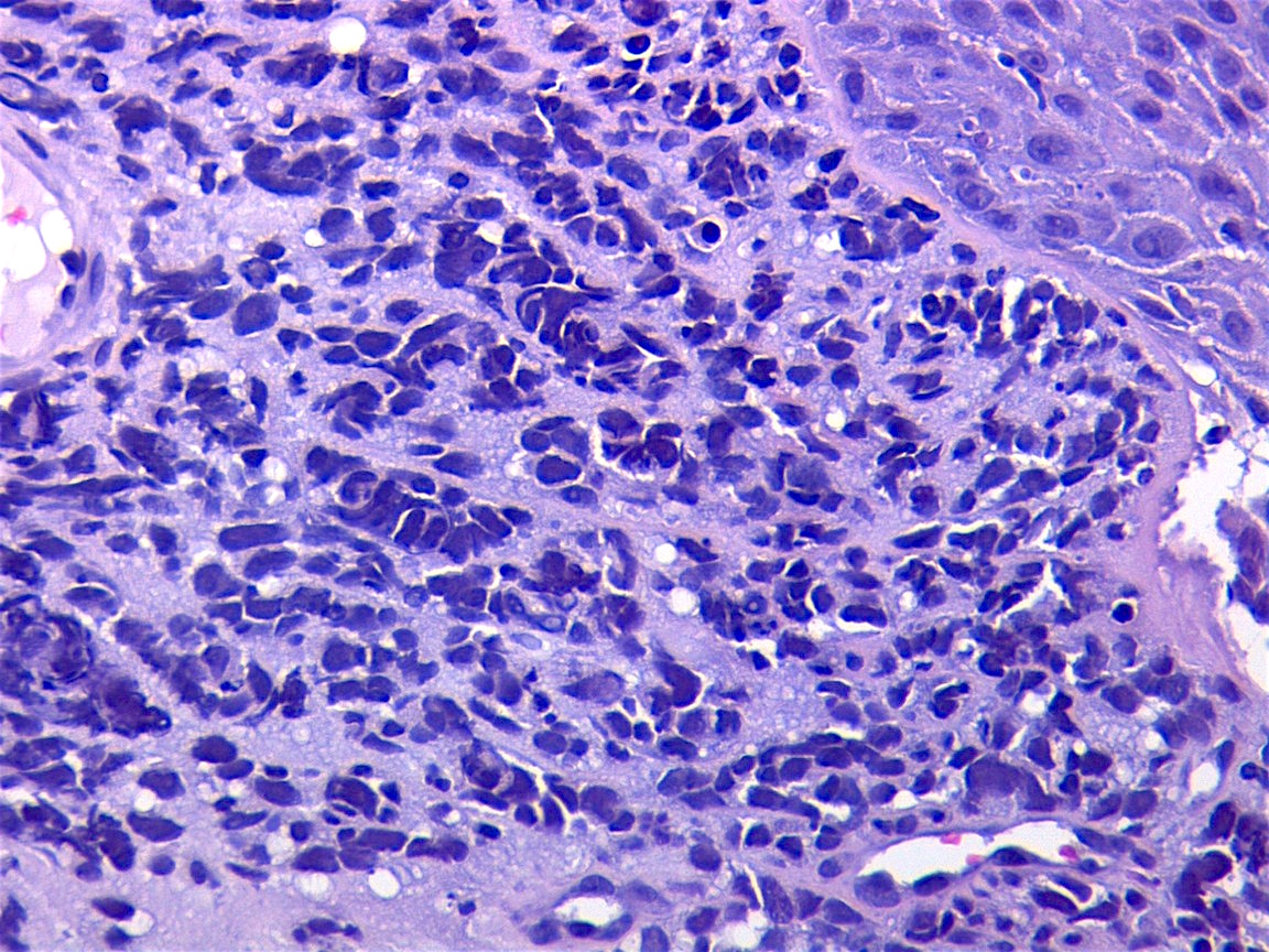
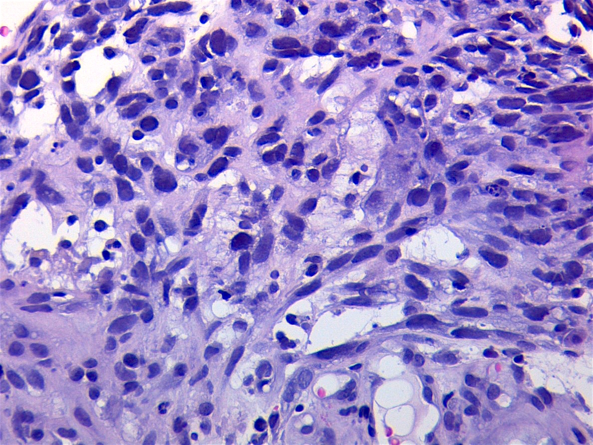
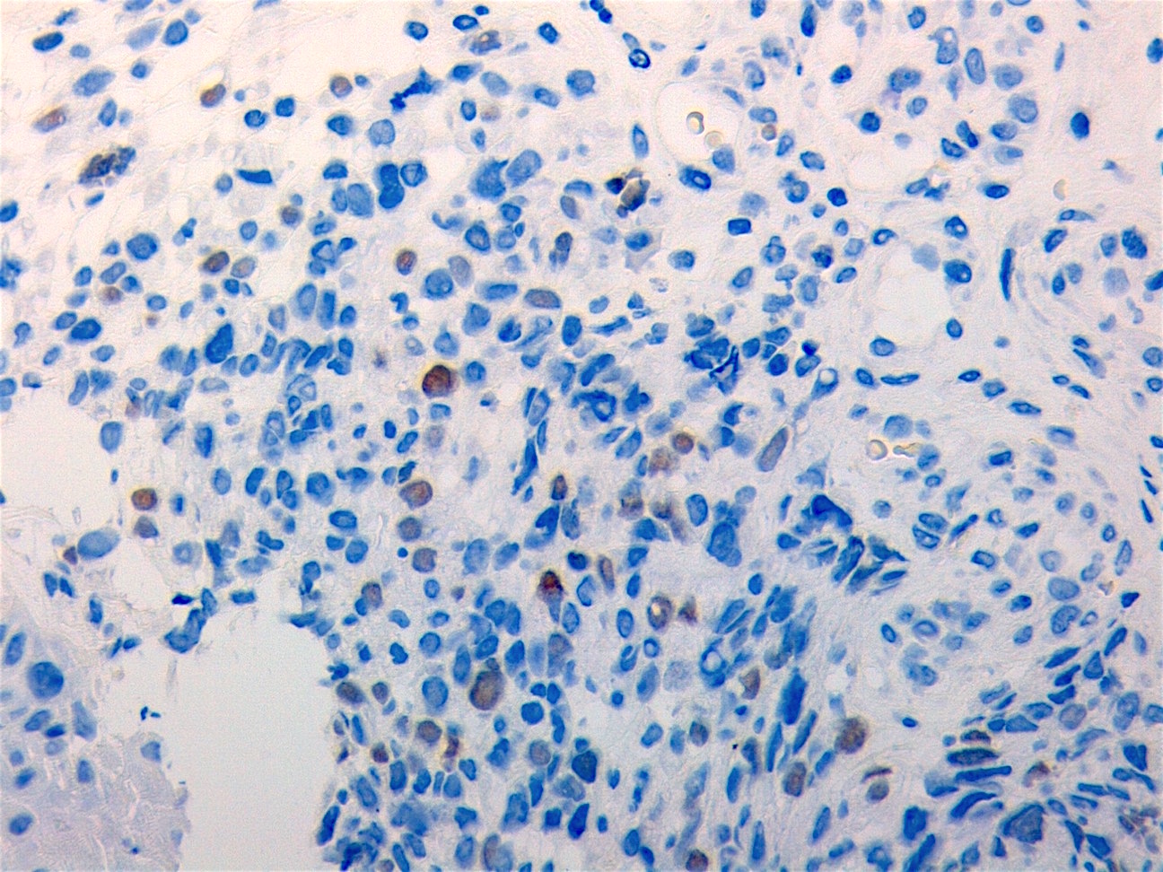
Histopathology images:

What is your diagnosis?
Diagnosis: Embryonal rhabdomyosarcoma
Stains:
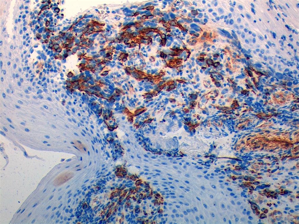
Actin
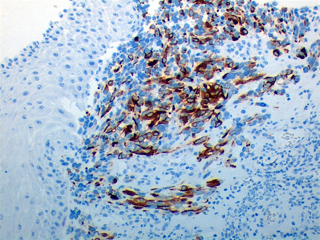
Desmin
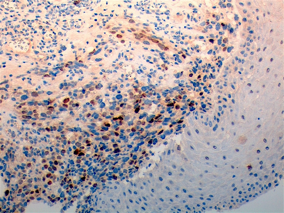
Myogenin
myoD1
Discussion:
Embryonal rhabdomyosarcoma is a primitive soft tissue sarcoma with small blue cells resembling embryonic skeletal muscle. It is the most common rhabdomyosarcoma (RMS) subtype (65% of RMS cases), and usually occurs in children ages 3-10 years in the head and neck (sinonasal tract, oral cavities, orbit, ear, periorbital region and pharynx), prostate or paratesticular regions. Subtypes include embryonal-NOS, anaplastic and botryoid. Embryonal-NOS is the most common subtype (75%), usually affecting boys. It peaks at ages 0-4 years (US National Cancer Institute).
The tumor consists of dense zones of undifferentiated, differentiating and well differentiated cells. A cambium layer (cells immediately beneath the epithelium) ) may be present especially in the botryoid variant. Undifferentiated cells are blue cells with minimal wispy cytoplasm but no skeletal differentiation, and central nuclei. Differentiating cells have moderate amphophilic or eosinophilic cytoplasm, often fibrillar. They may have a tadpole shape, often with nuclei arranged in tandem (so called strap cells). Some differentiated cells may have cytoplasmic cross-striations.
Tumors cells, as in this case, are immunoreactive for actin, desmin, myogenin and myoD1. There may occasionally show aberrant immunoreactivity for S100 and CAM5.2. The differential diagnosis consists of other small blue cell tumors, such as lymphoma and neuroblastoma. All children with rhabdomyosarcoma require multimodality therapy with systemic chemotherapy and either surgery or radiation therapy. Overall 5 year survival is over 80%, although it is much less for those with metastatic disease (eMedicine).
