
8 November 2012 - Case #257
All cases are archived on our website. To view them sorted by case number, diagnosis or category, visit our main Case of the Month page. To subscribe or unsubscribe to Case of the Month or our other email lists, click here.
Thanks to Dr. Josemari Arrinda Yeregui, Bidasoa Hospital (Spain), for contributing this case and the discussion.
Advertisement
Case #257
Clinical history:
A 50 year old man presented with watery diarrhea for 12 months. Endoscopy was suggestive of Crohn's disease of the right colon.
Microscopic images:
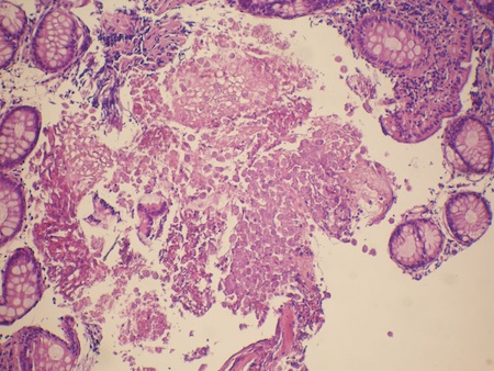
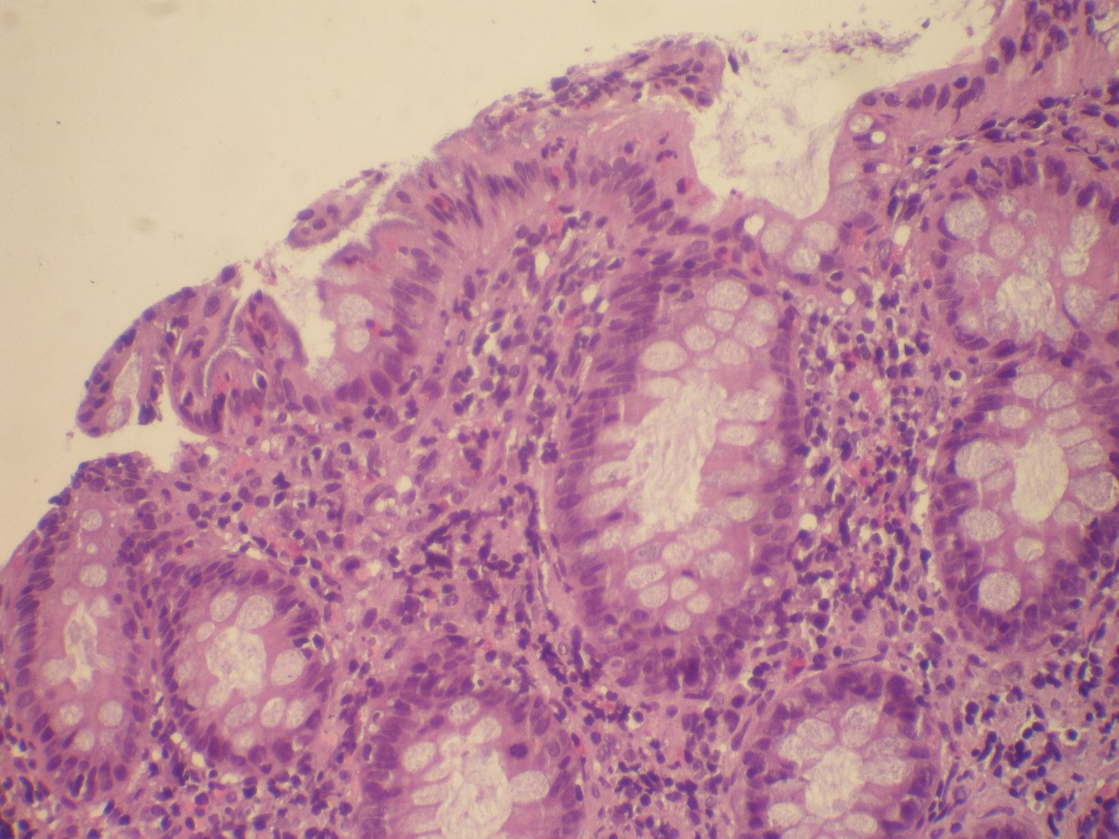
What is your diagnosis?
Diagnosis: Intestinal spirochetosis and amebiasis
Immunostains:


Discussion:
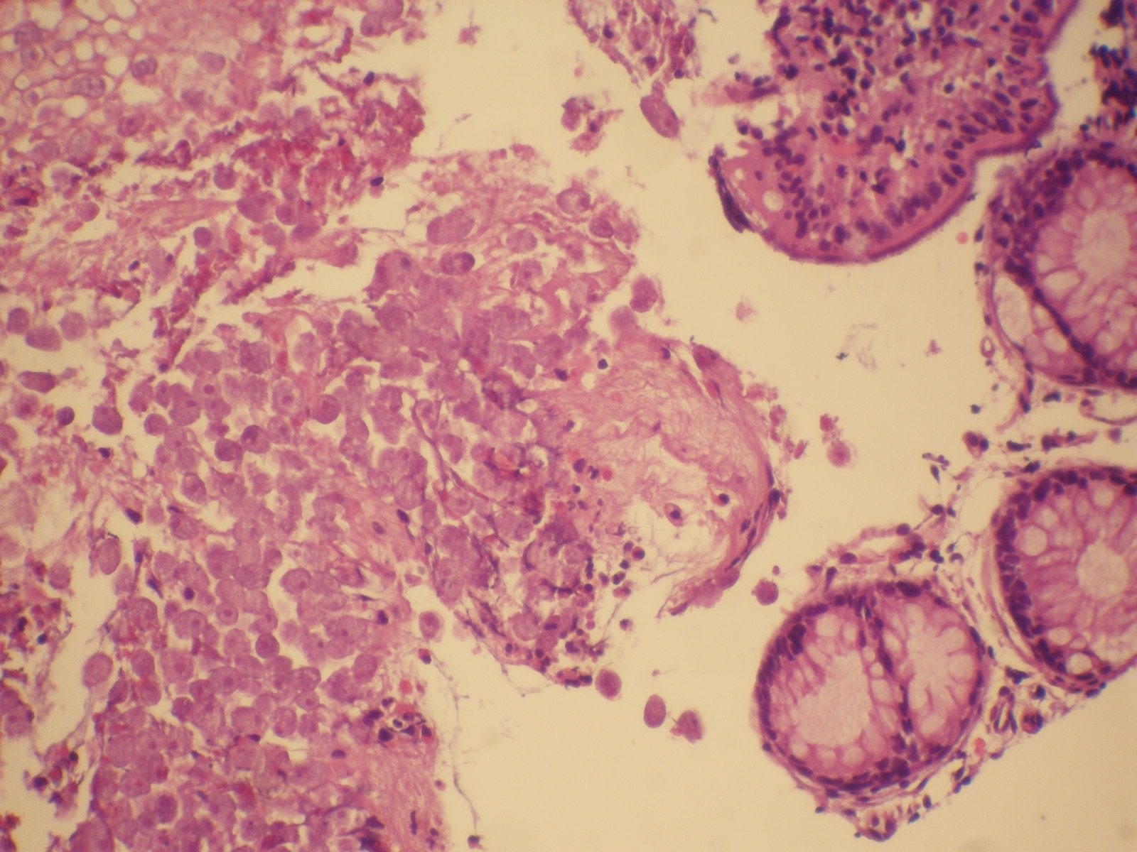
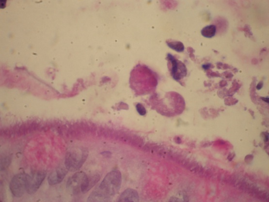
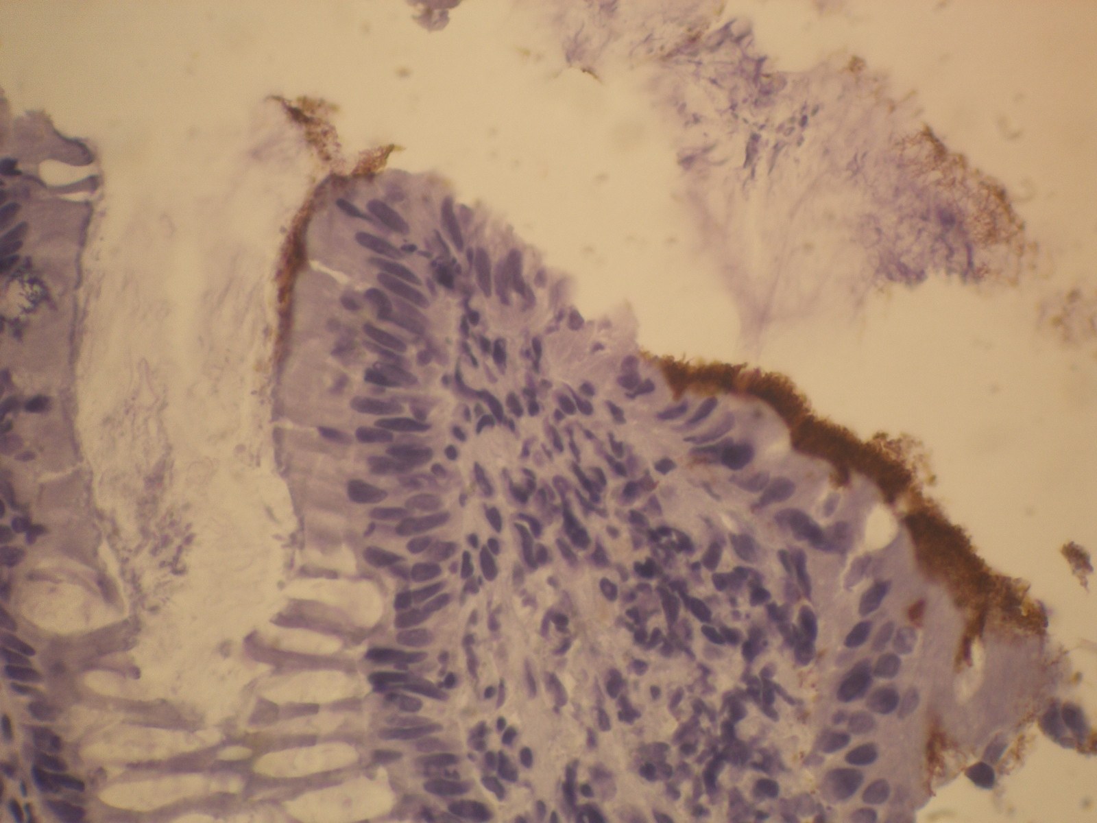
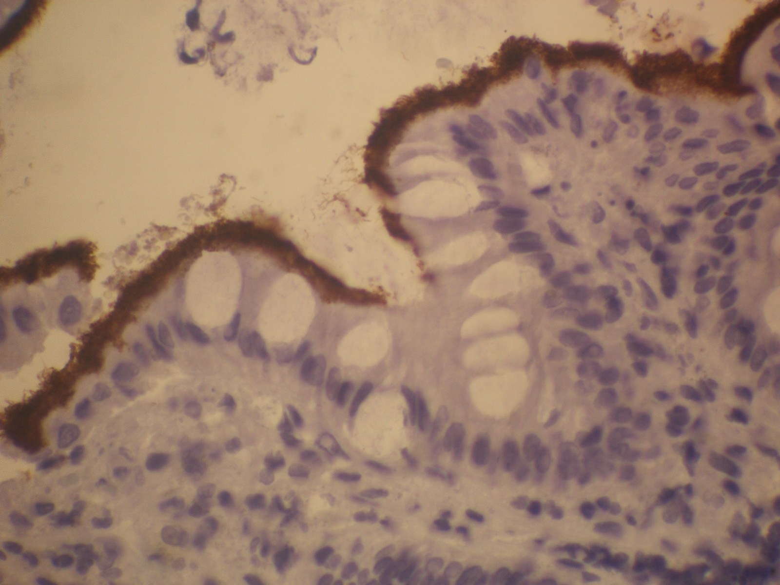
Microscopically, many Entamoeba histolytica trophozoites are seen, as well as a blue hazy barrier at the mucosa. This barrier is highlighted with PAS and with anti-Treponemal pallidum antibody (above).
Human intestinal spirochetosis is defined histologically by the presence of spirochetal microorganisms attached to the apical cell membrane of colorectal epithelium (Ger Med Sci 2010;8:Doc01).
Human intestinal spirochetes include Brachyspira aalborgi and Brachyspira pilosicoli. Incidence is common in poorly developed areas, but low where living standards are high. Homosexuals and HIV+ individuals are at high risk. Most patients are asymptomatic, but children, homosexual and HIV+ men are more likely to be symptomatic regardless of invasion. The bacteria can be highlighted using silver stains, PAS, Giemsa, Alcian blue (pH 2.5) and by immunohistochemistry.
Intestinal spirochetosis often coexists with other enteric pathogens, including Entamoeba histolytica (J Gastroenterol Hapatol 2007;22:64), Enterobius vermicularis, Helicobacter pylori, Shigella flexneri and Neisseria gonorrhoeae. Amebic colitis (amebiasis) resembles inflammatory bowel disease but is caused by Entamoeba histolytica protozoa. It may cause dysentery, colonic stricture, ulceration and perforation, liver abscesses up to 10 cm or be asymptomatic. Transmission is by fecal oral spread, with increased incidence in homosexuals, patients with AIDS or from the tropics (Wikipedia: Entamoeba Histolytica [Accessed 10 April 2024]).
The patient was treated with metronidazole, which covers both pathogens, which eliminated the diarrhea. The patient was subsequently diagnosed as HIV+.
All cases are archived on our website. To view them sorted by case number, diagnosis or category, visit our main Case of the Month page. To subscribe or unsubscribe to Case of the Month or our other email lists, click here.
Thanks to Dr. Josemari Arrinda Yeregui, Bidasoa Hospital (Spain), for contributing this case and the discussion.

Introducing New EP Clones
As the Rabbit Monoclonal Antibody (RabMAb) technology leader, Epitomics is pleased to announce new antibodies for anatomic pathology.
We are now offering 28 new EP Clones with antibodies for Colon, Germ Cell Tumor, and Lung Cancer targets including: AFP, Napsin A, and PLAP, among others.
Click here to view our new products
or visit us at epitomics.com/diagnostics for our complete product listing.
EP Clones™ are a line of high quality antibodies for anatomical pathology. Each antibody is generated using Epitomics' patented rabbit hybridoma technology offering superior binding affinity and specificity.
Website news:
(1) Our Feature Page for November highlights Consumable Lab Products / Clinical Lab Analyzers, and includes Leica Microsystems, Sakura Finetek USA and Ventana Medical. We also have a new Mystery Case on the right side of the Home Page.
(2) We have updated the Fibrohistiocytic section of the Soft Tissue chapter, based on reviews by Vijay Shankar, M.D.
(3) We have changed the name of a chapter from Management-Laboratory to Laboratory Administration to avoid confusion with the Management of pathology practices chapter.
Visit and follow our Blog to see recent updates to the website.
Case #257
Clinical history:
A 50 year old man presented with watery diarrhea for 12 months. Endoscopy was suggestive of Crohn's disease of the right colon.
Microscopic images:
What is your diagnosis?
Click here for diagnosis and discussion:
Diagnosis: Intestinal spirochetosis and amebiasis
Immunostains:
Discussion:
Microscopically, many Entamoeba histolytica trophozoites are seen, as well as a blue hazy barrier at the mucosa. This barrier is highlighted with PAS and with anti-Treponemal pallidum antibody (above).
Human intestinal spirochetosis is defined histologically by the presence of spirochetal microorganisms attached to the apical cell membrane of colorectal epithelium (Ger Med Sci 2010;8:Doc01).
Human intestinal spirochetes include Brachyspira aalborgi and Brachyspira pilosicoli. Incidence is common in poorly developed areas, but low where living standards are high. Homosexuals and HIV+ individuals are at high risk. Most patients are asymptomatic, but children, homosexual and HIV+ men are more likely to be symptomatic regardless of invasion. The bacteria can be highlighted using silver stains, PAS, Giemsa, Alcian blue (pH 2.5) and by immunohistochemistry.
Intestinal spirochetosis often coexists with other enteric pathogens, including Entamoeba histolytica (J Gastroenterol Hapatol 2007;22:64), Enterobius vermicularis, Helicobacter pylori, Shigella flexneri and Neisseria gonorrhoeae. Amebic colitis (amebiasis) resembles inflammatory bowel disease but is caused by Entamoeba histolytica protozoa. It may cause dysentery, colonic stricture, ulceration and perforation, liver abscesses up to 10 cm or be asymptomatic. Transmission is by fecal oral spread, with increased incidence in homosexuals, patients with AIDS or from the tropics (Wikipedia: Entamoeba Histolytica [Accessed 10 April 2024]).
The patient was treated with metronidazole, which covers both pathogens, which eliminated the diarrhea. The patient was subsequently diagnosed as HIV+.
