
22 August 2012 - Case #249
All cases are archived on our website. To view them sorted by case number, diagnosis or category, visit our main Case of the Month page. To subscribe or unsubscribe to Case of the Month or our other email lists, click here.
Thanks to Dr. Beata Maksymiuk, Institute of Tuberculosis and Lung Diseases (Poland), for contributing this case.
Advertisement
Case #249
Clinical history:
A 32 year old woman had surgery for a gangrenous appendicitis. During surgery, a 3 x 2.5 cm pedunculated tumor was discovered in the cecal region. Grossly it was compact, well circumscribed and unencapsulated with a white whirled cut surface.
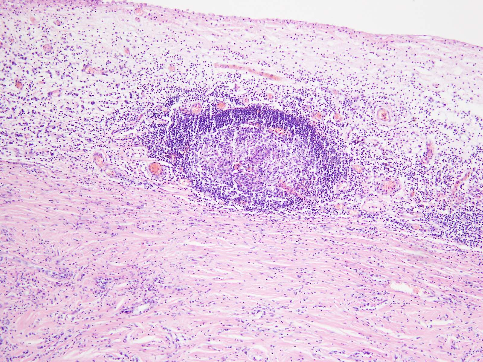
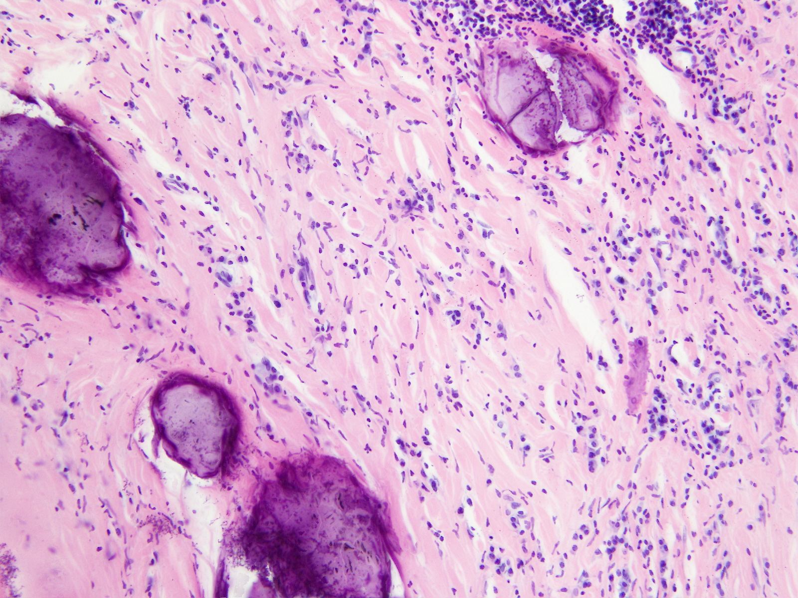
Microscopic images:


What is your diagnosis?
Diagnosis: Calcifying fibrous tumor of omentum
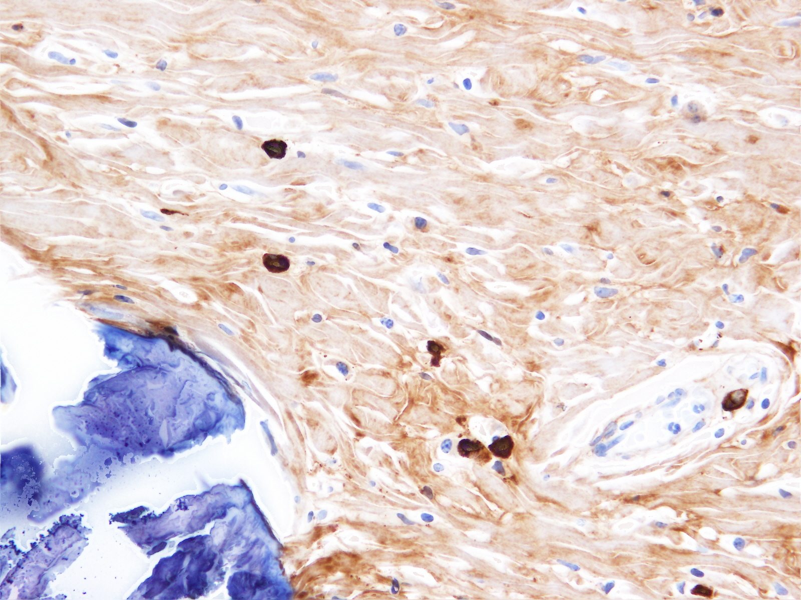
Immunostains:

Discussion:
The tumor was hypocellular with sclerotic fibrous tissue with occasional fibroblastic cells with bland nuclei. Focally, psammomatous and dystrophic calcifications were seen. Focal lymphoplasmacytic infiltrates with lymphoid aggregates were also seen.
Calcifying fibrous tumor is a soft tissue or visceral mass that occurs in children and young adults (extremities and trunk) or in adults (pleura, peritoneum, mediastinum, adrenal gland, lung). Pleural and peritoneal tumors are often multiple. There is no gender predilection.
Calcifying fibrous tumor may be due to prior inflammation and trauma. It overlaps with IgG4 related sclerosing diseases and may be related. In this case, tumor cells were occasionally IgG4 immunoreactive.
Treatment is simple excision and the prognosis is excellent but recurrence can occur.
References: J Med Case Rep 2011 Sep 28;5:487, J Cancer Res Ther 2011;7:500, Indian J Radiol Imaging 2011;21:306
All cases are archived on our website. To view them sorted by case number, diagnosis or category, visit our main Case of the Month page. To subscribe or unsubscribe to Case of the Month or our other email lists, click here.
Thanks to Dr. Beata Maksymiuk, Institute of Tuberculosis and Lung Diseases (Poland), for contributing this case.

Optimize your laboratory workflow with the
most powerful high-throughput slide scanner in anatomic pathology:
the VENTANA iScan HT scanner.
Transform your practice by delivering results faster with greater confidence:
● Gain time each day
● Improve workflow
● Engage instant remote consultation
Enhanced slide scanning and viewing is only the beginning. The expanding family of cutting-edge digital pathology solutions by Ventana empowers you to deliver the right test, to the right patient, at the right time.
Don't lose another minute of productive time click here to discover how the VENTANA iScan HT scanner can help you elevate the standard of patient care.
Website news:
(1) The Forensics chapter is now complete, written by Lindsey Harle, M.D.
(2) Our Feature Page for the month highlights Books and Journals and includes Laboratory Investigation, Lippincott, Williams & Wilkins and Modern Pathology.
Visit and follow our Blog to see recent updates to the website.
(1) The Forensics chapter is now complete, written by Lindsey Harle, M.D.
(2) Our Feature Page for the month highlights Books and Journals and includes Laboratory Investigation, Lippincott, Williams & Wilkins and Modern Pathology.
Visit and follow our Blog to see recent updates to the website.
Case #249
Clinical history:
A 32 year old woman had surgery for a gangrenous appendicitis. During surgery, a 3 x 2.5 cm pedunculated tumor was discovered in the cecal region. Grossly it was compact, well circumscribed and unencapsulated with a white whirled cut surface.
Microscopic images:
What is your diagnosis?
Click here for diagnosis and discussion:
Diagnosis: Calcifying fibrous tumor of omentum
Immunostains:
Discussion:
The tumor was hypocellular with sclerotic fibrous tissue with occasional fibroblastic cells with bland nuclei. Focally, psammomatous and dystrophic calcifications were seen. Focal lymphoplasmacytic infiltrates with lymphoid aggregates were also seen.
Calcifying fibrous tumor is a soft tissue or visceral mass that occurs in children and young adults (extremities and trunk) or in adults (pleura, peritoneum, mediastinum, adrenal gland, lung). Pleural and peritoneal tumors are often multiple. There is no gender predilection.
Calcifying fibrous tumor may be due to prior inflammation and trauma. It overlaps with IgG4 related sclerosing diseases and may be related. In this case, tumor cells were occasionally IgG4 immunoreactive.
Treatment is simple excision and the prognosis is excellent but recurrence can occur.
References: J Med Case Rep 2011 Sep 28;5:487, J Cancer Res Ther 2011;7:500, Indian J Radiol Imaging 2011;21:306
