
23 May 2012 - Case #243
All cases are archived on our website. To view them sorted by case number, diagnosis or category, visit our main Case of the Month page. To subscribe or unsubscribe to Case of the Month or our other email lists, click here.
Thanks to Dr. Saroona Haroon, The Aga Khan University Hospital (Pakistan), for contributing this case.
Advertisement
Case #243
Clinical history:
A 28 year old man presented with a firm, painless submandibular swelling for 3 months. The swelling was excised and measured 4.3 x 2.3 cm.
Microscopic images:

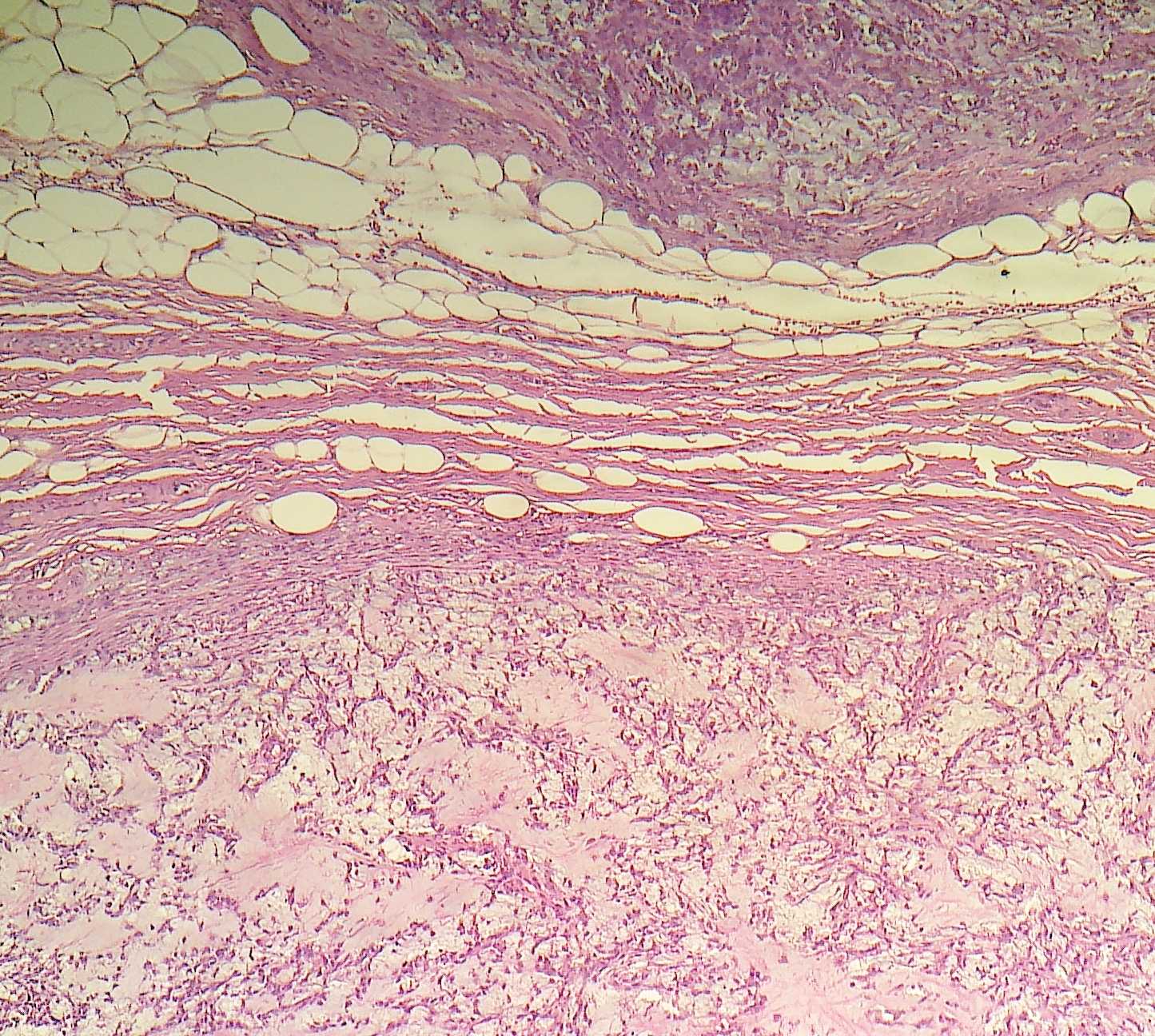
What is your diagnosis?
Diagnosis: Pleomorphic adenoma with satellite nodules
Discussion:
Pleomorphic adenoma (benign mixed tumor) is the most common tumor of salivary glands. It often occurs in women in their thirties but can present at any age as a painless, slow growing tumor. Most (90%) occur in the parotid gland, with 10% in submandibular gland and only rare tumors in the sublingual gland.
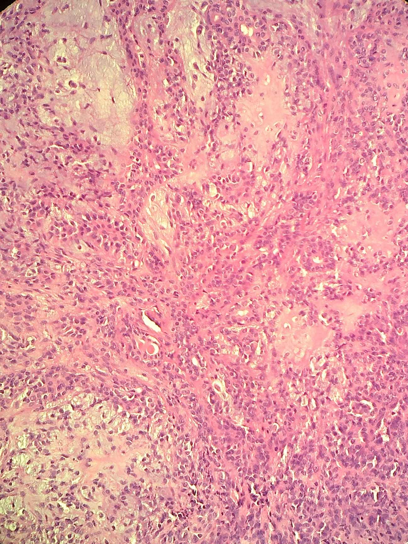
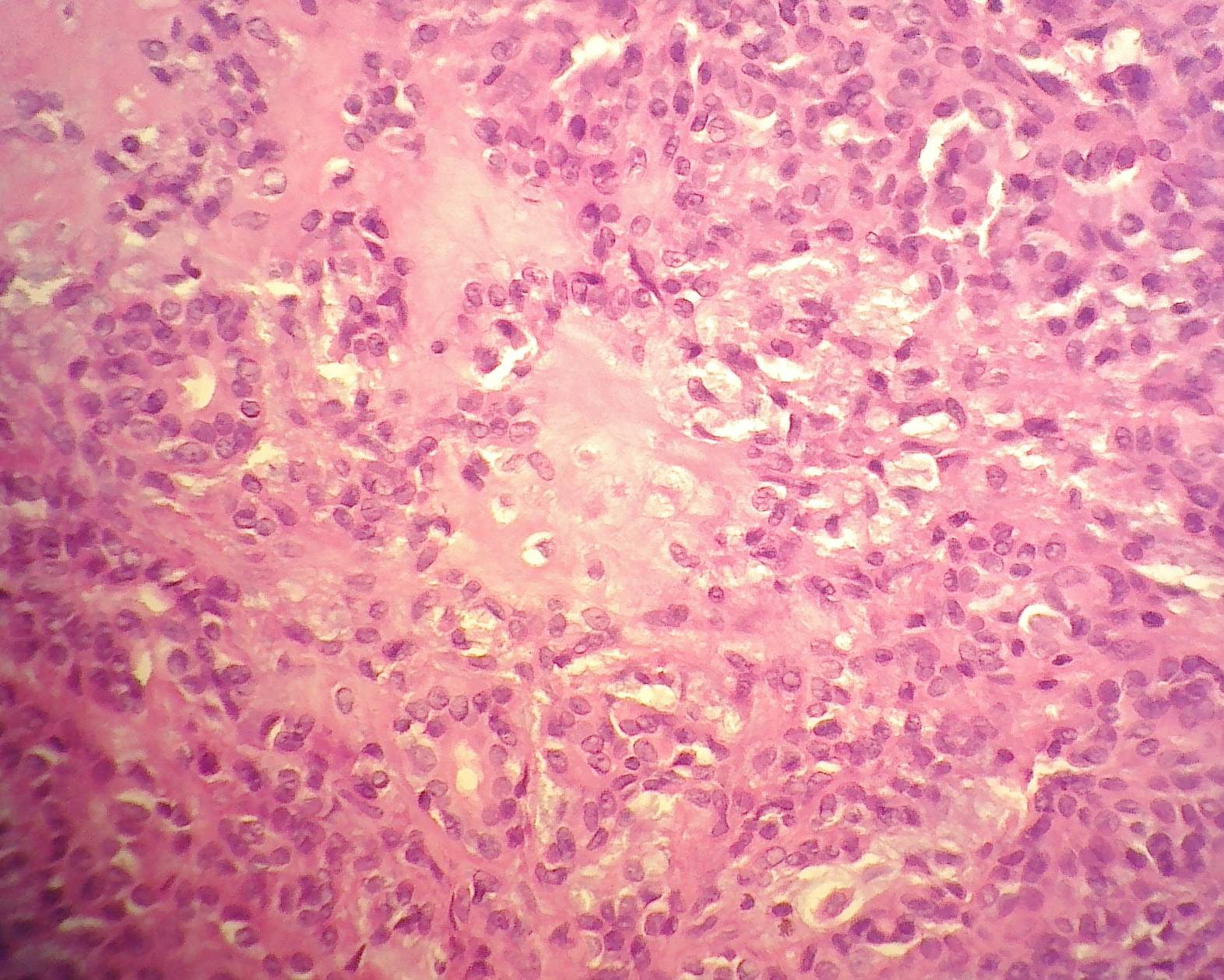
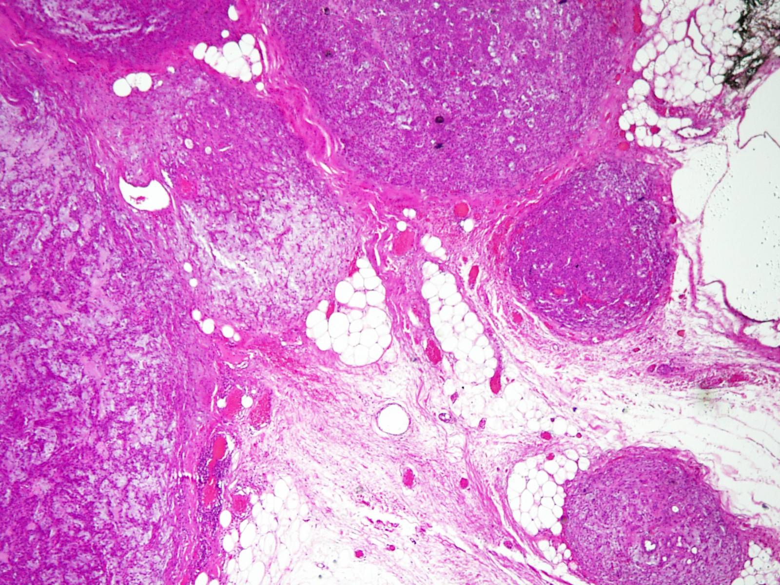
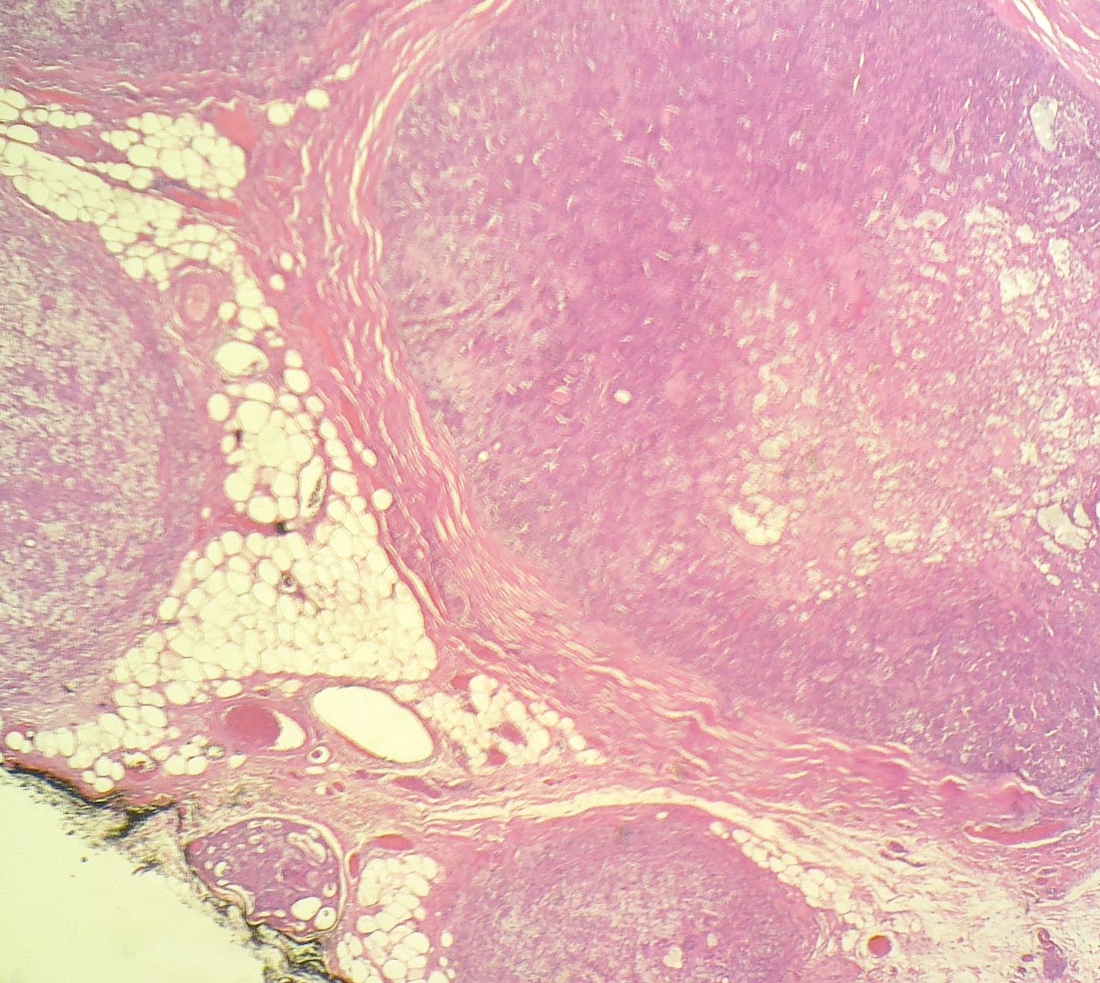
Grossly, they are well demarcated, partially encapsulated, with a gray-white cut surface. Tumor extension into adjacent tissue may be subtle. Histologically, there are often tongue-like protrusions into the surrounding salivary gland. In this case, satellite nodules were apparent. A thick capsule may be present; in this case, there were focal areas of capsular thinning. Pleomorphic adenoma is biphasic with epithelial and mesenchymal cells. The epithelial cells are usually glandular and occasionally squamous, spindle or oval, with large hyperchromatic nuclei. A myoepithelial layer may be present. The stroma is usually myxochondroid or hyaline. Mucin is often present. There is no atypia, no mitotic figures and no necrosis.
Pleomorphic adenomas of the salivary gland have been considered to be pure epithelial cell tumors arising from myoepithelial or ductal reserve cells that can produce large amounts of matrix substances (Am J Surg Pathol 1984;8:803). The epithelial and mesenchymal cells often arise from the same cell clone (Hum Pathol 2000;31:498).
Treatment has traditionally been wide local excision including all of the submandibular gland to avoid recurrent tumors, which are frequently multifocal and difficult to excise completely (Eur Arch Otorhinolaryngol 2007;264:1447). However, endoscopic resection and gland preserving surgery have recently been described (Laryngoscope 2010;120:970, Br J Surg 2008;95:1252). Risk factors for malignant transformation include a submandibular location, older age, larger size, prominent hyalinization and increased mitotic rate.
All cases are archived on our website. To view them sorted by case number, diagnosis or category, visit our main Case of the Month page. To subscribe or unsubscribe to Case of the Month or our other email lists, click here.
Thanks to Dr. Saroona Haroon, The Aga Khan University Hospital (Pakistan), for contributing this case.

A new Mouse on Mouse IHC kit (ab127055) offering you:
• The freedom and peace of mind to stain mouse tissues using mouse antibodies with no background from endogenous IgG
• Confidence to produce publication quality images the first time with a sensitive polymer detection system
• The flexibility to study biotin containing tissues with a biotin-free detection system
• Easy staining experiments with a simple and reproducible protocol
Further enhance your staining experiments with EXPOSE IHC detection systems offering reduced background and greater sensitivity than polymer detection systems. In addition, discover extensive resources to troubleshoot your IHC experiments.
Website news:
(1) Our Feature Page for the month highlights Computer software and systems, and includes Milestone Medical and Voicebrook. Also check out our new monthly Mystery Image, on the right side of the Home Page.
(2) We are constantly looking for new Reviewers for our 7,000 topics, which we want to update every 1-2 years. Visit our newly updated Author Instructions page for more information.
(3) The Table of Contents of our Stains chapter now includes all 360 stains / biomarkers described in the chapter. The CD Markers chapter contains an additional 260 markers. We update these pages regularly but let us know of any new markers we should include or other changes we should make.
Visit and follow our Blog to see recent updates to the website.
Case #243
Clinical history:
A 28 year old man presented with a firm, painless submandibular swelling for 3 months. The swelling was excised and measured 4.3 x 2.3 cm.
Microscopic images:
What is your diagnosis?
Click here for diagnosis and discussion:
Diagnosis: Pleomorphic adenoma with satellite nodules
Discussion:
Pleomorphic adenoma (benign mixed tumor) is the most common tumor of salivary glands. It often occurs in women in their thirties but can present at any age as a painless, slow growing tumor. Most (90%) occur in the parotid gland, with 10% in submandibular gland and only rare tumors in the sublingual gland.
Grossly, they are well demarcated, partially encapsulated, with a gray-white cut surface. Tumor extension into adjacent tissue may be subtle. Histologically, there are often tongue-like protrusions into the surrounding salivary gland. In this case, satellite nodules were apparent. A thick capsule may be present; in this case, there were focal areas of capsular thinning. Pleomorphic adenoma is biphasic with epithelial and mesenchymal cells. The epithelial cells are usually glandular and occasionally squamous, spindle or oval, with large hyperchromatic nuclei. A myoepithelial layer may be present. The stroma is usually myxochondroid or hyaline. Mucin is often present. There is no atypia, no mitotic figures and no necrosis.
Pleomorphic adenomas of the salivary gland have been considered to be pure epithelial cell tumors arising from myoepithelial or ductal reserve cells that can produce large amounts of matrix substances (Am J Surg Pathol 1984;8:803). The epithelial and mesenchymal cells often arise from the same cell clone (Hum Pathol 2000;31:498).
Treatment has traditionally been wide local excision including all of the submandibular gland to avoid recurrent tumors, which are frequently multifocal and difficult to excise completely (Eur Arch Otorhinolaryngol 2007;264:1447). However, endoscopic resection and gland preserving surgery have recently been described (Laryngoscope 2010;120:970, Br J Surg 2008;95:1252). Risk factors for malignant transformation include a submandibular location, older age, larger size, prominent hyalinization and increased mitotic rate.
