
Home
Case
of Week Home
Jobs
Conferences
Fellowships
Books
4 August 2011 Case of the Week #212
This email is only sent to subscribers. To subscribe or unsubscribe to this or our other email
lists, click
here.
All cases are archived on our website. To view them sorted by number, diagnosis or category,
visit our Home Page and click on the Case of the Week button on the left hand side.
Thanks to Rachiel Oakley, M.D., Ephrata Hospital (Pennsylvania, USA), for contributing this case and the discussion.
To contribute a Case of the Week, follow the guidelines on our Case of the Week page.
[#2664]
Advertisement
Website news:
(1) As we begin our 11th year, we thank you for your support. This year, we plan to increase the frequency of chapter updates, add more images and videos, and start adding smart phone / iPad apps. Let us know if you have any other comments or suggestions.
(2) We are looking for reviewers for part/all of these chapters: Coagulation, Salivary Glands, Stains, Uterus; also Fallopian Tubes, Lung-tumor, Ureters, Urethra, Vagina, Vulva. If interested, contact Liz at eapathology@gmail.com, and send a copy to NatPernick@hotmail.com.
(3) We have updated the Skin-nontumor chapter based on reviews by Ha Kirsten Do, M.D., IUPUI; Mowafak Hamodat, MB.CH.B, MSc., FRCPC, Eastern Health, St. Johns (Canada); Nat Pernick, M.D., PathologyOutlines.com, Inc. and Cecilia Rosales, M.D., Baylor College. Over the next several months, we will be adding more images and references to these topics.
Case of the Week #212
Clinical History:
A 70 year old man had an enlarging left thyroid nodule. Thyroid ultrasound demonstrated a lobulated hypoechoic solid mass with some vascularity. After a fine needle aspiration, the mass was resected.
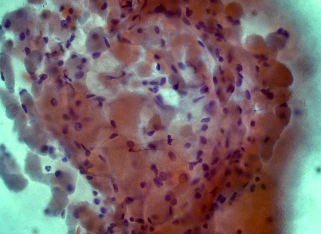
Micro images:
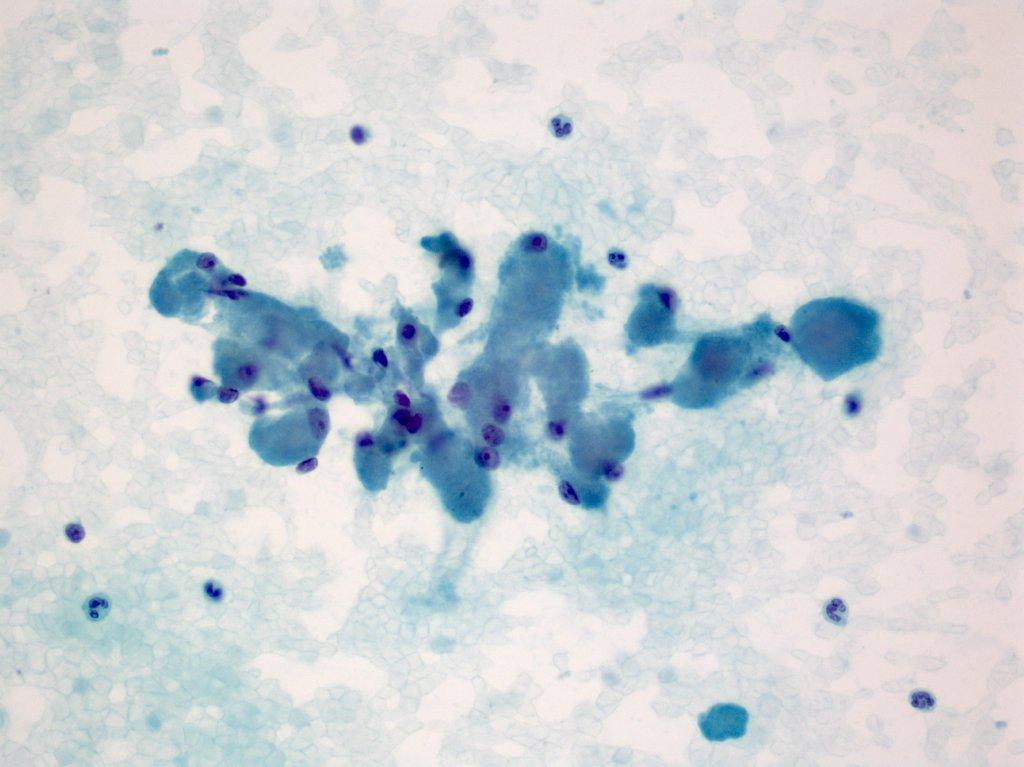
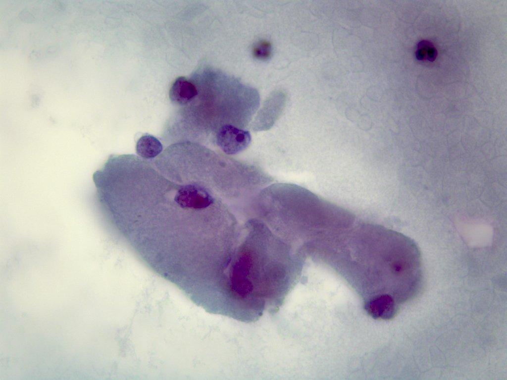

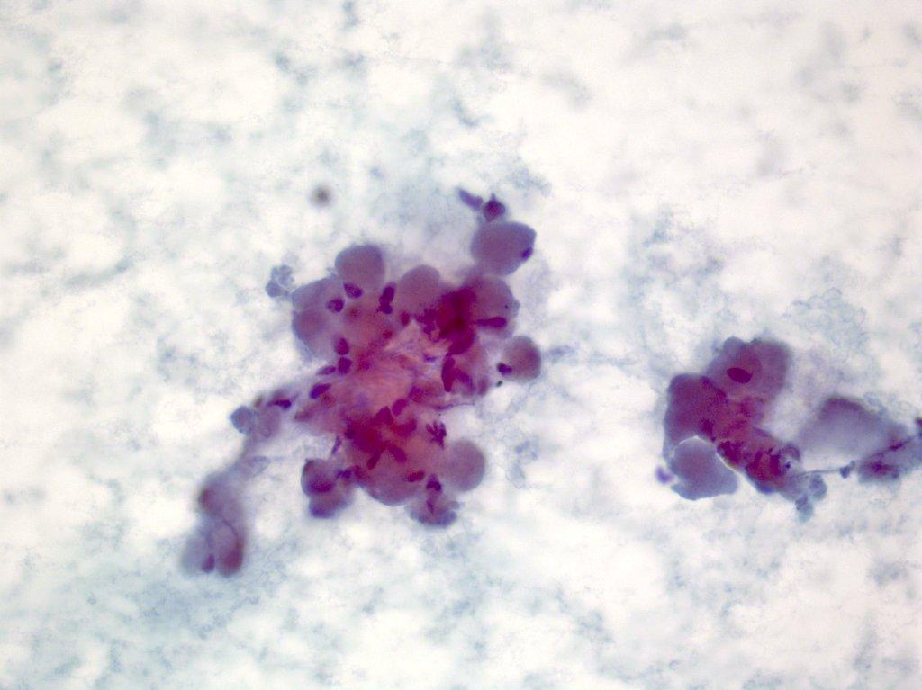
FNA - cell block on right
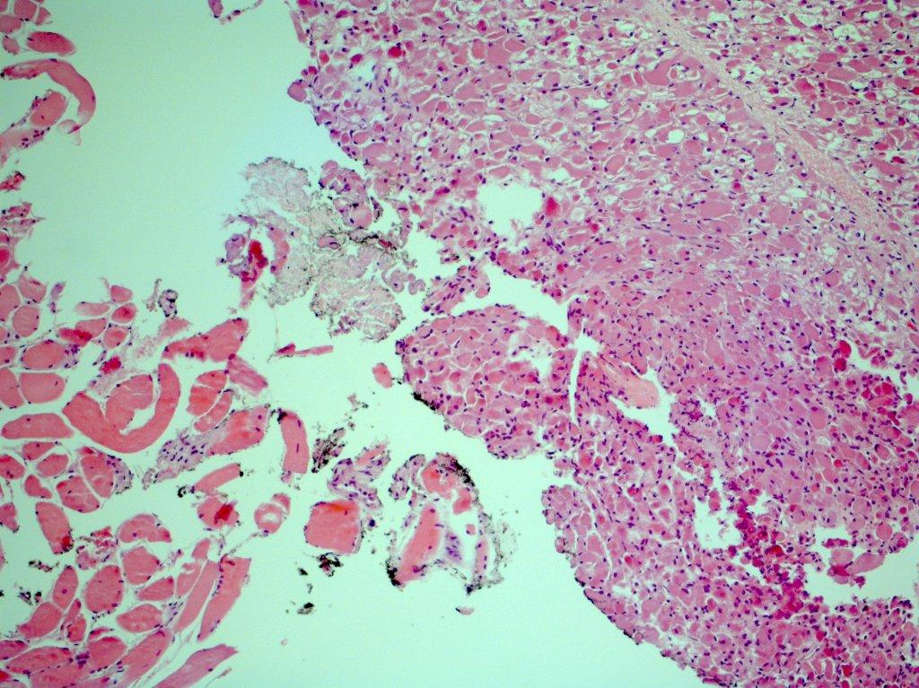
Excision of mass - H&E
What is your diagnosis?
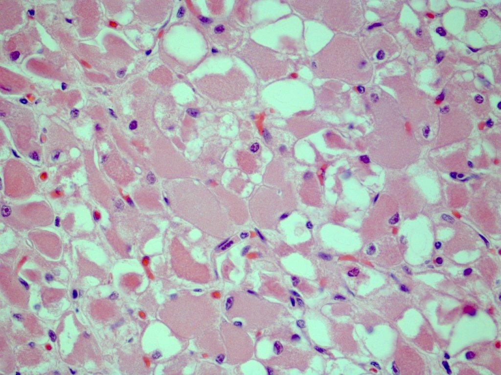
Diagnosis:
Rhabdomyoma
Discussion:
The initial FNA impression was oncocytoma (Hurthle cell neoplasm). The lesion has fibrillary eosinophilic cytoplasm but no nuclear atypia, no necrosis and no mitotic activity. The lesion was immunoreactive for desmin and actin, supporting the diagnosis of a rhabdomyoma.
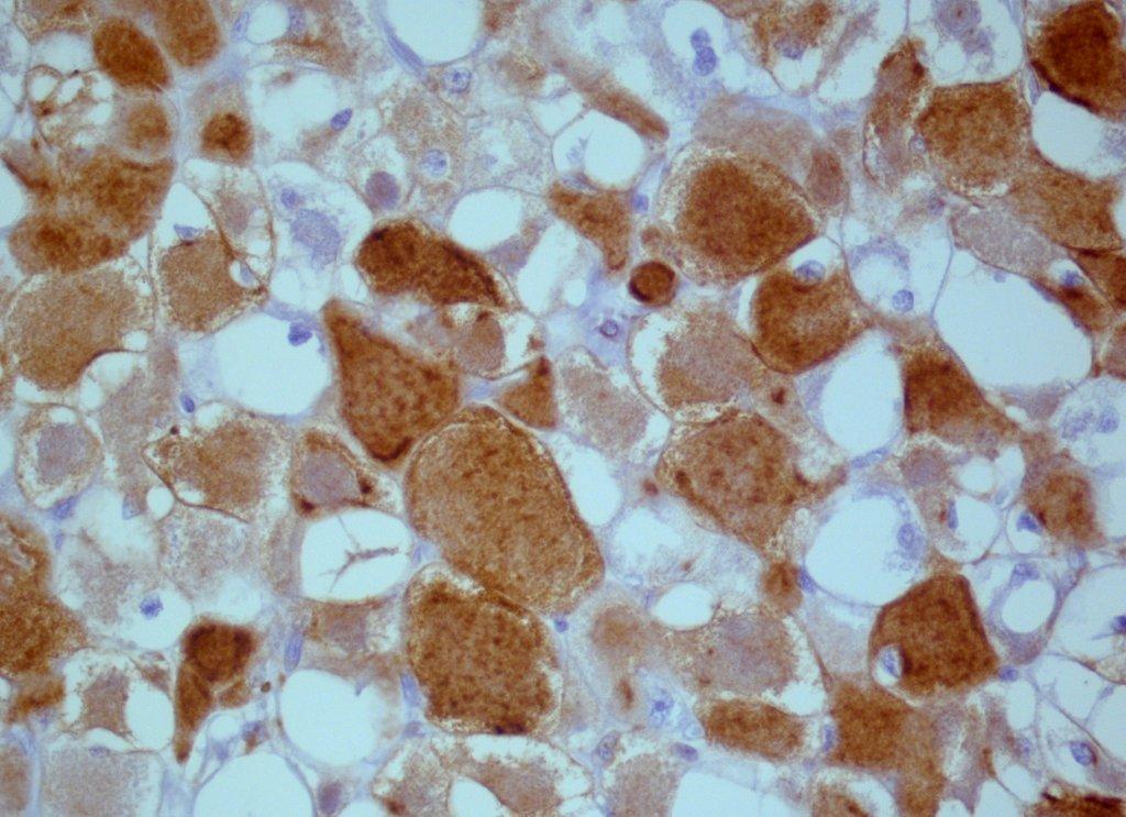
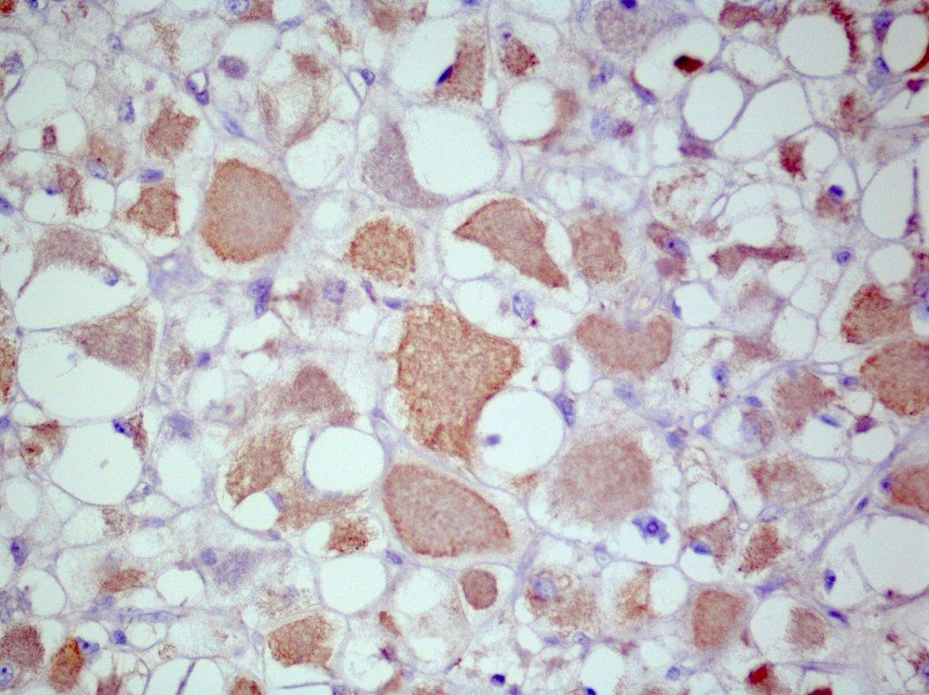
Left: desmin; right: actin
Rhabdomyomas are rare benign tumors with skeletal muscle differentiation (Eur J Pediatr Surg 2001;11:66). Rhabdomyomas are divided into cardiac and extracardiac types. Cardiac rhabdomyomas are associated with tuberous sclerosis. Extracardiac rhabdomyomas are further divided into fetal and adult types, based on the maturity of the muscle fibers. Both forms of rhabdomyomas tend to occur in the head and neck regions and in males more than females, but a genital type also occurs in the vulva and vagina of middle-aged females.
The malignant counterpart, rhabdomyosarcoma, is much more common than rhabdomyoma and is one of the more common malignant soft tissue tumors in children and young adults.
The differential diagnosis for rhabdomyoma includes granular cell tumor, hibernoma, paraganglioma and oncocytoma, but all of these are negative for muscle specific actin. Also in the differential diagnosis is muscle hypertrophy or accessory muscles (Acta Cytol 2000;44:223) but location, adjacent bone overgrowth and age can aid in the correct diagnosis.
Nat Pernick, M.D., President
and Liz Parker, B.A., Associate Medical Editor
PathologyOutlines.com, Inc.
30100 Telegraph Road, Suite 408
Bingham Farms, Michigan (USA) 48025
Telephone: 248/646-0325
Email:
NatPernick@Hotmail.com
Alternate email: NatPernick@gmail.com
