
Home
Case
of Week Home
Jobs
Conferences
Fellowships
Books
11 May 2011 Case of the Week #206
This email is only sent to subscribers. To subscribe or unsubscribe to this or our other email
lists, click
here.
All cases are archived on our website. To view them sorted by number, diagnosis or category,
visit our Home Page and click on the Case of the Week button on the left hand side.
Thanks to Ana Martinez-Peuela, Hospital de Navarra (Spain), for contributing this case.
To contribute a Case of the Week, follow the guidelines on our Case of the Week page.

Modern Pathology:
The Long Course Issue
A comprehensive update in thyroid and endocrine pathology for practicing pathologists and pathologists-in-training.
The Long Course Issue is comprised of lectures reviewing practical diagnostic and differential diagnostic issues and when appropriate, emphasizing the role of contemporary ancillary techniques. Access select articles from the Long Course Issue for the next three weeks only.
Click here for more information.
Advertisement
Website news:
(1) We are currently updating these chapters:
Bladder,
Cervix-Cytology,
Drugs of interest to pathologists and
Stains.
(2) We are always looking for reviewers of our chapters, or parts of chapters. If interested, email us your CV and the chapters / topics you are interested in reviewing. More information about reviewing is found here.
Case of the Week #206
Clinical History
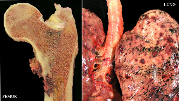
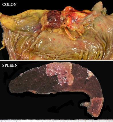
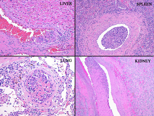
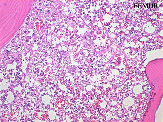
An 80 year old man was admitted for anemia and angina. Workup revealed hepatosplenomegaly and multiple splenic infarctions. He got worse, and a splenectomy was performed. Further studies showed multiple lesions in bones, the gastrointestinal tract, pancreas, liver, peritoneum, lungs and both kidneys. Two days after the surgery, the patient died. An autopsy was performed.
Gross images:
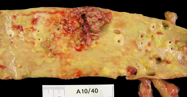

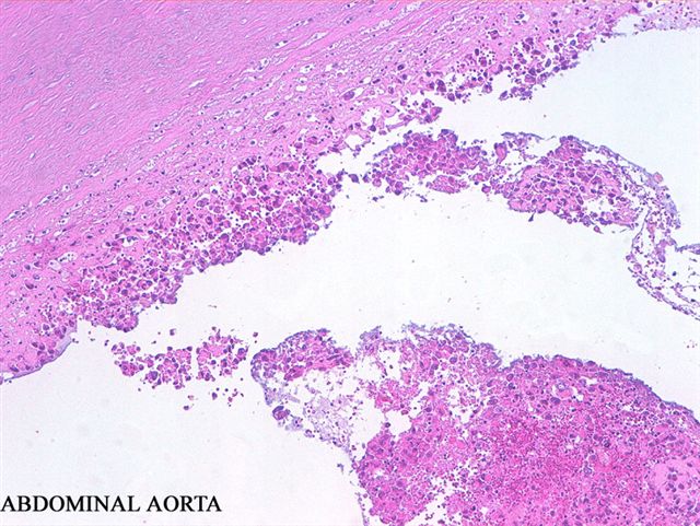
The left two images are from the abdominal aorta
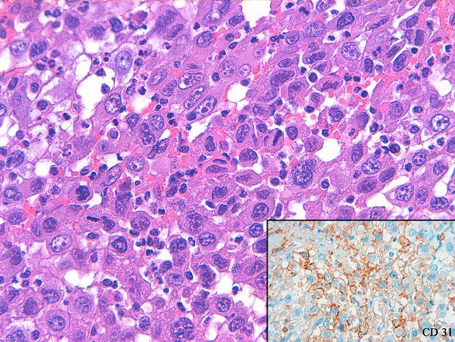
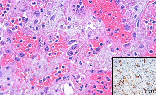
Micro images:
What is your diagnosis?
Diagnosis:
Intimal sarcoma of abdominal aorta with multiple tumor emboli and metastasis with angiosarcomatous differentiation
Discussion:
Intimal sarcoma is defined as a malignant tumor arising in the tunica intima of large blood vessels. Most cases develop in the aorta, and are undifferentiated. In undifferentiated cases, there is no gender preference, and mean age is 66 years (Am J Surg Pathol 2005;29:1184). Tumors are typically largely necrotic, with poorly differentiated epithelioid and pleomorphic cells associated with the tunica intima. Tumors are considered to be of endothelial origin, due to immunoreactivity for CD31 (Am J Surg Pathol 1988;12:798) and FLI1, with variable staining of other markers.
Metastatic tumors tend to have different morphology from the primary tumor, including angiosarcomatous (as in this case), osteosarcomatous or rhabdomyosarcomatous differentation.
Most patients have metastatic disease and die within 1 year of diagnosis. Tumors typically present with thromboemboli (J Bras Pneumol 2009;35:814), and post-mortem diagnosis is common (Hum Pathol 1997;28:1306)
Nat Pernick, M.D., President
and Liz Parker, B.A., Associate Medical Editor
PathologyOutlines.com, Inc.
30100 Telegraph Road, Suite 408
Bingham Farms, Michigan (USA) 48025
Telephone: 248/646-0325
Email:
NatPernick@Hotmail.com
Alternate email: NatPernick@gmail.com