![]()
31 August 2007 – Case of the Week #94
This and other cases can be accessed by visiting our Home Page at www.PathologyOutlines.com and clicking on the Case of the Week button on the left hand side. To view the images or references, you must click on the links in blue. Links in green are to journals with free full text-no registration.
This email is sent only to subscribers. To subscribe or unsubscribe, email info@PathologyOutlines.com, indicating subscribe or unsubscribe to Case of the Week. We do not sell, share or use your email address for any other purpose. We also have emails for Pathologist-PhD jobs (biweekly), Other laboratory jobs (biweekly), website news (monthly), new books (monthly), and a newsletter (twice a year). You must subscribe or unsubscribe separately to these email lists.
In September, we will attend the Beaumont Hospital DNA Conference in Troy, Michigan and the CAP Annual Meeting in Chicago. In October, we will attend the Wayne State University annual conference in Dearborn, Michigan and the ASCP Annual Meeting in New Orleans. Conference information for all but CAP is on our Conferences page (or click here). If you will be attending any of these conferences, stop by our booth and say hello.
We thank Dr. Juan José Segura Fonseca, Departamento de Patología, Hospital San Juan de Dios, San José, Costa Rica, for contributing this case and the discussion. To contribute a Case of the Week, please email info@PathologyOutlines.com with attachments of microscopic images (any size, we will shrink if necessary) in JPG, GIF or TIFF format, a clinical history, your diagnosis and any other images (gross, immunostains, etc.) that may be helpful or interesting. We will write the discussion (unless you want to), list you as the contributor, and send you a check for $35 (US) for your time after we send out the case. Please only send cases with a definitive diagnosis.
Case of the Week #94
Clinical History
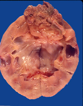
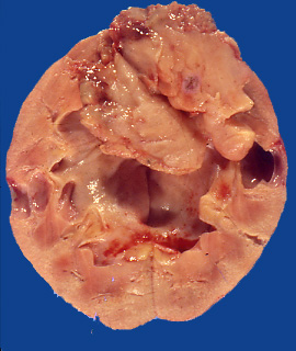
A 57 yr old Caucasian man was admitted for gross hematuria and flank pain. An intravenous pyelogram disclosed a large filling defect in the renal pelvis due to tumor. A nephrectomy was performed. On opening the kidney, a large solid tumor filled the entire renal pelvis, and was associated with dilated calyces. The tumor was polypoid and did not adhere to the inner surface of the urothelium. It was attached by a broad base to the superior border of the pelvis. It exhibited hemorrhagic necrosis and was composed of soft tan tissue (Figure 1, Figure 2).
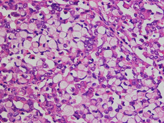
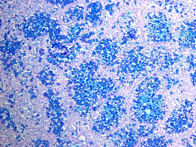
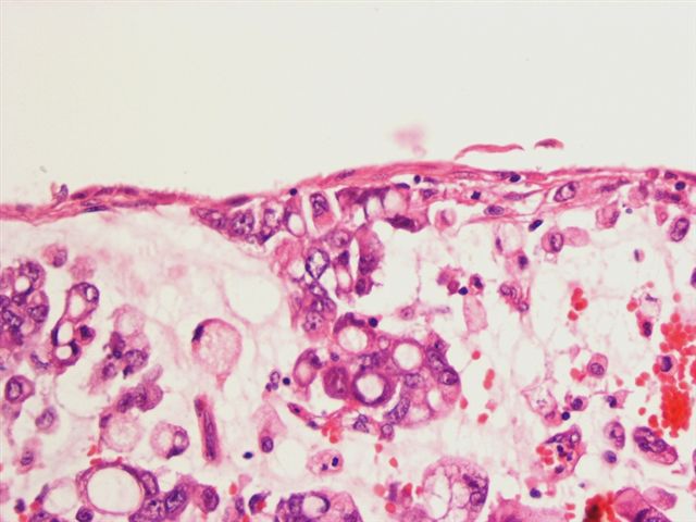
Microscopy showed numerous cells with pink, rhabdoid features and abundant intracytoplasmic lumina (Figure 3). Some areas were composed almost entirely of signet-ring cells (Figure 4) that demonstrated abundant intracytoplasmic mucin with the PAS-Alcian-blue stain (Figure 5). No areas of urothelial (transitional cell) carcinoma were identified.
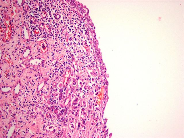
There was also severe chronic inflammation of the renal pelvis with atrophic urothelium and superficial invasion by tumor cells (Figure 6), as well as superficial goblet cell metaplasia of the urothelium (Figure 7).
What is your diagnosis?
Diagnosis:
Signet-ring cell adenocarcinoma of the renal pelvis
Discussion
Primary neoplasms of the renal pelvis are rare, and account for 7% of all renal tumors. Most are urothelial (transitional cell) carcinomas (1-3). The urothelium can display a wide variety of differentiation and metaplastic changes, particularly in high-grade neoplasms. These changes have been described in detail in the urinary bladder (4), but are less well known in the renal pelvis.
Among the high-grade carcinomas of the renal pelvis, mucinous adenocarcinoma is very uncommon, representing less than 1% of renal pelvic neoplasms (5). Most are pure adenocarcinomas with gland formation and mucin production. Only rarely are they associated with conventional urothelial carcinoma (5, 6).
Mucinous carcinomas are usually large, infiltrating tumors which fill a dilated renal pelvis and calyces, and invade the adjacent renal parenchyma (7-10). The signet ring pattern is rare (9), and may be associated with collagenous spherulosis (11). Intestinal, mucinous, cystitis cystica and goblet cell metaplasia of the urothelium, present in this case, have been implicated as precursor lesions (12).
The differential diagnosis includes high grade urothelial carcinoma, which may show foci of signet rings (3), and the rare lipoid-cell variant of urothelial carcinoma (13). The signet ring cells in both variants are mucin negative, and the tumors also have areas of usual type high grade urothelial carcinoma. Metastases from the stomach, breast or other organs must also be ruled out.
References
1. Droller MJ. Transitional Cell Carcinoma: Upper tracts and bladder. In: Walsh PC, Grittes RF, Perlmutter AD, Stamey TA (eds). Campbell´s Urology. WB Saunders: Philadelphia, 1986, pp 1343-1440
2. Olgac S, Mazumdar M, Dalbagni G, Reuter VE. Urothelial carcinoma of the renal pelvis: a clinicopathologic study of 130 cases. Am J Surg Pathol 2004;28:1545
3. Perez-Montiel D, Wakely PE, Hes O, Michal M, Suster S. High grade urothelial carcinoma of the renal pelvis: clinicopathologic study of 108 cases with emphasis on unusual morphologic variants. Mod Pathol 2006;19:494
4. Bostwick SM, Eble JN. Renal pelvis and ureter. In Bostwick DM, Eble JN (eds). Urologic Surgical Pathology. Mosby: St. Louis. Missouri (USA), 1977, pp 149-165
5. Stein A, Sova Y, Lurie M, Lurie A. Adenocarcinoma of the renal pelvis. Report of two cases, one with simultaneous transitional cell carcinoma of the bladder. Urol Int 1988;43:299
6. Takehara K, Nomata K, Eguchi J, et al. Mucinous adenocarcinoma of the renal pelvis associated with transitional cell carcinoma in the renal pelvis and the bladder. Int J Urol 2004;11:1016
7. Ueda T, Okumi M, Ichimaru N, et al. Mucinous adenocarcinoma of the renal pelvis in the horseshoe kidney: a case report. Hinyokika Kiyo 2002; 48:187
8. Watanabe S. Tanaka M, Nishizawa K, Moriguchi R. Mucinous adenocarcinoma of the renal pelvis: a case report. Hinyokika Kiyo 1997;43:727
9. Shibahara N, Okada S, Onishi S, et al. Primary mucinous carcinoma of the renal pelvis. Pathol Res Pract 1993;189:946
10. Wan J, Ohl DA, Weatherbee L. Primary mucinous adenocarcinoma of renal pelvis in solitary pelvic kidney. Urology 1993; 41:292
11. Hes O, Curik R, Mainer K, Michal M. Urothelial signet-ring cell carcinoma of the renal pelvis with collagenous spherulosis: a case report. Int J Surg Pathol 2005;13:375
12. Raghavendran M, Rastogi A, Dubey D, et al. Stones associated renal pelvic malignancies. Indian J Cancer 2003;40:108.
13. Leroy X, Gonzalez S, Zini L, Aubert S. Lipoid-cell variant of urothelial carcinoma: a clinicopathologic and immunohistochemical study of five cases. Am J Surg Pathol 2007;31:770).
Nat Pernick, M.D., President
PathologyOutlines.com, Inc.
30100 Telegraph Road, Suite 404
Bingham Farms, Michigan (USA) 48025
Telephone: 248/646-0325
Fax: 248/646-1736
Email: NPernick@PathologyOutlines.com