![]()
12 July 2007 – Case of the Week #89
These cases can also be accessed by clicking on the Case of the Week button on the left hand side of our Home Page at www.PathologyOutlines.com. To view the images or references, you must click on the links in blue. Links in green are to journals with free full text-no registration.
This email is sent only to subscribers. To subscribe or unsubscribe, email info@PathologyOutlines.com, indicating subscribe or unsubscribe to Case of the Week. We do not sell, share or use your email address for any other purpose. We also have emails for Pathologist jobs (biweekly), non-pathologist laboratory jobs (biweekly), website news (monthly), new books (monthly), and a newsletter (twice a year). You must subscribe or unsubscribe separately to these email lists.

Visit our New Products and Services page regularly (from the Home Page, click on the button on the left hand side that says New Products or click on the banner above). This page has announcements of interest to pathology personnel, and includes pictures, links and contacts. This is a great way to stay up to date.
We thank Dr. Julie E. Vitko, Southbay Hospital, SunCity Center, Florida (USA), for contributing this case. To contribute a Case of the Week, please email info@PathologyOutlines.com with attachments of microscopic images (any size, we will shrink if necessary) in JPG, GIF or TIFF format, a clinical history, your diagnosis and any other images (gross, immunostains, etc.) that may be helpful or interesting. We will write the discussion (unless you want to), list you as the contributor, and send you a check for $35 (US) for your time after we send out the case. Please only send cases with a definitive diagnosis.
Case of the Week #89
Clinical History
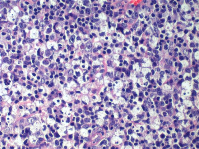
A 22 year old Caucasian woman presented with right cervical adenopathy. Her medical history was otherwise unremarkable. A lymph node was excised.
Micro Images: image #1; #2; #3; #4; #5; #6
AFB and GMS stains were negative for fungi or microorganisms. Serology was negative for toxoplasmosis, EBV or cat-scratch disease.
What is your diagnosis?
Diagnosis:
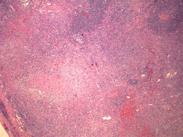
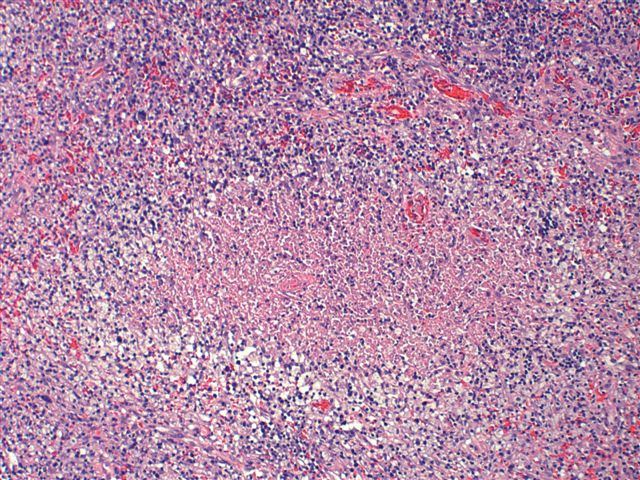
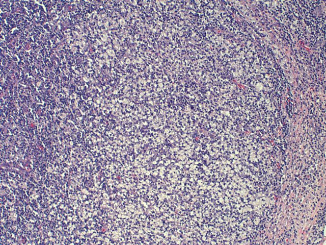
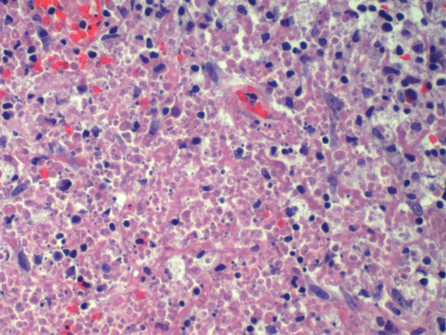
Kikuchi-Fujimoto’s lymphadenopathy
Discussion
This disorder, also called histiocytic necrotizing lymphadenitis and Kikuchi’s disease, was initially described in Japan and other Asian countries, but now is found worldwide. It primarily affects young women, who present with cervical lymphadenopathy (tender or painless), fever and often leukopenia. The etiology is unknown, although the prominent necrosis is due to apoptosis mediated by cytotoxic T cells (Mod Path 1997;10:231)
The microscopic picture is dominated by well circumscribed necrotic lesions, often with a starry sky appearance. There is also karryorhexis, fibrin deposits, phagocytic and foamy histiocytes, plasmacytoid monocytes (two to three times larger than small lymphocytes with variable cytoplasm, round nuclei with open chromatin, small nucleoli, CD4+) and T cells (CD8+, cytotoxic phenotype). There are no/rare plasma cells, neutrophils, follicular hyperplasia or atypia. Occasionally, there is no overt necrosis (AJSP 1990;14:514). Fine needle aspiration shows phagocytic histiocytes with peripheral nuclei and plasmacytoid monocytes.
The differential diagnosis includes lymphoma with necrosis, cat-scratch disease and lupus lymphadenitis. Lymphoma may have prominent necrosis, but atypical cells should be present, and plasmacytoid monocytes are lacking. Cat-scratch disease, caused by Bartonella henselae, has a clinical history of cat exposure with an initial skin papule. Lymph nodes show prominent neutrophils and follicular hyperplasia, not present in Kukuchi’s disease. Lupus lymphadenitis has paracortical necrosis and hematoxylin bodies, as well as a clinical history of lupus (Hum Path 1989;20:295).
Kikuchi’s disease is usually benign and self-limited (Ann Saudi Med 2005;25:319), although 3% of cases recur (AJSP 1995;19:798).
Additional references: AJSP 1994;18:219, Hum Path 1993;24:1114 (plasmacytoid monocytes), J Clin Pathol 1985;38:1252, eMedicine
Nat Pernick, M.D., President
PathologyOutlines.com, Inc.
30100 Telegraph Road, Suite 404
Bingham Farms, Michigan (USA) 48025
Telephone: 248/646-0325
Fax: 248/646-1736
Email: NPernick@PathologyOutlines.com