![]()
3 May 2007 Case of the Week #82
These cases can also be accessed by clicking on the Case of the Week button on the left hand side of our Home Page at www.PathologyOutlines.com. This email is sent only to subscribers. To view the images or references, you must click on the links in blue.
To subscribe or unsubscribe, email info@PathologyOutlines.com, indicating subscribe or unsubscribe to Case of the Week. We do not sell, share or use your email address for any other purpose. We also have emails for Pathologist jobs (biweekly), Other Laboratory jobs (biweekly), website news (monthly), new books (monthly), and a newsletter (twice a year). You must subscribe or unsubscribe separately to these email lists.

Peloris rapid tissue processing
This case is sponsored by Vision BioSystems and its Peloris rapid tissue processing. Peloris is a rapid tissue processor that creates high quality results for any tissue. It has a xylene-free option and a unique ActivFlo system that generates ideal processing conditions. A range of Peloris consumables adds extra value with cassettes that eliminate biopsy pads, solid Parablocks wax for easy, safe wax transfer, and reagents to completely eliminate xylene. With Peloris, any laboratory can enjoy the confidence of high quality results while meeting new workflow challenges like reduced turnaround times, Lean production and same day diagnosis.
Peloris is part of Vision BioSystems Lean Histology range and it is ideal for laboratories pursuing Lean and six sigma principles.
For more information on the Peloris system, visit our website www.vision-bio.com.
Visit our Stains chapter for our recently expanded coverage of all of the cytokeratins, with hundreds of new references and image links. We also recently added a new article on "Pricing for Pathology billing" to the Management chapter.
We thank Dr. Ankur Sangoi, Stanford University, Stanford, California (USA) for contributing this case. We invite you to contribute a Case of the Week by emailing info@PathologyOutlines.com with microscopic images (any size, we will shrink if necessary) in JPG or TIFF format, a clinical history, your diagnosis and any other images (gross, immunostains, EM, etc.) that may be helpful or interesting. We will write the discussion (unless you want to), list you as the contributor, and send you a check for $35 (US dollars) for your time after we send out the case. Please only send cases with a definitive diagnosis.
Case of the Week #82
Clinical History
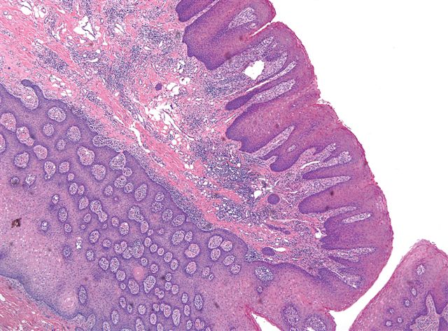
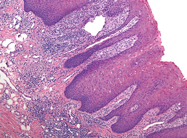
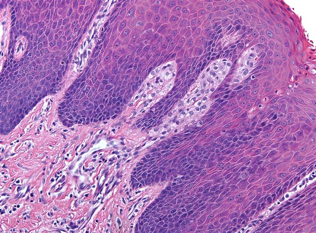
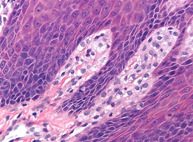
A 57 year old man had papillary lesions on his palate, which were excised:
Micro images: image #1; #2; #3; #4
What is your diagnosis?
Diagnosis:
Verruciform xanthoma
Discussion
Verruciform xanthoma is a rare lesion of the oral mucosa that usually occurs in the gingiva or alveolar ridge. It occurs less commonly on the penis or scrotum. It is considered reactive (AJSP 1998;22:479), although a multifocal lesion was reported in a child with a systemic lipid disorder (AJSP 1989;13:309). It is probably not HPV related (APMIS 2005;113:629, Archives 2005;129:e62, but see Am J Dermatopathol 2000;22:447).
Grossly, verruciform xanthoma appears as a raised granular or verrucous lesion. Histologically, the characteristic finding is foamy macrophages within dermal papillae. The foam cells are immunoreactive for CD68, vimentin and Factor XIIIa, and negative for S100 and weak/negative for keratin. The overlying epidermis is either verruciform, papillary or flat (Oral Oncol 2003;39:325). No epithelial atypia is present.
The differential diagnosis includes squamous cell carcinoma, verrucous carcinoma and condyloma acuminatum. All of these entities lack prominent foamy macrophages. In addition, the lack of atypia rules out squamous cell carcinoma. Verrucous carcinoma is composed of lobules of mature squamous epithelium with minimal atypia, but they are ulcerating or fungating. Condyloma acuminatum has prominent koilocytosis in the upper epidermis that is not found in verruciform xanthoma.
Excision is adequate treatment, and these tumors do not recur (J Formos Med Assoc 2007;106:141).
Nat Pernick, M.D., President
PathologyOutlines.com, Inc.
30100 Telegraph Road, Suite 404
Bingham Farms, Michigan (USA) 48025
Telephone: 248/646-0325
Fax: 248/646-1736
Email: NPernick@PathologyOutlines.com