![]()
12 April 2007 – Case of the Week #80
These cases can also be accessed by clicking on the Case of the Week button on the left hand side of our Home Page at www.PathologyOutlines.com. This email is sent only to subscribers. To view the images or references, you must click on the links in blue.
To subscribe or unsubscribe, email info@PathologyOutlines.com, indicating subscribe or unsubscribe to Case of the Week. We do not sell, share or use your email address for any other purpose. We also have emails for new Pathologist jobs (biweekly), new Laboratory but not pathologist jobs (biweekly), website news (monthly), new books (monthly), and a newsletter (twice a year). You must subscribe or unsubscribe separately to these email lists.
In response to many inquiries, we will never charge our users or require registration for PathologyOutlines.com. We believe the Internet should be free to all, and in fact, are trying to convince more pathology journals to make their content “free full text” without registration, at least for the older issues. We recently started to emphasize references to journals with free full text-no registration, which are highlighted in green.
Share your tips on using PathologyOutlines.com by entering our Contest (click on the Contests button on the left side of the Home Page) to win $50 for the best tip.
We thank Dr. Alia Albawardi, Faculty of Medicine & Health Sciences, United Arab Emirates University (UAE), for contributing this case and some of the discussion. We invite you to contribute a Case of the Week by emailing info@PathologyOutlines.com with microscopic images (any size, we will shrink if necessary) in JPG format, a clinical history, your diagnosis and any other images (gross, immunostains, EM, etc.) that may be helpful or interesting. We will write the discussion (unless you want to), list you as the contributor, and send you a check for $35 (US dollars) for your time after we send out the case. Please only send cases with a definitive diagnosis.
Case of the Week #80
Clinical History
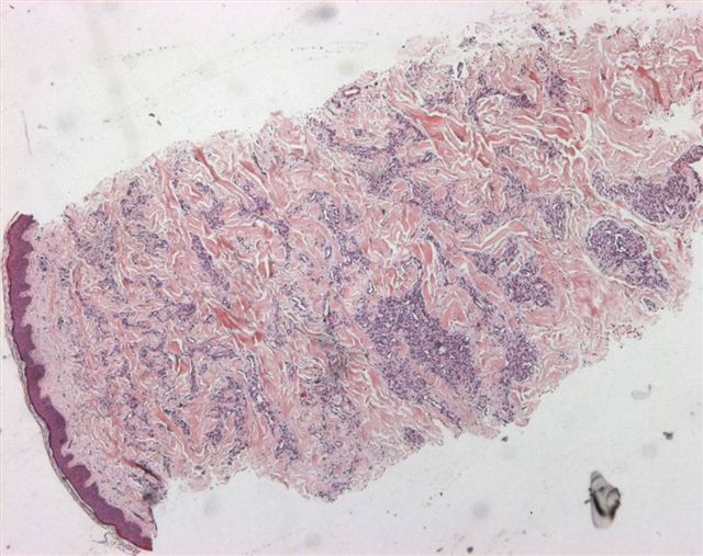
A 28 year old man had a red nodule on the left thigh for 6 months that was slowly growing. The clinical suspicion was Kaposi’s sarcoma.
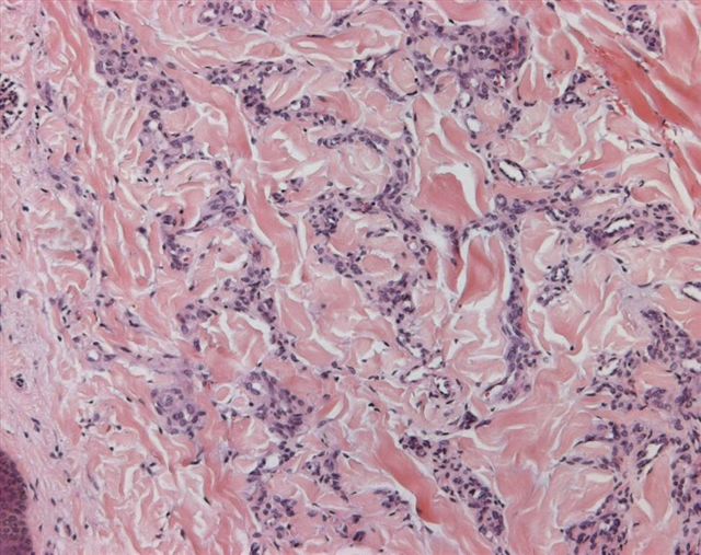
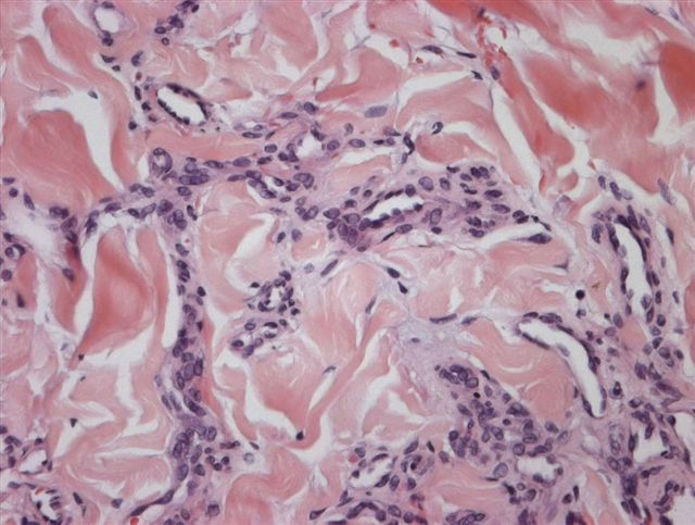
Micro images: image #1; #2; #3
What is your diagnosis?
Diagnosis:
Microvenular hemangioma
Discussion
Microvenular hemangioma was first described by Hunt in 1991 (J Cutan Pathol 1991;18:235). It is rare, with less than 50 cases reported. It presents as a slow growing, solitary, asymptomatic, purple-red papule or plaque in young to middle-aged adults, often on the trunk or limbs. The histology shows a dermal proliferation of small, irregular branching capillaries and venules with inconspicuous lumina. Endothelial cells may be plump, but no atypia is present. The stroma is collagenous. No spindle cells are present.
The endothelial cells are immunoreactive for CD34 and Factor VIII related antigen. Smooth muscle actin highlights pericytes surrounding the endothelial cells.
The main differential diagnosis is patch stage Kaposi's sarcoma, which also has irregular vascular spaces. However, they are anastomosing and not collapsed, and are accompanied by atypical endothelial cells, eosinophilic hyaline globules, plasma cells and fascicles of spindle cells. There may be irregular dissection of collagen bundles by vessels. The spindle cells are HHV8 positive (Am J Clin Pathol 2004;121:335), and the patients are HIV positive, which is usually helpful. However, there was a recent report of HHV8 positive microvenular hemangioma (image-see fig c, d) in an unusual case of POEMS syndrome (Arch Pathol Lab Med 2003;127:1034)
Kaposiform hemangioendothelioma also has slit-like lumina, although they are due to nodules and sheets of compact spindle cells. It affects the skin or retroperitoneum of infants and children, and may be associated with severe coagulopathy.
Microvenular hemangioma is a benign lesion. To date, excision appears to be curative.
Case reports: 40 year old woman (Dermatology 2003;206:161), 23 year old Japanese woman (Pathol Int 1998;48:237)
Nat Pernick, M.D., President
PathologyOutlines.com, Inc.
30100 Telegraph Road, Suite 404
Bingham Farms, Michigan (USA) 48025
Telephone: 248/646-0325
Fax: 248/646-1736
Email: NPernick@PathologyOutlines.com