![]()
9 March 2007 Case of the Week #76
These cases can also be accessed by clicking on the Case of the Week button on the left hand side of our Home Page at www.PathologyOutlines.com. This email is sent only to those who subscribe in writing or by email. To view the images or references, you must click on the links in blue.
To subscribe or unsubscribe, email info@PathologyOutlines.com, indicating subscribe or unsubscribe to Case of the Week. We do not sell, share or use your email address for any other purpose. We also have a biweekly email of new Pathologist jobs added to our Jobs pages, a biweekly email of new Laboratory jobs added to our Jobs pages, and a monthly email of news about the website. You must subscribe or unsubscribe separately to these email lists.
We just added two new Email lists - to receive a list of new pathology books once a month and to receive our newsletter twice a year. The new books email will list new books added to our books pages with links to more information. It will also describe book discounts. To subscribe, email info@pathologyoutlines.com with subscribe to books email in the subject line. Our newsletter will be sent out by email or regular mail, initially twice a year, and will contain descriptions of website features, tips and other helpful information. To subscribe, email info@pathologyoutlines.com with either subscribe to newsletter by email or subscribe to newsletter by regular mail. For the latter, please include a mailing address.
We thank Dr. J. Ross Slemmer, SkyRidge Medical Center, Cleveland, Tennessee (USA) for contributing this case. We invite you to contribute a Case of the Week by emailing NPernick@PathologyOutlines.com with microscopic images (any size, we will shrink if necessary) in JPG format, a clinical history, your diagnosis and any other images (gross, immunostains, EM, etc.) that may be helpful or interesting. We will write the discussion (unless you want to), list you as the contributor, and send you a check for $35 (US) for your time after we send out the case. Please only send cases with a definitive diagnosis.
Case of the Week #76
Clinical history
A 49 year old man had a left inguinal mass. A CT scan revealed bulky retroperitoneal lymphadenopathy, and his serum PSA was 6.5. Serum CA125 was not obtained. He had a two year history of sporadic hematuria, but cystoscopy showed only a slight irregularity or granularity of the prostatic urethra, and his serum PSA was normal.
At pelvic ultrasound, the urologist reported that he could not identify the prostate, which was apparently obliterated by a neoplastic process. He obtained blind core biopsies for frozen section to determine if he was in or near the prostate. An enlarged femoral lymph node was also excised and sent for frozen section.
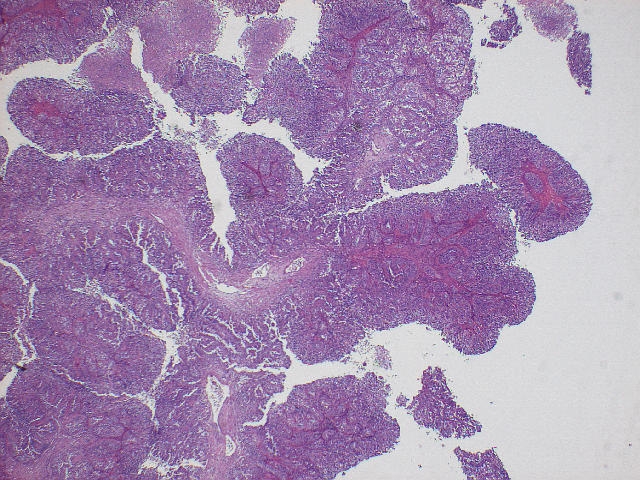
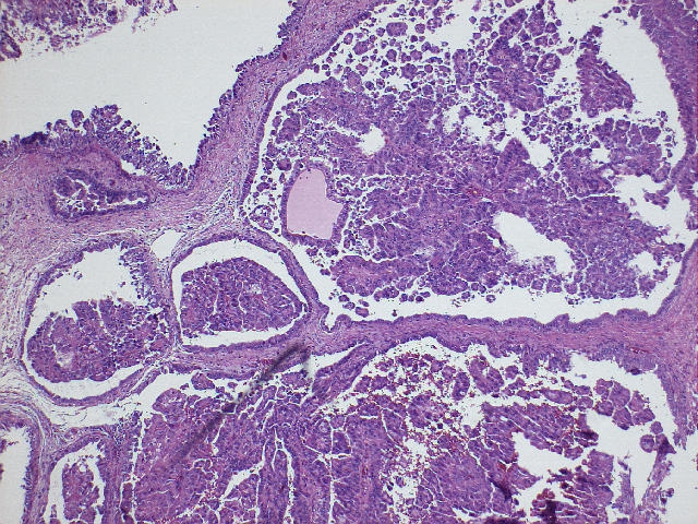
Micro images: low power - #1; #2
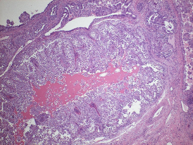
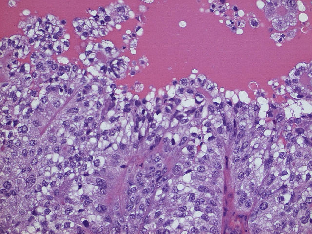
high power - #5; #6; #7; #8; #9; #10; #11; #12; #13
What is your diagnosis?
(scroll down to continue)
Diagnosis:
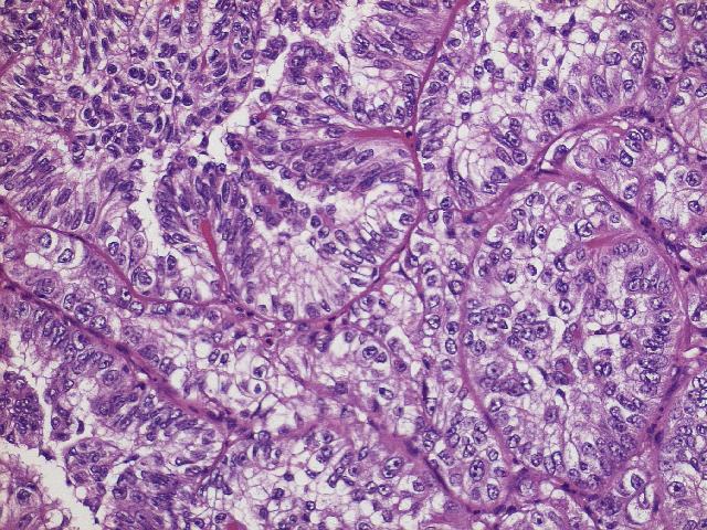
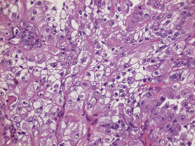
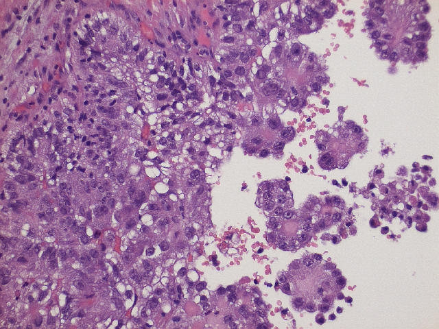
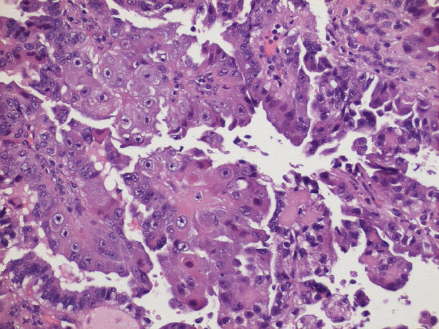
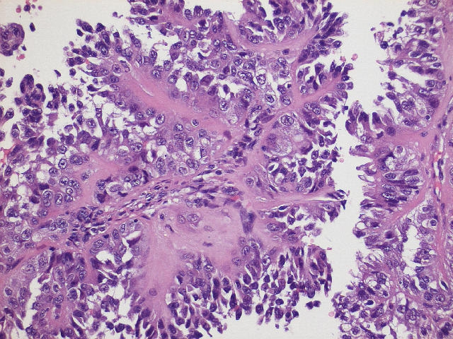
Clear cell adenocarcinoma of prostate with tubulocystic growth and metastasis to femoral lymph node
Discussion
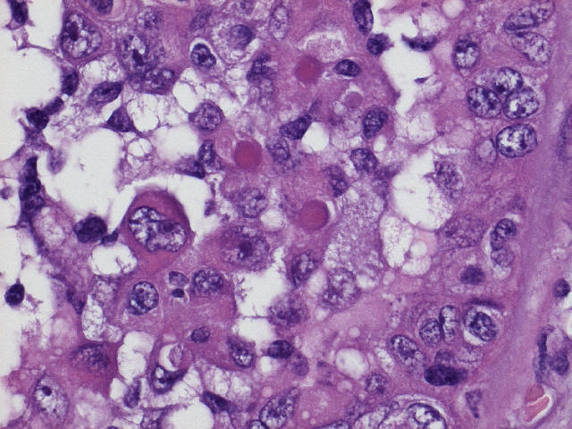
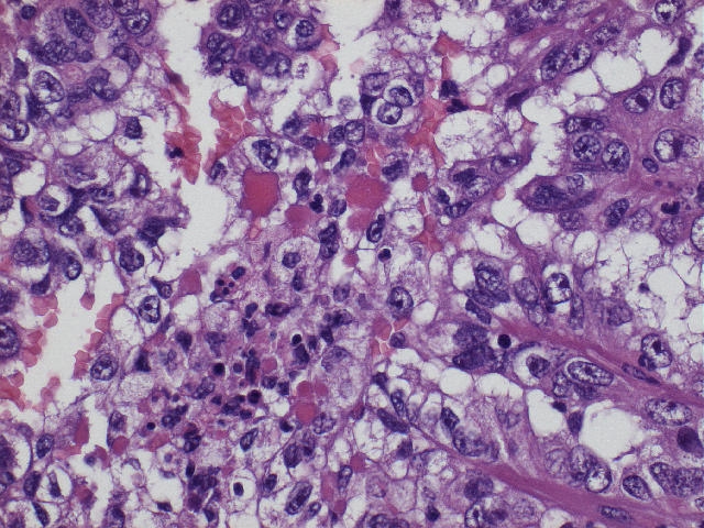
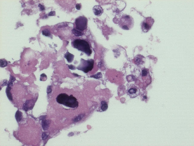
This is a very rare tumor of the prostate, whose diagnosis was confirmed by Dr. Jonathan Epstein. A similar case was reported from Taiwan, published by Dr. Epstein et. al. (AJSP 2000;24:1433). In both cases, the tumor was composed of papillary and tubulocystic glands lined by cuboidal and hobnail cells with clear to eosinophilic cytoplasm. The nuclei were mildly pleomorphic and had prominent nucleoli. Psammoma bodies were occasionally seen (image #13). There was no/minimal mitotic activity. Hyaline globules were present in this case (image #11; #12), but not reported in the published case.
The tumor cells in the published case were immunoreactive for pan-cytokeratin, low molecular weight cytokeratin and EMA, and focally positive for high molecular weight keratin (34betaE12). They were negative for PSA and PAP. The patient in the published case had an elevated serum CA125, but normal serum PSA and CEA.
Clear cell adenocarcinomas, although common in women, are very rare in the urinary tract of men, and occur predominantly in the urethra or urinary bladder. Other reported cases in the prostate include a clear cell carcinoma of the prostatic utricle in a 16 year old boy reported from Uruguay (Ann Diagn Pathol 2005;9:153) and a renal cell type of prostatic carcinoma which lacked hobnail cells and had staining patterns similar to renal cell carcinoma (AJSP 2003;27:407).
The differential diagnosis is limited. Some prostatic ductal carcinomas or Gleason pattern 4 adenocarcinomas have focal papillary formation. However, there are no tubulocystic or papillary structures lined by clear or hobnail cells. These tumors also have the usual staining pattern of PSA+, PAP+ for prostatic adenocarcinoma. Other clear cell tumors include nephrogenic adenoma (Hum Path 1994;25:390), clear cell urothelial carcinoma and metastatic renal cell carcinoma.
Nat Pernick, M.D., President
PathologyOutlines.com, Inc.
30100 Telegraph Road, Suite 404
Bingham Farms, Michigan (USA) 48025
Telephone: 248/646-0325
Fax: 248/646-1736
Email: NPernick@PathologyOutlines.com